Use of flow cytometry to develop and characterize a set of monoclonal antibodies specific for rabbit leukocyte differentiation molecules
16
0
0
전체 글
(2) 52 William C. Davis et al.. mAbs submitted to the Animal Homologues Section of the HLDA8 have been specific for major histocompatibility (MHC) I and II molecules, CD7, CD9, CD14, CD21, CD11a, CD18, CD44, CD45RB, CD49d, CD209 [54]. In light of these findings, it is apparent that a more direct approach will be required to identify mAbs for research in rabbits. As part of our continued effort to develop mAbs critical to our research efforts in ruminants, we have developed a flow cytometric approach for initial identification and characterization of mAbs to LDM [11]. Previous studies have shown that two parameter single fluorescence flow cytometry can be used to cluster mAb that recognize the same or different epitopes on the same LDM, based on the pattern of expression of the molecule on one or more lineages of leukocytes [11,16,35]. Comparative studies have shown this method can also be used to identify and tentatively cluster mAbs that recognize epitopes on orthologous LDM based on the similarity of the pattern of expression of the LDM on leukocytes in different species. Our studies have revealed the pattern of expression of many orthologous LDM has been conserved cross species. This observation has proven useful, especially in the characterization of mAbs specific for LDM in less well studied species [13-15,59,60]. It has also proven useful in determining whether mAbs that cross react with LDM in one or more species recognize an epitope conserved on bona fide LDM orthologues. Specificity has also been documented by cloning and expression of LDM initially identified with cross reactive mAbs [59]. To aid others as well as ourselves, we have also developed a web based program, the Taxonomic Key Program (TKP; College of Veterinary Medicine, Washington State University, USA), to facilitate characterization of mAbs generated against LDM in less well studied species. The program contains a searchable database on known CD molecules and a database containing a catalog of mAbs known to react with LDM in one or more of the less well studied species. In the present report we summarize the results we have obtained thus far, in our efforts to develop mAbs for use in immunological investigations in the rabbit. Information on the mAbs recognizing rabbit LDMs are listed under reactivity of antibodies in the TKP program (NCBI Rabbit Genome Resources, USA).. Materials and Methods Animals Rabbits being used in other studies were used as a source of blood and tissues. They were housed and maintained according to the Institutional Animal Care and Use committee guidelines and Association for Assessment and Accreditation of Laboratory Animal Care (USA). Both male and female rabbits were used since initial studies did not reveal any apparent differences in the frequency of. leukocyte subsets. The age of the rabbits varied from six months to about two years.. Preparation of leukocytes for flow cytometry Because of the tendency for T lymphocytes to bind to erythrocytes, separation medium could not be used to isolate leukocytes. Whole blood, collected in anti- coagulant citratedextrose (ACD), was used with a fix-lyse solution to obtain leukocytes for analysis. For single color flow cytometry (FC), 50 µl of blood was distributed in conical bottom 96-well microtiter plates (Corning, USA) containing 50 µl of optimally diluted mouse mAbs and then incubated for 15 min on ice. Following centrifugation, the supernatants were removed by aspiration. The lymphocytes were subjected to 3 cycles of centrifugation and washing in FC first wash buffer (FWB, PBS co ntaining 20% ACD and 0.5% horse serum) and then incubated with a second step fluorescein conjugated polyclonal goat anti-mouse IgG/IgM second step reagent (Caltag Laboratories, USA) for an additional 15 min. Following 2 cycles of centrifugation and washing in FC second wash buffer (PBS-20% ACD) the lymphocytes were resuspended in FACS lysing solution (Becton Dickinson, USA) to lyse erythrocytes. The lymphocytes were then centrifuged and resuspended in 2% PBSbuffered formaldehyde and kept in the refrigerator until examined. For multi-color FC, blood was distributed in microtiter plates containing 2 or 3 mAbs and incubated as described. Following centrifugation and 3 cycles of washing, the lymphocytes were incubated with second step reagents. For most of the studies, combinations of mAbs of different isotype were used with isotype specific goat anti-mouse immunoglobulins conjugated with fluorescein (FL), phycoerythrin (PE), PE-Cy5, or Cy5 (Caltag Laboratories, USA). Where the mAbs of interest were the same isotype, Zenon Fab fragments of goat isotype specific anti-mouse antibody, conjugated with different fluorochromes, were used according to the manufacturers' instructions (Invitrogen, USA). One µg of each mAb in 20 µl of FWB were incubated separately with 5 µl of Zenon-Fab reagent conjugated with different fluorochromes (FL, PE, PE-Cy5, or Cy5) for 5 min at room temperature as recommended by the manufacturers. The mixtures were then incubated with 5 µl of blocking reagent (mouse immunoglobulin) for an additional 5 min. The labeled antibodies were then added to the lymphocyte preparations under study. Following 15 min of incubation on ice, the lymphocytes were processed as described and fixed in 2% buffered formaldehyde. Peripheral blood mononuclear lymphocytes (PBMC) and spleen lymphocytes stimulated with concanavalin A (ConA) were used for immunization and identification of mAbs that recognize molecules upregulated on activated lymphocytes (rabbit activation molecules, RACT). To simplify initial screening of supernatants from primary cultures of hybridomas for the presence of a mAb that.
(3) Monoclonal antibodies specific for rabbit leukocyte differentiation molecules. recognizes a RACT, spleen lymphocytes stimulated with ConA (5 µg/ml) for 24 to 48 h were incubated with hydroethidine (250 µg/ml in tissue culture medium), a vital dye that is selectively taken up by live cells. Hydroethidine (Polysciences, USA) intercalates into DNA similar to propidium iodide. It is excited at 488 nm and emits at high wave lengths (580 nm and higher). Following 8 min o incubation at 37 C, the cells were subjected to 2 cycles of washing by centrifugation and re-suspension in medium and then added to an equivalent concentration of unstimulated cells. The mixed populations of cells were then incubated with tissue culture supernatants on ice as described and prepared for FC. Screening was performed with live cells immediately after labeling. For further analysis of the pattern of expression of mAbdefined LDM, cells were obtained from thymus, spleen, and appendix at the time of necropsy. Cells from the respective tissues were isolated by mincing the tissues with a scissors and then passing the tissue preparation through a 100 mesh stainless steel sieve and suspended in PBS. Cells were used immediately or cryopreserved for later use. For 7 8 cryopreservation, 10 to 10 cells were resuspended in bovine calf serum containing 10% DMSO and kept in a liquid nitrogen freezer.. Development of mAbs to rabbit LDM Five fusions were made with groups of 5 mice hyperimmunized with thymus (RT and RTH), ConA stimulated spleen cells (ISC), resting and ConA stimulated spleen and PBMC (MRB), or ConA stimulated PBMC (RACT) as previously described [22]. The general protocol was to 6 immunize mice 5 times subcutaneously with ∼5 × 10 cells per mouse. Seventy two hours before fusion, mice were injected i.v. through the tail vein with approximately 3 × 6 10 cells. After 72 h, spleen cells were harvested and 8 7 pooled. 10 lymphocytes were fused with 4 × 10 X63 myeloma cells as previously described [22] and then distributed into ten 96 well culture plates. The rest of the lymphocytes were cryopreserved for use in additional fusions. At 8 days, supernatants were collected and screened by FC for the presence of antibody, using blood or unstimulated and ConA stimulated spleen cells as described above. Supernatants from primary cultures of hybridomas were screened for the presence of mAb specific for LDM using FC with whole blood. Positive cultures were expanded in 12 well culture plates. Supernatants were collected for further analysis and the cells cryopreserved. Since there was limited information on the pattern of reactivity of known LDM expressed on rabbit leukocytes, all hybridomas producing mAb were cryopreserved. This included hybridomas identified in screening experiments where hydroethidine was used to identify hybridomas producing mAbs to activation molecules.. 53. Antibodies Cross reactive and new mAbs developed in our laboratory are shown in Table 1. mAbs specific for CD4 (Ken4) [31], CD11b (mAb 198) [65], CD11c (mAb 3/22, no longer listed by AbD Serotec [NC]), CD45 (mAb L12/201) [65], CD58 (VC21) [64] were purchased from AbD Serotec (USA). A mAb thought to react with rabbit CD5 (Ken5; BioSource, USA) [31]. CD8 (12.C7) [18] was purchased from Abcam (USA). mAbs specific for CD11a (Ken11) [31] and CD25 (Kei-α1) [32] were purchased from BD Pharmingen (USA). Fluorescein conjugated anti-rabbit Ig was purchased from Zymed (USA). Clustering and Characterization of mAbs All hybridomas producing mAbs to LDM expressed on lymphocytes or granulocytes were cloned. Hybridomas producing mAbs to LDM expressed on multiple lineages of leukocytes were first clustered based on the unique patterns of expression of the molecule on leukocytes, as detected by 2 parameter FC (SSC vs fluorescence). Two or three hybridomas were selected from each distinct cluster for cloning and further analysis. Hybridomas producing mAbs that yielded profiles similar to MHC class I and II molecules were set aside for later analysis. For further characterization, FC dot plot profiles of whole blood preparations of leukocytes labeled with new mAbs were compared to each other and with profiles obtained with the cross reactive mAbs or commercially available mAbs specific for rabbit LDM. Two color FC analysis was performed to determine whether mAbs in a cluster recognized the same or different molecules. Two mAbs were considered to recognize the same molecule if one of the mAb blocked labeling by the other or if the mAbs being compared yielded a diagonal pattern of labeling [11,34,35]. Pairs of mAbs yielding a diffuse pattern of labeling were considered to recognize different molecules on the same population of lymphocytes. Flow cytometry A Becton Dickinson FACSort equipped with a MAC computer and Cell Quest software (BD Immunocytometry Systems, USA) were used to collect data. Data analysis Cell Quest and FCS Express software (DeNovo Software, USA) were used to analyze the data.. Results Identification of cross-reactive mAbs that recognize conserved epitopes expressed on orthologous LDM in rabbits At the initiation of the study, we screened sets of mAbs we developed against LDM in cattle, goats, sheep, horses,.
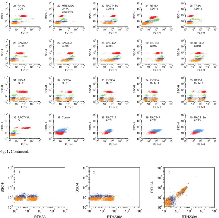
(4) 54 William C. Davis et al.. pigs, cats, and dogs for mAb that cross reacted with rabbit LDM. We also screened additional sets of mAbs we developed during the course of the study for cross reactivity. Several strategies were used to increase the potential of generating mAbs that react with conserved determinants. These included hyperimmunization with leukocytes from multiple species and then selecting a single species to screen supernatants from primary cultures of freshly prepared hybridomas, hyperimmunization with leukocytes from a single species and screening for mAbs reactive with leukocytes from another species of interest, hyperimmunizing with leukocytes from a single species and screening for all mAbs that reacted w ith LD M from the sam e species and then screening for cross reactivity with LDM in other species. Although not used extensively for identification of cross reactive mAbs, simultaneous examination of primary cultures for mAb that recognized epitopes conserved on LDM in two species, using hydroethidine to mark one set of cells, showed that cross reactive mAbs could be identified directly. Cross reactive mAbs to bovine, caprine, and ovine CD4, CD8, CD45R, and CD45R0 were identified by this method [11,28]. Regardless of the strategy used for immunization, the most frequently encountered cross reactive mAbs were specific for MHC class I and II molecules. Other mAbs of interest that were identified by single fluorescence analysis recognized epitopes only conserved on orthologous molecules in closely related species e.g.: epitopes conserved on orthologous LDM in bison, water buffalo, Cape buffalo, goats, and sheep, with highest conservation noted between orthologues in cattle and bison [43]. Some of the epitopes recognized by mAbs were highly conserved and expressed on LDM in closely and distantly related species[12,54] (Table 1). The screening of several hundred mAbs developed in our laboratory yielded 13 mAbs that recognize conserved epitopes expressed on rabbit LDM. The specificity of 10 of the mAbs (RH1A and LT86A [CD9]; HUH73A [CD11a]; CAM36A [CD14]; H20A, BAQ30A, and HUH82A [CD18]; and BAG40A and LT41A [CD44] ) was validated in the Animal Homologues section of the HLDA8 (Table 1, Fig. 1) [12,54]. Two additional mAbs, RACT48A and GBSP71A submitted to the workshop reacted with molecules expressed on multiple lineages of leukocytes in humans and other species. No clear match was obtained with standard panels of human leukocytes or cell lines transfected with known CD molecules. BAQ44A and CADO34A were not submitted to the HLDA8 workshop since they did not react with leukocytes from humans. However, the mAb-defined epitope recognized by BAQ44A is expressed on B lymphocytes in multiple species of ruminants. The epitope recognized by CADO34A is expressed on granulocytes, B lymphocytes and subsets of T lymphocytes in dogs and cats. Multiple mAbs were identified that reacted with rabbit MHC I and II molecules. The best characterized. mAbs are listed in Table 1. Analysis of the specificities of TH14B and TH81A5 have shown they recognize epitopes conserved on the orthologues of HLA-DR and HLA-DQ, respectively [1].. Identification of mAbs that recognize LDM expressed on T lymphocytes Screening of the mAb sets obtained from the different fusions yielded multiple mAbs that recognize LDM expressed on all lymphocytes or subsets of lymphocytes. These were further analyzed to determine which mAbs detected LDM expressed on T lymphocytes, B lymphocytes, or T and B lymphocytes using 2 color FC. Fluorescein conjugated anti-rabbit Ig was used to identify mAbs recognizing LDM on B lymphocytes. Ken4 (CD4) and Ken5 (pan T) were used to identify mAbs recognizing LDM on T lymphocytes. 12.C7 (CD8) was used to verify specificity of mAbs reacting with CD8 [18]. As summarized in Tables 1 and 2 and fig. 1A, 1B, 8 mAbs were identified that recognize LDM expressed on all T lymphocytes (MRB61A, RT22A, RTH2A, RTH21A, RTH26A, RTH65A, RTH230A, and RACT53A). Cross comparison of the patterns of reactivity of the mAbs using 2 color FC showed RTH2A and RTH230A; RT21A and RTH21A; and RTH26A, RTH65A, and Ken5; recognize Pan T1, Pan T2, and Pan T4 LDM respectively. Zenon second step antibodies were used to demonstrate RTH2A (IgG1) and RTH230A (IgG1) recognize the same LDM (Fig. 2). RACT53A (PanT5) recognizes an additional molecule expressed on all T cells (Fig. 3). Analysis of the reactivity of MRB61A (Pan T3) revealed it detects a LDM expressed on all T lymphocytes and basophils (Fig 1 #7, two color labeling not shown). Seven mAbs were identified that recognize LDM expressed on T lymphocyte subsets. Comparison of labeling with Ken 4 and 12.C7 demonstrated that RTH1A recognizes CD4 [41] and that ISC16A, ISC27A, ISC29A, ISC38A, and RT1A recognize CD8 [18] (Table 1, Fig. 1 #9 & #10, FC two color comparisons not shown). No information was obtained on whether the CD8 mAbs recognize epitopes on CD8α or CD8β. Comparison of labeling with RACT19A (Fig. 1 #12) with PanT1, RTH1A and ISC38A revealed the molecule detected is expressed on a large subset of CD4 and the majority of CD8 lymphocytes (Fig. 4). Identification of mAbs that recognize LDM expressed on B lymphocytes Eleven mAbs were identified that recognize LDM expressed on B lymphocytes (Tables 1 and 2, Fig. 1 #16, #17 & #18). Comparison of labeling with fluorescein conjugated polyclonal anti-rabbit immunoglobulin (Ig), RACT30A (Fig. 5) and PanT5 (Fig. 3) were used to demonstrate that MRB25, MRB29A, and MRB143A recognize one or more molecules expressed on all B.
(5) Monoclonal antibodies specific for rabbit leukocyte differentiation molecules. Table 1. Monoclonal antibodies reactive with rabbit mhc and leukocyte differentiation molecules MoAb H1A H58A TH14B TH81A5 RTH2A RTH230A RTH21A RT22A MRB61A RTH26A RTH65A RACT53A RTH1A RTH192A ISC16A ISC27A ISC29E ISC38A RT1A RACT19A RACT20A MRB120A RACT14A RACT21A RACT30A MRB25A MRB29A MRB143A BAQ44A CADO34A RT19A MRB107A MRB102A RTH186A RH1A LT86A RACT48A HUH73A RTH161A RT18A RT3A CAM36A H20A HUH82A BAQ30A 25-32 BAG40A LT41A ISC18A. Ig isotype IgG2a IgG2a IgG2a IgG2a IgG1 IgG1 IgG1 IgM IgG1 IgG2a IgM IgG1 IgG1 IgG1 IgM IgG2a IgG1 IgG1 IgM IgM IgG1 IgG1 IgM IgM IgM IgM IgM IgM IgM IgM IgM IgG1 IgM IgG1 IgG3 IgG2a IgG1 IgG1 IgG1 IgM IgM IgG1 IgG1 IgG2a IgG1 IgG1 IgG3 IgG2a IgG2a. Specificity MHC CL I MHC CL I MHC CL II HLA-DR equivalent MHC CL II HLA-DQ equivalent (polymorphic determinant) Pan T1 = Pan T1 Pan T2 = Pan T2 (blocked by RTH21A) Pan T3 (also expressed on basophils) Pan T4 = Serotec Ken 5 (diagonal co-labeling) Pan T4 = RTH26A (diagonal co-labeling) Pan T5 CD4 = Serotec Ken 4 (diagonal co-labeling) CD5 (inferred from pattern of FC labeling) CD8 (diagonal co-labeling with ISC27A, ISC29A, ISC38A) CD8 (diagonal co-labeling with12.C7, ISC29A, ISC38A, RT1A) CD8 CD8 CD8 CD4 and CD8 subpopulations Basophils and subpopulation of CD4+ T lymphocytes Granulocytes, basophils, and monocytes Subpopulations of B and T lymphocytes Subpopulations of B and T lymphocytes Pan B (expressed on some T lymphocytes?) Pan B Pan B Pan B Pan B and subpopulation of CD4 and CD8 T lymphocytes Pan B and subpopulation of CD4 and CD8 T lymphocytes B subpopulation B subpopulation Pan lymphocyte Pan lymphocyte CD9 CD9 CD11a = Serotec CD11a CD11a = RACT48A CD11a = HUH73A = RACT48A CD11b = Serotec CD11b CD11c = Serotec CD11c CD14 CD18 CD18 CD18 CD44 CD44 CD44 = BAG40A CD45 = Serotec. 55.
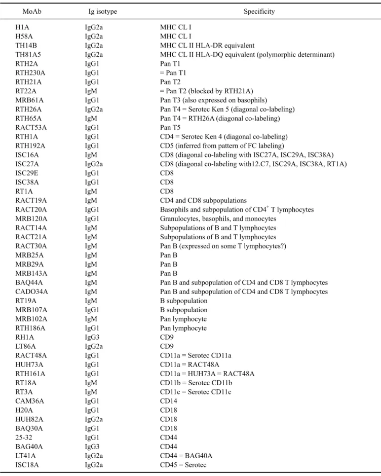
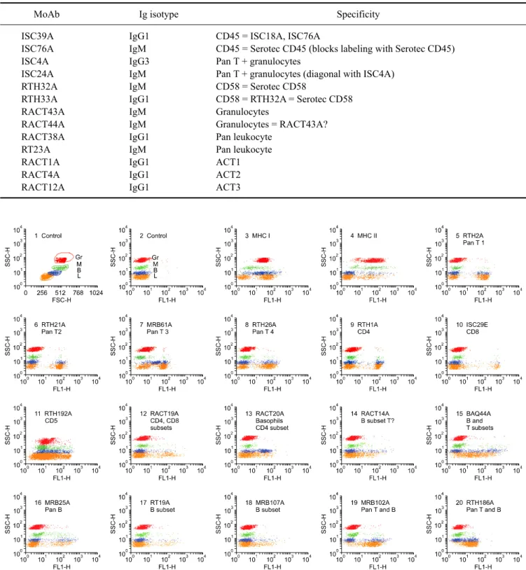
(6) 56 William C. Davis et al.. Table 1. Continued MoAb ISC39A ISC76A ISC4A ISC24A RTH32A RTH33A RACT43A RACT44A RACT38A RT23A RACT1A RACT4A RACT12A. Ig isotype IgG1 IgM IgG3 IgM IgM IgG1 IgM IgM IgG1 IgM IgG1 IgG1 IgG1. Specificity CD45 = ISC18A, ISC76A CD45 = Serotec CD45 (blocks labeling with Serotec CD45) Pan T + granulocytes Pan T + granulocytes (diagonal with ISC4A) CD58 = Serotec CD58 CD58 = RTH32A = Serotec CD58 Granulocytes Granulocytes = RACT43A? Pan leukocyte Pan leukocyte ACT1 ACT2 ACT3. Fig. 1. Representative dot plot profiles of peripheral blood leukocytes labeled with the mAbs indicated. A single representative profile is shown for mAbs that recognize the same or different epitopes on the same subset of cells. A side light scatter (SSC) vs forward light scatter dot plot was used to gate and color code the major populations of leukocytes: red for granulocytes, green for monocytes, blue for basophils, and orange for lymphocytes. Note that in contrast to other species, rabbits have a relatively large population of basophils in blood. It was necessary to label leukocytes in blood and use a fix lyse solution to isolate and analyze the composition leukocytes in peripheral blood. T lymphocytes bind to erythrocytes and are lost when leukocytes are separated using density gradient separation media..
(7) Monoclonal antibodies specific for rabbit leukocyte differentiation molecules. 57. Fig. 1. Continued.. Fig. 2. Two color FC analysis of labeling with mAbs that recognize different epitopes expressed on the same molecule. mAbs that recognize epitopes on the same molecule yield a diagonal pattern of labeling if the epitopes are sterically distant from each other. If the mAbs recognize the same epitope or epitopes that are sterically close, labeling with one mAb will block labeling with the second mAb. RTH2A and RTH230A recognize different epitopes expressed on a molecule expressed on all T lymphocytes.. lymphocytes (dot plots not shown). Comparison of labeling of MRB107A with MRB25A and BAQ44A demonstrated that MRB107A recognizes a LDM expressed on a subset of B lymphocytes. The molecule detected is only + expressed on a subset of MRB25 B lymphocytes. The whole population is included in the BAQ44A positive. population of B lymphocytes (Fig. 6). As shown in Table 2, the level of expression of the pan B mAb-defined LDM(s) were similar in peripheral blood, thymus and spleen. However, other mAbs that recognize LDM expressed on subsets of B lymphocytes exhibited different patterns of expression (Table 2, FC for thymus and spleen not shown)..
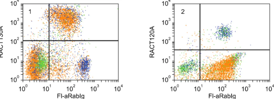
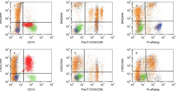
(8) 58 William C. Davis et al.. Fig. 3. Two color FC analysis showing the pattern of labeling obtained with mAbs that recognize different molecules only expressed on T lymphocytes. The subsets labeled with anti-CD4 and CD8 mAbs are included in the population labeled with a pan T mAb, panels 1 and 2. Labeling with the two anti-pan T mAbs yields a diffuse pattern of labeling, panel 3. Mutually exclusive populations of lymphocytes are labeled with mAbs specific for T and B lymphocytes, panel 4. The example presented here suggests a small subset of B lymphocytes may express the pan T4-defined T lymphocyte molecule.. Fig. 4. Two color FC analysis showing RACT19A recognizes a molecule expressed on a major subset of T lymphocytes, panel 1. mAbs specific for PanT1, CD4, and CD8 were combined to show the molecule is expressed on a large subset of CD4 T lymphocytes and most CD8 lymphocytes. The level of expression of the RACT19A-defined molecule on CD4 lymphocytes is less than the level of expression on CD8 lymphocytes.. Fig. 5. Two color FC analysis demonstrating that RACT30A recognizes a molecule expressed on all B cells, panel 1. As shown in panel 2, immunoglobulin detected with polyclonal anti-rabbit Ig is also present on basophils. RACT20A recognizes a molecule expressed on basophils and a subset of CD4 T lymphocytes.. The subset of B lymphocytes detected with RACT14A and RACT21A was in low frequency in peripheral blood and in high frequency in spleen and appendix. The subset detected with RTH72A was low in peripheral blood, thymus, and appendix and high in spleen. The subset detected with mAbs RT19A and RTH172A was low in peripheral blood but high in thymus, spleen, and appendix.. Identification of mAbs that recognize LDM expressed on T and B lymphocytes Four mAbs were identified that recognize LDM expressed on T and B lymphocytes, MRB102A, RTH186A, RTH192A, and BAQ44A (a cross reactive mAb) (Fig. 1 #19, #20, #11 & #15, respectively). The level of expression of the LDM detected with MRB102A on lymphocytes was higher than.
(9) Monoclonal antibodies specific for rabbit leukocyte differentiation molecules. Table 2. Reactivity of monoclonal antibodies with leukocyte from blood and primary and secondary lymphoid organs mAb H1A H58A TH14B TH81A5 RTH2A RTH230A RTH21A RT22A MRB61A RTH26A RTH65A RACT53A RTH1A RTH192A ISC16A ISC27A ISC29E ISC38A RT1A RACT19A RACT20A MRB120A RACT30A MRB25A MRB29A MRB143A RACT14A RACT21A RT19A RTH72A RTH172A. %+ Peripheral blood. %+ Thymus. 86 82 57 58 27 28 26 27 40 28 26 30 24 50 4 4 4 4 4 8 18 22 26 14 15 15 9 11 5 3 7. 57 46 63 52 37 37 92 97 99 99 99 28 87 16 83 84 75 86 84 6 2 1 11 10 6 5 14 13 50 8 59. %+ %+ Spleen Appendix 79 75 50 47 37 40 45 44 45 48 36 59 20 57 9 12 9 9 10 15 4 6 45 44 45 38 45 48 45 31 44. 8 7 99 99 4 4 4 4 8 5 5 11 3 97 1 1 1 1 1 13 3 20 94 82 77 77 69 69 56 5 61. 59. Table 2. Continued mAb MRB107A MRB102A RTH186A BAQ44A CADO34A RACT48A HUH73A RTH161A RT18A RT3A MRB128A CAM36A H20A HUH82A BAQ30A BAG40A LT41A ISC18A ISC39A ISC76A ISC4A ISC24A ISC26A ISC36A ISC90A RT15A RTH33A RACT43A RACT44A RACT38A RT23A. %+ Peripheral blood. %+ Thymus. 7 38 40 34 12 99 99 99 28 7 8 8 99 99 99 93 99 99 99 99 20 15 34 24 28 33 99 32 27 99 99. 4 75 14 40 3 97 NT 99 5 1 2 1 97 NT 99 15 NT 90 NT NT 49 91 97 98 97 97 96 9 9 99 95. %+ %+ Spleen Appendix 18 69 76 59 64 99 NT 99 53 20 36 5 73 NT 76 85 NT 92 NT NT 54 48 81 70 82 85 99 26 28 88 95. 3 79 8 82 90 99 NT 99 4 4 7 2 99 NT 99 99 NT 97 NT NT 5 5 52 9 93 87 31 5 5 99 96. Fig. 6. Two color FC analysis of the expression of a molecule detected with MRB107A that is expressed on a subset of B lymphocytes. The molecule is expressed on a subset of MRB25A+ B lymphocytes, panel 1. All the MRB107A+ lymphocytes co-express the molecule detected with BAQ44A, panel 2..
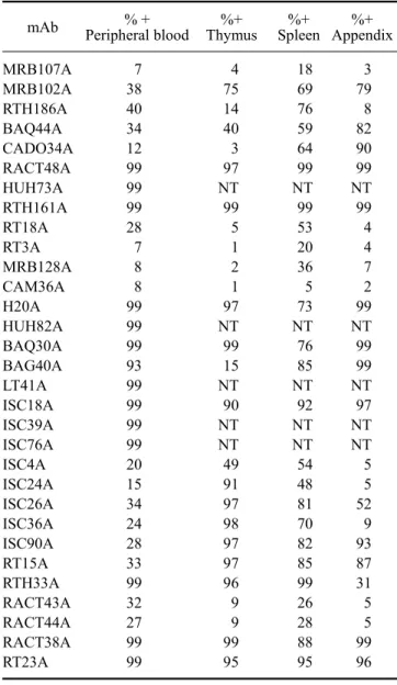
(10) 60 William C. Davis et al.. the LDM detected with RTH186A. However, two color analysis showed the level of expression of both LDMs on CD4 and CD8 T and B lymphocyte subsets was similar (FC not shown). The level of expression of the MRB102Adefined LDM was also high on lymphocytes in the thymus, spleen, and appendix. In contrast, the RTH186A defined LDM was only expressed on a few lymphocytes in the thymus and appendix. It was expressed on a large population of lymphocytes in the spleen (Table 2, FC not shown). The level of expression of the LDM detected with RTH192A and BAQ44A differed on CD4 and CD8 T and B lymphocytes. The level of expression of the RTH192A-. +. defined LDM was variable on PanT 1 lymphocytes (Fig. 7). It was low on CD4 T and B lymphocytes (Fig. 7). It was high on CD8 T lymphocytes (Fig. 7). It was only expressed on a few thymocytes. It was expressed at a high level on about 50% of lymphocytes in the spleen and essentially all lymphocytes in the appendix (Table 2, FC not shown). The pattern of expression on T and B lymphocytes suggests the LDM detected is CD5 [41,49-51]. The level of expression of the LDM detected with BAQ44A also differed on CD4 and CD8 T and B lymphocytes (Fig. 8). Simultaneous labeling with Pan T1, CD4, and CD8 mAbs and anti-rabbit Ig demonstrated that the LDM is. Fig. 7. Two color FC analysis of the expression of RTH192A on T and B lymphocytes. The level of expression of PanT4 on RTH192A+ lymphocytes was variable from high to low, panel 1. Expression of CD4 and the MRB25A-defined B molecule were also low, panels 2 and 4. Expression of the TH192A-defined molecule was invariably higher on CD8 lymphocytes than expression on the other mAb-defined populations, panel 3.. Fig. 8. Two color FC analysis of the expression of BAQ44A- and CADO34A-defined molecules. The BAQ44A-defined molecule was not expressed on granulocytes or monocytes. Comparison of labeling with BAQ44A in combination with mAbs to PanT1, CD4, and CD8 showed subsets of CD4 and CD8 co-expressed the BAQ44A-defined molecule, panel 2. The molecule was not expressed on + basophils, panel 4. The pattern of labeling indicate a subset of Pan T CD4 , CD8 also express the BAQ44A-difined molecule. B cells also co-expressed the molecule. The molecule was not expressed on basophils, panel 3. A similar pattern of labeling was observed with the CADO34A-defined molecule, Panels 2 and 3. The molecule was also expressed on granulocytes, panel 1..
(11) Monoclonal antibodies specific for rabbit leukocyte differentiation molecules. 61. +. highly expressed on B lymphocytes, a subset of CD4 and CD8 negative T lymphocytes, a large subset of CD4 T lymphocytes and the majority of CD8 T lymphocytes. The BAQ44A-defined LDM was expressed on large populations of lymphocytes in the thymus, spleen and appendix (Table 2, FC not shown).. basophils and a subset of CD4 lymphocytes (Fig. 9). Expression on CD8 lymphocytes is low or absent. Two color analyses were not performed to determine whether expression of the RACT20A-defined LDM on small populations of cells detected in the thymus, spleen and appendix were basophils or T lymphocytes (Table 2).. Identification of a mAb that recognizes a LDM expressed on granulocytes, T and B lymphocytes One cross reactive mAb, CADO34A, was identified that recognizes a LDM expressed on granulocytes as well as T and B lymphocytes (Fig. 8). Comparison of labeling with CADO34A to labeling with BAQ44A revealed the pattern of labeling is similar to the labeling pattern obtained with BAQ44A for T and B lymphocytes. Simultaneous labeling with Pan T1, CD4, and CD8 mAbs demonstrated the LDM is expressed on a subset of CD4 and CD8 negative lymphocytes, a large subset of CD4 and the majority of CD8 T lymphocytes. The LDM was only expressed on a few thymocytes. It was expressed on a large population of lymphocytes in the spleen and most of the lymphocytes in the appendix (Table 2, FC not shown).. Identification of mAbs recognizing LDM expressed on granulocytes Two mAbs (RACT43A and RACT44A Table 1, Fig. 1 #36) were identified that recognize a LDM expressed on granulocytes. The similarity of the pattern of labeling with the mAbs suggests they may recognize the same LDM.. Identification of a mAbs that recognize a LDM expressed on granulocytes and monocytes, granulocytes, monocytes and basophils, or basophils Three mAb were identified that detect LDM expressed on granulocytes and monocytes, granulocytes, monocytes and basophils or basophils (CAM36A, MRB120A, and RACT20A, Fig. 1 #26, #22 & #13, respectively and Fig. 9). CAM36A recognizes a conserved epitope expressed on CD14. In contrast to some species, expression of CD14 is high on rabbit granulocytes. Two color analyses showed CD14 is not expressed on basophils (Fig. 9). Two color analyses showed the MRB120A recognizes a LDM expressed on granulocytes, monocytes and basophils while the RACT20A recognizes a LDM only expressed on. Identification of mAbs recognizing CD9 Comparison of the labeling patterns with cross reactive RH1A and LT86A (Table 1, Fig. 1 #21) showed they recognize CD9 in rabbits. Identification of mAbs recognizing CD11a, CD11b, CD11c, and CD18 mAbs that recognize CD11a, CD11b, CD11c, and CD18 (Table 1, Fig. 1 #23, #24, #25 & #27 respectively) were identified by two color FC with mAbs that recognize epitopes conserved on orthologues in one or more species or with commercially available mAbs generated against the rabbit orthologues. Comparison of the patterns of labeling obtained with RACT48A and RTH161A with commercially available anti-CD11a (Ken11) [31] suggested these mAbs recognize CD11a. Subsequent comparative two color FC analysis with HUH73A, a mAb demonstrated to recognize a conserved epitope on the CD11a orthologue in the Animal Homologues section of the HLAD8 workshop [54], verified these mAbs recognize CD11a (Fig. 10). The studies also demonstrated RTH161A recognizes a species restricted epitope. Comparison of the patterns of labeling obtained with RT18A with anti-CD11b (198) and subsequent. Fig. 9. Two color analysis of the expression of LDM detected with RACT20A and MRB120A on basophils, monocytes, and CD4 T lymphocytes. mAbs to CD14 and Pan T1 were combined and used to distinguish monocytes and T lymphocytes simultaneously. MRB102A was used to identify all T and B lymphocytes. Polyclonal anti-Ig was used to distinguish B lymphocytes. Panel 1 shows the populations present in PBMC: basophils, lower left quadrant; monocytes, upper left quadrant; T lymphocytes, upper right quadrant; B lymphocytes, lower right quadrant. Panel 2 shows the LDM recognized by RACT20A is only expressed on basophils, upper right quadrant. As noted, Ig is present on basophils. Panel 3 shows the LDM recognized by MRB120A is expressed on monocytes, upper left quadrant and basophils, upper right quadrant. Panel 4 shows the LDM recognized by RACT20A is expressed on basophils, lower right quadrant and also a subset of CD4 T lymphocytes, upper right quadrant..
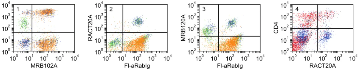
(12) 62 William C. Davis et al.. Fig. 10. Two color analysis of HUH73A and MRB161A. The similarity in the pattern of labeling obtained with both mAbs indicates they recognize the same molecule, panels 1 and 2. The diagonal pattern of labeling indicates the mAbs recognize epitopes on the same molecule. Based on findings with HUH73A in the animals homologues section of the 8th human leukocyte antigen workshop, the molecule identified is the rabbit orthologue of CD11a.. 2 color FC showed RT18A recognizes CD11b (Fig. 1 #24). Similar studies comparing RT3A with 3/22 showed RT3A recognizes CD11c (Fig. 1 #25). Three mAbs demonstrated to recognize conserved epitopes on orthologues of CD18 (Fig. 1 #27), in 2 or more species were shown to recognize rabbit CD18. The pattern of expression of the rabbit orthologues for each molecule was similar to that noted in other species.. Identification of mAbs that recognize CD44, CD45, and CD58 Screening of a large series of mAbs that recognize conserved epitopes on CD44 in 2 or more species, including humans, showed 25-32 [40], BAG40A, and LT41A (Table 1, Fig. 1 #28) recognize epitopes expressed on rabbit CD44 [14,24,54]. The pattern of expression of rabbit CD44 was similar to that noted in other species. Comparison of the pattern of labeling obtained with L12/201 (CD45) with the panels of mAbs developed against rabbit LDM revealed several mAbs yielded similar patterns of labeling (Table 1, Fig. 1 #29). Two color flow cytometry yielded diagonal patterns of labeling or blocking, indicating the mAbs recognize rabbit CD45 (FC not shown). Comparison of the labeling pattern obtained with VC21 (CD58) revealed two mAbs (RTH32A, RTH33A, Table 1, Fig. 1 #30) yielded similar patterns of labeling. Two color comparisons yielded diagonal patterns of labeling indicating the mAbs recognize CD58 (FC not shown). Identification of mAbs that recognize LDM expressed on granulocytes and lymphocytes Six mAbs were identified that recognize LDM expressed on granulocytes and lymphocytes ISC4A, ISC24A, ISC26A, ISC36A, ISC90A, and RT15A (Table 1, Fig. 1 #31, #26, #36, #90 and #35). Two color analysis showed ISC4A and ISC24A recognize the same molecule (FC not shown). The others identified different molecules. The molecules identified with ISC90A and RT15A are also expressed on. some monocytes. The molecule identified with RT15A is highly expressed on thymocytes, spleen lymphocytes, and lymphocytes in the appendix (Table 2). Two color analysis with anti-CD4 and -CD8 mAbs indicate none of the mAbs recognize CD45R0. The studies completed thus far indicate there are no clearly defined subsets of CD4 and CD8 negative for these LDM (FC not shown).. Identification of mAbs that recognize LDM expressed on all leukocytes Three mAbs under investigation identify LDM expressed on all leukocytes RACT38A, RT23A, and GBSP71A (Table 1, FC not shown). Comparison of the flow cytometric profiles with those of known CD molecules has thus far not suggested which molecules are recognized by these mAbs. Identification of mAbs that recognize LDM expressed on activated lymphocytes Three mAbs were identified that recognize mAbs upregulated on ConA activated lymphocytes RACT1A, RACT4A, RACT12A (Table 1, Fig. 1 #38, #39 & #40). Two color analysis with mAbs specific for ConA and CD25 [32] demonstrated the mAbs recognize different LDM (FC not shown).. Discussion Cumulative data obtained from international workshops convened to complete characterization of mAbs to LDM in humans have shown flow cytometry can be used to compare and cluster mAbs that appear to recognize the same LDM for further analysis [34]. Our studies and studies conducted as part of workshops convened to characterize mAb-defined LDM in ruminants, swine, horses, and dogs have shown that the pattern of expression of many orthologous of CD molecules is conserved cross species [11]. These findings have afforded an opportunity to devise a strategy for characterizing mAb to LDM in.
(13) Monoclonal antibodies specific for rabbit leukocyte differentiation molecules. additional less well studied species. mAb that identify LDM with patterns of expression similar to the patterns of expression of known Human Cell Differentiation Molecules (HCDM) can be clustered for further analysis. The TKP can be used to facilitate determining whether a mAb recognizes a new or known HCDM where flow cytometric data are not conclusive. Where cross reactive mAbs are identified, they can be used to validate the specificity of the mAbs recognizing species restricted epitopes using two color FC. A diagonal pattern of labeling implies the epitopes detected are present on the same molecule. Complete or partial blocking of labeling with one of the mAbs indicates the epitopes detected are sterically close on the same molecule, with the binding of one mAb interfering with the binding of the second mAb. These methods of analysis have been used effectively to develop a set of mAbs for use in alpacas and llamas [14] and, as demonstrated in the present report, rabbits. Cross reactive mAbs allowed us to identify mAbs specific for MHC class I and II and several CD molecules early in the course of the studies. Two color analyses with commercially available mAbs to some rabbit LDM facilitated validation of the specificity of additional mAbs generated in our laboratory. To date, we have identified mAbs to MHC class I and II molecules and 16 known LDM. Additional mAbs to T and B lymphocytes have been identified that require further characterization to determine their relation to known mAb-defined HCDM. Until now the lack of mAbs to rabbit LDM and MHC has made it difficult to compare the immune system of rabbits to those characterized in other species. Comparative studies have shown the immune systems of different orders and species of vertebrates are similar but not identical. Differences have been noted in the evolution and expression of immunoglobulin genes with expansion of the IgA genes a unique feature of rabbits [41] and the development of IgG heavy chain genes a unique feature of camelids [21,41]. The expression of αβ CD4 and CD8 T cell subsets have appeared similar with an exception in swine. There is a large population of CD4/CD8 double positive cells in peripheral blood. Analysis has shown the proportion of double positive cells increases with age and is correlated with the appearance of the majority of memory T cells in this population [67,68]. The most striking difference noted is in the abundance of γδ T cells in some species [20]. γδ T cells comprise a high proportion of lymphocytes in peripheral blood of chickens [5,9], swine [2,3,17] and ruminants [19,25,39]. The relation between the large population of γδ T cells that have evolved in chickens and the one in pigs and ruminants is not clear. However, recent studies have provided an explanation for the abundance in the latter species. Abundance is attributable to the presence of a unique subset of γδ T cells that has only been found in artiodactyla. The subset is characterized by the expression. 63. of a molecule referred to as workshop cluster 1 (WC1) in + ruminants [42]. The WC1 population may comprise 50% or more of T lymphocytes in the blood of young ruminants [62,63]. A WC1- population is also present but comprises ~5% of γδ T cells in blood [10]. The large subset identified in swine expresses the orthologue of WC1 [7,17]. The subset expressing the orthologue has been identified in camelids also [14]. An additional difference noted in the comparative studies is in the expression of MHC II. In humans, mice, and ruminants MHC II is expressed primarily on monocytes and B lymphocytes in blood. MHC II is upregulated on T cells following activation. MHC II is expressed on monocytes, B cells and resting T cells in horses [37], swine [48], dogs [8], and cats (personal observation). As mentioned, the flow cytometric approach to identifying and characterizing mAbs to rabbit LDMs has shown the pattern of expression of rabbit orthologues of known hCDLDMs have proven, thus far, to be similar. The composition of lymphocyte subsets also appear more similar to that of humans than ruminants and swine. Analysis of T lymphocytes in the rabbit shows αβ T cells are the major population present in blood. Comparison of the percentages of cells expressing LDM present on all T cells with subsets of cells expressing CD4 or CD8 in two color flow cytometry has not revealed any CD4 CD8 double negative subset. Likewise comparison of the percentages of lymphocytes expressing LDM on T cells with those expressing LDM on B cells has not revealed the presence of a clearly defined subset of lymphocytes negative for T or B cell LDM. The findings suggest γδ T cells comprise a small percentage of lymphocytes in blood. Further studies are needed to identify rabbit γδ T cells. The unique differences that have been noted are with the expression of MHC II on granulocytes, expression of certain mAb-defined LDM, and presence of immunoglobulin on basophils. Multiple mAbs of different isotype to MHC II were used to confirm expression of MHC II on granulocytes. Both IgG1 and IgG2a isotype mAbs yielded identical patterns of labeling. As in humans and mice, expression of MHC II on monocytes and B cells is similar. It is upregulated on activated T cells. Basophils comprise 5% to 30% of blood leukocytes in rabbits. Two mAbs were identified that identify molecules expressed on all T cells and basophils (MRB61A) and basophils and a subset of CD4 T cells (RACT20A). No information has been obtained on the functional activity of these LDM. Two color analyses have demonstrated that immunoglobulin is present on basophils. This finding is in agreement with an earlier study demonstrating the presence of multiple immunoglobulin isotypes on basophils, presumably binding to basophils through Fc receptors [6]. In summary, the use of flow cytometry has provided an approach to the identification and characterization of.
(14) 64 William C. Davis et al.. mAbs to MHC I and MHC II molecules and LDM in less well studied species. mAbs that recognize the same LDM can be clustered based on the similarity of the pattern of expression and further characterized by comparing the pattern of expression with that of known LDM. When available, identity can be verified by comparison with a mAb that recognizes a conserved epitope on orthologous molecules. Where needed, mAb cluster-defined LDM can be subjected to immunoprecipitation and micro sequencing. We have used this approach successfully when participating in the international workshops of LDM in ruminants [26,27,46], swine [23,38,53], horses [33,36], dogs [8], the homologues section of the HLDA8 [54,66] and independently in the characterization of mAbs to LDM in lamas [14] and rabbits. The mAbs characterized here should facilitate characterization of the immune system in rabbits and the use of rabbits in immunological investigations.. Acknowledgments Thank you is extended to the technical staff that assisted in the conduct of the studies over the past years and to Betty Davis for recording information into the TKP. The studies were supported in part by the Department of Veterinary Microbiology and Pathology and the Washington State University Monoclonal Antibody Center. mAbs to rabbit MHC I and II and LDM are available through VMRD, Inc. (www.vmrd.com). Unlicensed mAbs are available through the WSU Monoclonal Antibody Center. The website of TKP, NCBI Rabbit Genome Resources and HCDM are www.vetmed.wsu.edu/tkp, www3.niaid.nih.gov/research/ resources/ri, www.medicine.uiowa.edu/cigw/rabbit.htm and www.hcdm.org, respectively.. References 1. Ababou A, Goyeneche J, Davis WC, Lévy D. Evidence for the expression of three different BoLa-class II molecules on the bovine BL-3 cell line: determination of a non-DR non-DQ gene product. J Leukoc Biol 1994, 56, 182-186. 2. Binns RM. The null/γδTCR+ T cell family in the pig. Vet Immunol Immunopathol 1994, 43, 69-77. 3. Binns RM, Duncan IA, Powis SJ, Hutchings A, Butcher GW. Subsets of null and γδ T-cell receptor + T lymphocytes in the blood of young pigs identified by specific monoclonal antibodies. Immunology 1992, 77, 219-227. 4. Brodersen R, Bijlsma F, Gori K, Jensen KT, Chen W, Dominguez J, Haverson K, Moore PF, Saalmuller A, Sachs D, Slierendrecht WJ, Stokes C, Vainio O, Zuckermann F, Aasted B. Analysis of the immunological cross reactivities of 213 well characterized monoclonal antibodies with specificities against various leucocyte surface antigens of human and 11 animal species. Vet Immunol Immunopathol 1998, 64, 1-13. 5. Bucy RP, Chen CH, Cooper MD. Analysis of γδ T cells in. the chicken. Semin Immunol 1991, 3, 109-117. 6. Cabana VG, Teodorescu M, Dray S. Identification of basophils as the cells bearing both allelic immunoglobulin allotypes among white blood cells from the peripheral blood of heterozygous rabbits. J Immunol 1980, 124, 2268-2280. 7. Carr MM, Howard CJ, Sopp P, Manser JM, Parsons KR. Expression on porcine γδ lymphocytes of a phylogenetically conserved surface antigen previously restricted in expression to ruminant γδ T lymphocytes. Immunology 1994, 81, 36-40. 8. Cobbold S, Metcalfe S. Monoclonal antibodies that define canine homologues of human CD antigens: summary of the First International Canine Leukocyte Antigen Workshop (CLAW). Tissue Antigens 1994, 43, 137-154. 9. Cooper MD, Chen C-LH, Bucy RP, Thompson CB. Avian T cell ontogeny. Adv Immunol 1991, 50, 87-117. 10. Davis WC, Brown WC, Hamilton MJ, Wyatt CR, Orden JA, Khalid AM, Naessens J. Analysis of monoclonal antibodies specific for the γδ TcR. Vet Immunol Immunopathol 1996, 52, 275-283. 11. Davis WC, Davis JE, Hamilton MJ. Use of monoclonal antibodies and flow cytometry to cluster and analyze leukocyte differentiation molecules. In: Davis WC (ed.). Monoclonal Antibody Protocols. pp. 149-167, Humana Press, Totowa, 1995. 12. Davis WC, Drbal K, Mosaad AE, Elbagory AR, Tibary A, Barrington GM, Park YH, Hamilton MJ. Use of flow cytometry to identify monoclonal antibodies that recognize conserved epitopes on orthologous leukocyte differentiation antigens in goats, lamas, and rabbits. Vet Immunol Immunopathol 2007, 119, 123-130. 13. Davis WC, Hamilton MJ. Use of flow cytometry to characterize immunodeficiency syndromes in camelids. Small Rumin Res 2006, 61, 187-193. 14. Davis WC, Heirman LR, Hamilton MJ, Parish SM, Barrington GM, Loftis A, Rogers M. Flow cytometric analysis of an immunodeficiency disorder affecting juvenile llamas. Vet Immunol Immunopathol 2000, 74, 103-120. 15. Davis WC, Khalid AM, Hamilton MJ, Ahn JS, Park YH, Cantor GH. The use of crossreactive monoclonal antibodies to characterize the immune system of the water buffalo (Bubalus bubalus). J Vet Sci 2001, 2, 103-109. 16. Davis WC, Marusic S, Lewin HA, Splitter GA, Perryman LE, McGuire TC, Gorham JR. The development and analysis of species specific and cross reactive monoclonal antibodies to leukocyte differentiation antigens and antigens of the major histocompatibility complex for use in the study of the immune system in cattle and other species. Vet Immunol Immunopathol 1987, 15, 337-376. 17. Davis WC, Zuckermann FA, Hamilton MJ, Barbosa JIR, Saalmuller A, Binns RM, Licence ST. Analysis of monoclonal antibodies that recognize γδ T/null cells. Vet Immunol Immunopathol 1998, 60, 305-316. 18. De Smet W, Vaeck M, Smet E, Brys L, Hamers R. Rabbit leukocyte surface antigens defined by monoclonal antibodies. Eur J Immunol 1983, 13, 919-928. 19. Goddeeris BM. Immunology of cattle. In: Pastoret PP, Griebel P, Bazin H, Govaerts A (eds.). Handbook of Vertebrate Immunology. pp. 439-484, Academic Press, San Diego, 1998..
(15) Monoclonal antibodies specific for rabbit leukocyte differentiation molecules. 20. Haas W, Pereira P, Tonegawa S. Gamma/delta cells. Annu Rev Immunol 1993, 11, 637-685. 21. Hamers R, Muyldermans S. Immunology of camels and llamas. In: Pastoret PP, Griebel P, Bazin H, Govaerts A (eds.). Handbook of Vertebrate Immunology. pp. 421-438, Academic Press, San Diego, 1998. 22. Hamilton MJ, Davis WC. 1995. Culture conditions that optimize outgrowth of hybridomas. In: Davis WC (ed.). Monoclonal Antibody Protocols. pp. 17-28, Humana Press, Totowa, 1995. 23. Haverson K, Saalmuller A, Alvarez B, Alonso F, Bailey M, Bianchi ATJ, Boersma WJA, Chen Z, Davis WC, Dominguez J, Engelhardt H, Ezquerra A, Grosmaire LS, Hamilton MJ, Hollemweguer E, Huang CA, Khanna KV, Kuebart G, Lackovic G, Ledbetter JA, Lee R, Llanes D, Lunney JK, McCullough KC, Molitor T, Nielsen J, Niewold TA, Pescovitz MD, Perez de la Lastra J, Rehakova Z, Salmon H, Schnitzlein WM, Seebach J, Simon A, Sinkora J, Sinkora M, Stokes CR, Summerfield A, Sver L, Thacker E, Valpotic I, Yang H, Zuckermann FA, Zwart R. Overview of the Third International Workshop on Swine Leukocyte Differentiation Antigens. Vet Immunol Immunopathol 2001, 80, 5-23. 24. Hein WR, Dudler L, Mackay CR. Surface expression of differentiation antigens on lymphocytes in the ileal and jejunal Peyer's patches of lambs. Immunology 1989, 68, 365-370. 25. Hein WR, Mackay CR. Prominence of γδ T cells in the ruminant immune system. Immunol Today 1991, 12, 30-34. 26. Hopkins J, Ross A, Dutia BM. Summary of workshop findings of leukocyte antigens in sheep. Vet Immunol Immunopathol 1993, 39, 49-59. 27. Howard CJ, Leibold W. Individual antigens of cattle. Bovine CD5 (BoCD5). Vet Immunol Immunopathol 1991, 27, 55-60. 28. Howard CJ, Morrison WI. Comparison of reactivity of monoclonal antibodies on bovine, ovine and caprine tissues and on cells from other animal species. Vet Immunol Immunopathol 1991, 27, 32-34. 29. Howard CJ, Morrison WI, Bensaid A, Davis WC, Eskra L, Gerdes J, Hadam M, Hurley D, Leibold W, Letesson JJ, MacHugh N, Naessens J, O'Reilly K, Parsons KR, Schlote D, Sopp P, Splitter G, Wilson R. Summary of workshop findings for leukocyte antigens of cattle. Vet Immunol Immunopathol 1991, 27, 21-27. 30. Howard CJ, Naessens J. Summary of workshop findings for cattle (tables 1 and 2). Vet Immunol Immunopathol 1993, 39, 25-47. 31. Kotani M, Yamamura Y, Tamatani T, Kitamura F, Miyasaka M. Generation and characterization of monoclonal antibodies against rabbit CD4, CD5 and CD11a antigens. J Immunol Methods 1993, 157, 241-252. 32. Kotani M, Yamamura Y, Tsudo M, Tamatani T, Kitamura F, Miyasaka M. Generation of monoclonal antibodies to the rabbit interleukin-2 receptor alpha chain (CD25) and its distribution in HTLV-1-transformed rabbit T cells. Jpn J Cancer Res 1993, 84, 770-775. 33. Kydd J, Antczak DF, Allen WR, Barbis D, Butcher G, Davis W, Duffus WPH, Edington N, Grunig G, Holmes. 65. MA, Lunn DP, McCulloch J, O'Brien A, Perryman LE, Tavernor A, Williamson S, Zhang C. Report of the First International Workshop on equine leucocyte antigens, Cambridge, UK, July 1991. Vet Immunol Immunopathol 1994, 42, 3-60. 34. Lanier LL, Allison JP, Phillips JH. Correlation of cell surface antigen expression on human thymocytes by multi-color flow cytometric analysis: implications for differentiation. J Immunol 1986, 137, 2501-2507. 35. Lanier LL, Engleman EG, Gatenby P, Babcock GF, Warner NL, Herzenberg LA. Correlation of Functional Properties of Human Lymphoid Cell Subsets and Surface Marker Phenotypes Using Multiparameter Analysis and Flow Cytometry. Immunol Rev 1983, 74, 143-160. 36. Lunn DP, Holmes MA, Antczak DF, Agerwal N, Baker J, Bendali-Ahcene S, Blanchard-Channell M, Byrne KM, Cannizzo K, Davis W, Hamilton MJ, Hannant D, Kondo T, Kydd JH, Monier MC, Moore PF, O'Neil T, Schram BR, Sheoran A, Stott JL, Sugiura T, Vagnoni KE. Report of the second equine leucocyte antigen workshop, Squaw Valley, California, July 1995. Vet Immunol Immunopathol 1998, 62, 101-143. 37. Lunn DP, Holmes MA, Duffus WPH. Equine T-lymphocyte MHC II expression: variation with age and subset. Vet Immunol Immunopathol 1993, 35, 225-238. 38. Lunney JK, Walker K, Goldman T, Aasted B, Bianchi A, Binns R, Licence S, Bischof R, Brandon M, Blecha F, Kielian TL, McVey DS, Chu RM, Carr M, Howard C, Sopp P, Davis W, Dvorak P, Dominguez J, Canals A, Sanchez Vizcaino JM, Kim YB, Laude H, Mackay CR, Magnusson U, McCullough K, Misfeldt M, Murtaugh M, Molitor T, Choi C, Pabst R, Parkhouse RM, Denham S, Yang H, Pescovitz M, Pospisil R, Tlaskalova H, Saalmueller A, Weiland E, Salmon H, Sachs D, Arn S, Shimizu M, Stokes C, Stevens K, Valpotic I, Zuckermann F, Husmann R. Overview of the First International Workshop to define swine leukocyte cluster of differentiation (CD) antigens. Vet Immunol Immunopathol 1994, 43, 193206. 39. Mackay CR, Hein WR. A large proportion of bovine T cells express the γδ T cell receptor and show a distinct tissue distribution and surface phenotype. Int Immunol 1989, 1, 540-545. 40. Mackay CR, Maddox JF, Wijffels GL, MacKay IR, Walker ID. Characterization of a 95,000 molecule on sheep leucocytes homologous to murine Pgp-1 and human CD44. Immunology 1988, 65, 93-99. 41. Mage RG. 1998. Immunology of lagomorphs. In: Pastoret PP, Griebel P, Bazin H, Govaerts A (eds.). Handbook of Vertebrate Immunology. pp. 223-260, Academic Press, San Diego, 1998. 42. Morrison WI, Davis WC. Individual antigens of cattle. Differentiation antigens expressed predominantly on CD4CD8-T lymphocytes (WC1, WC2). Vet Immunol Immunopathol 1991, 27, 71-76. 43. Mossad AA, Elbagoury AR, Khalid AM, Waters WR. Tibary A, Hamilton MJ, Davis WC. Identification of monoclonal antibody reagents for use in the study of immune response in camel and water buffalo. Proc Int Sci Conf.
(16) 66 William C. Davis et al.. Camels 2006, 2391-2411. 44. Muriuki SP, Olaho-Mukani W, Naessens J. A panel of monoclonal antibodies that cross-react with leukocyte differentiation antigens from dromedary camel (Cameluus dromedarius). J Camel Prac Res 1998, 5, 179-185. 45. Naessens J. Characterisation of lymphocyte populations in African buffalo (Syncerus caffer) and waterbuck (Kobus defassa) with workshop monoclonal antibodies. Vet Immunol Immunopathol 1991, 27, 153-162. 46. Naessens J, Hopkins J. Introduction and summary of workshop findings. Vet Immunol Immunopathol 1996, 52, 213-235. 47. Naessens J, Olubayo RO, Davis WC, Hopkins J. Cross-reactivity of workshop antibodies with cells from domestic and wild ruminants. Vet Immunol Immunopathol 1993, 39, 283-290. 48. Pescovitz MD, Lunney JK, Sachs DH. Preparation and characterization of monoclonal antibodies reactive with porcine PBL. J Immunol 1984, 133, 368-375. 49. Pospisil R, Fitts MG, Mage RG. CD5 is a potential selecting ligand for B cell surface immunoglobulin framework region sequences. J Exp Med 1996, 184, 1279-1284. 50. Pospisil R, Obiakor H, Newman BA, Alexander C, Mage RG. Stable expression of the extracellular domains of rabbit recombinant CD5: development and characterization of polyclonal and monoclonal antibodies. Vet Immunol Immunopathol 2005, 103, 257-267. 51. Raman C, Knight KL. CD5+ B cells predominate in peripheral tissues of rabbit. J Immunol 1992, 149, 3858-3864. 52. Saalmueller A, Denham S, Haverson K, Davis WC, Dominguez J, Pescovitz MD, Stokes CC, Zuckermann F, Lunney JK. The second International Swine CD Workshop. Vet Immunol Immunopathol 1996, 54, 155-158. 53. Saalmuller A, Aasted B, Canals A, Dominguez J, Goldman T, Lunney JK, Maurer S, Pescovitz MD, Pospisil R, Salmon H, Tlaskalova H, Valpotic I, Vizcaino JS, Weiland E, Zuckermann F. Summary of workshop findings for porcine T-lymphocyte antigens. Vet Immunol Immunopathol 1994, 43, 219-228. 54. Saalmuller A, Lunney JK, Daubenberger C, Davis W, Fischer U, Göbel TW, Griebel P, Hollemweguer E, Lasco T, Meister R, Schuberth HJ, Sestak K, Sopp P, Steinbach F, Xiao-Wei W, Aasted B. Summary of the animal homologue section of HLDA8. Cell Immunol 2005, 236, 51-58. 55. Saalmuller A, Pauly T, Lunney JK, Boyd P, Aasted B, Sachs DH, Arn S, Bianchi A, Binns RM, Licence S, Whyte A, Blecha F, Chen Z, Chu RM, Davis WC, Denham S, Yang H, Whittall T, Parkhouse RM, Dominguez J, Ezquerre A, Alonso F, Horstick G, Howard C, Sopp P, Kim YB, Lipp J, Mackay C, Magyar A, McCullough K, Arriens A, Summerfield A, Murtaugh M, Nielsen J, Novikov B, Pescovitz MD, Schuberth HJ, Leibold W, Schutt C, Shimizu M, Stokes C, Haverson K, Bailey M, Tlaskalova H, Trebichavsky I, Valpotic I, Walker J, Lee R, Zuckermann FA. Overview of the Second International Workshop to define swine cluster of differentiation (CD). antigens. Vet Immunol Immunopathol 1998, 60, 207-228. 56. Sopp P, Howard CJ. Cross-reactivity of monoclonal antibodies to defined human leucocyte differentiation antigens with bovine cells. Vet Immunol Immunopathol 1997, 56, 11-25. 57. Sopp P, Kwong LS, Howard CJ. Cross-reactivity with bovine cells of monoclonal antibodies submitted to the 6th International Workshop on Human Leukocyte Differentiation Antigens. Vet Immunol Immunopathol 2001, 78, 197206. 58. Sopp P, Redknap L, Howard C. Cross-reactivity of human leucocyte differentiation antigen monoclonal antibodies on porcine cells. Vet Immunol Immunopathol 1998, 60, 403- 408. 59. Tavernor AS, Deverson EV, Coadwell WJ, Lunn DP, Zhang C, Davis W, Butcher GW. Molecular cloning of equine CD44 cDNA by a COS cell expression system. Immunogenetics 1993, 37, 474-477. 60. Tumas DB, Brassfield AL, Travenor AS, Hines MT, Davis WC, McGuire TC. Monoclonal antibodies to the equine CD2 T lymphocyte marker, to a pan-granulocyte/monocyte marker and to a unique pan-B lymphocyte marker. Immunobiology 1994, 192, 48-64. 61. Vilmos P, Kurucz E, Ocsovszki I, Keresztes G, Ando I. Phylogenetically conserved epitopes of leukocyte antigens. Vet Immunol Immunopathol 1996, 52, 415-426. 62. Wijngaard PLJ, MacHugh ND, Metzelaar MJ, Romberg S, Bensaid A, Pepin L, Davis WC, Clevers HC. Members of the novel WC1 gene family are differentially expressed on subsets of bovine CD4-CD8-γδ T-lymphocytes. J Immunol 1994, 152, 3476-3482. 63. Wijngaard PLJ, Metzelaar MJ, MacHugh ND, Morrison WI, Clevers HC. Molecular characterization of the WC1 antigen expressed specifically on bovine CD4-CD8- ?? T lymphocytes. J Immunol 1992, 149, 3273-3277. 64. Wilkinson JM, Galea-Lauri J, Sellars RA, Boniface C. Identification and tissue distribution of rabbit leucocyte antigens recognized by monoclonal antibodies. Immunology 1992, 76, 625-630. 65. Wilkinson JM, Mcdonald G, Smith S, Galea-Lauri J, Lewthwaite J, Henderson B, Revell PA. Immunohistochemical identification of leucocyte populations in normal tissue and inflamed synovium of the rabbit. J Pathol 1993, 170, 315-320. 66. Zola H, Swart B, Nicholson I, Aasted B, Bensussan A, Boumsell L, Buckley C, Clark G, Drbal K, Engel P, Hart D, Horejsi V, Isacke C, Macardle P, Malavasi F, Mason D, Olive D, Saalmueller A, Schlossman SF, Schwartz-Albiez R, Simmons P, Tedder TF, Uguccioni M, Warren H. CD molecules 2005: human cell differentiation molecules. Blood 2005, 106, 3123-3126. 67. Zuckermann FA, Gaskins HR. Distribution of porcine CD4/CD8 double-positive T lymphocytes in mucosa-associated lymphoid tissues. Immunology 1996, 87, 493-499. 68. Zuckermann FA, Husmann RJ. Functional and phenotypic analysis of porcine peripheral blood CD4/CD8 doublepositive T cells. Immunology 1996, 87, 500-512..
(17)
수치
+5
관련 문서
The self-flushing pump head uses a self-flush seal and secondary set of check valves to create a continuous and positive flow in the area behind the high-pressure pump seal..
It considers the energy use of the different components that are involved in the distribution and viewing of video content: data centres and content delivery networks
After first field tests, we expect electric passenger drones or eVTOL aircraft (short for electric vertical take-off and landing) to start providing commercial mobility
• When a word w appears in a text, its context is the set of words that appear nearby (within a fixed-size window). • Use the many contexts of w to build up a
1 John Owen, Justification by Faith Alone, in The Works of John Owen, ed. John Bolt, trans. Scott Clark, "Do This and Live: Christ's Active Obedience as the
Use the average shear flow method to calculate the shear throughout each of the panels in the idealized tapered box beam pictured in Figure 4 9 13 Table 4 9 4 lists the
The dimension of a convex set is defined to be the the maximum number of its affinely independent vectors minus
and Sobanjo, J., “Use of a shared tuned mass damper (STMD) to reduce vibration and pounding in adjacent structures,” Earthquake Engineering and
