288 CASE REPORT
DOI 10.4070 / kcj.2009.39.7.288
Print ISSN 1738-5520 / On-line ISSN 1738-5555 Copyright ⓒ 2009 The Korean Society of Cardiology
A Case of Postcardiac Injury Syndrome Presenting as Acute Mediastinitis
Hong-Kyu Lim, MD, Young-Phil Bae, MD, Byeong-Do Lee, MD, Bong-Gun Kim, MD, Jong-Hwa Park, MD, Jun-Hyung Kim, MD and Jae-Sik Jang, MD
Department of Internal Medicine, Busan St. Mary’s Medical Center, Busan, Korea ABSTRACT
A 41-year-old man sought evaluation at the emergency department for pain in the anterior chest that had been ongoing for approximately 35 hours. The electrocardiogram showed marked ST segment elevation in the precor- dial leads. Cardiac biomarker levels were elevated. He subsequently underwent coronary angioplasty and stenting of the left anterior descending artery using two sirolimus-eluting stents. The following day, the patient complain- ed of severe pain in his chest and shoulders. Computed tomography (CT) of the chest showed small gas bubbles around the aortic wall and mild pericardial thickening with subtle air densities, suggesting acute mediastinitis.
With an impression of postcardiac injury syndrome and acute mediastinitis, he was treated with intravenous an- tibiotics and oral ibuprofen. Two days later, the patient had subjective improvement and the friction rub was no longer heard. (Korean Circ J 2009;39:288-291)
KEY WORDS: Mediastinitis; Myocardial infarction.
Introduction
Postcardiac injury syndrome (PCIS) refers to a gen- eralized inflammatory reaction to various types of my- ocardial injuries. The characteristic features of PCIS are pleuritic chest pain, low-grade fever, an abnormal chest X-ray, and the presence of exudative pericardial or pleu- ral effusions.1) PCIS includes postmyocardial infarction syndrome, postcommissurotomy syndrome, and postpe- ricardiotomy syndrome. This syndrome also has been observed after percutaneous intervention, pacemaker implantation, and radiofrequency catheter ablation.2)3)
Acute myocardial infarction (AMI) and percutaneous coronary intervention (PCI) are two causes of PCIS, but cases with presenting features of acute mediastinitis are extremely rare. We report a rare case of PCIS after PCI for AMI in which the presenting clinical features were characteristic of acute mediastinitis.
Case
A 41-year-old male with a history of cigarette smok- ing sought evaluation at the emergency room due to pain in the anterior chest and epigastrium of 35 hours duration. At the time of onset of the chest pain, he was isolated on an island and unable to access transporta- tion for transfer to the hospital. On registration in the emergency department, the patient’s vital signs were as follows: temperature, 37.1℃; heart rate, 116 beats per minute; blood pressure, 130/90 mmHg; and pulse ox- imetry, 98% on room air. The electrocardiogram (ECG) showed marked ST segment elevation in the precordial leads (Fig. 1). The results of laboratory tests were nota- ble for a white blood cell count of 1,7540/mm3 and el- evated cardiac biomarkers. The C-reactive protein was elevated to 151.54 mg/dL. An echocardiographic exam- ination revealed akinesia of the left ventricular septum and severe left ventricular dysfunction. He subsequently underwent coronary angioplasty and stenting of the left anterior descending artery using two sirolimus-elut- ing stents (Fig. 2).
One day after the PCI, the patient complained of se- vere pleuritic chest pain radiating to both shoulders and the lower jaw. On physical examination, pericardial fric- tion rubs and a crunching sound were heard over the precordium. The erythrocyte sedimentation rate was el-
Received: December 20, 2008 Revision Received: March 6, 2009 Accepted: April 21, 2009
Correspondence: Jae-Sik Jang, MD,Department of Internal Medicine, Bu-
san St. Mary’s Medical Center, 538-41 Yongho-dong, Nam-gu, Busan 608-090, Korea
Tel: 82-51-933-6898, Fax: 82-51-933-7243 E-mail: jsjang@medimail.co.kr
Hong-Kyu Lim, et al.·289
evated to 120 mm/h. A repeat echocardiogram was per- formed, and the results were unchanged. Computed to- mography (CT) showed small gas bubbles around the aortic wall and mild pericardial thickening with subtle air densities suggesting acute mediastinitis (Fig. 3). Based on these results, the patient was diagnosed with PCIS and acute mediastinitis, and was given oral ibuprofen and broad-spectrum intravenous antibiotics (ceftriaxone, ami- noglycoside, and lingcosimide). The patient began to feel some subjective improvement within 24 hours and the pericardial friction rub was no longer heard on phy- sical examination. The antibiotics were terminated on the 6th day. A chest CT was repeated; the previously not- ed soft tissue density lesion surrounding the ascending aortic wall and the subtle streaky fatty infiltrations with small gas bubbles had resolved. However, multiple low- density nodules were noted in the apex of the left ven- tricle, suggesting acute thrombi (Fig. 4). Thus, intrave-
nous heparin with oral warfarin treatment was started.
The patient was discharged from the hospital on the day 17 with persistent apical mural thrombi.
Discussion
PCIS was first described in 1953 following mitral com- missurotomy.4) In 1956, Dressler5) reported the develop- ment of PCIS in a patient after a myocardial infarction (MI). Dressler’s syndrome or postmyocardial infarction syndrome pertains to patients who develop PCIS after MI.5) Dressler’s syndrome is usually a late complication developing weeks-to-months after an acute MI, but rarely may be evident within the first week following a MI. A similar syndrome has been reported following PCIs, pe- ricardiotomy, radiofrequency catheter ablation, and chest trauma.2)3)6)7) Although there are some records to suggest an immunologic or viral origin for PCIS, the causes of PCIS remain uncertain.1) Immunologic factors are con- sidered to be of primary importance in PCIS.1)8)9) There- fore, the presumptive pathogenic sequence begins with a myocardial injury, which releases cardiac antigens and stimulates antibody formation. Immune complexes are generated and accumulate in the pericardium, pleura, and lungs. Finally, the inflammatory response occurs.
Kennedy et al.9) reported that studies in patients under- going cardiac surgery have revealed a statistically signif- icant correlation between the postoperative to preopera- tive ratios of actin and myosin antibodies and the clin- ical occurrence of PCIS.
The clinical manifestations of PCIS include malaise, pleuritic chest pain, a pericardial friction rub, and high fever. Pleural or pericardial effusions are characteristic findings and the erythrocyte sedimentation rate is gen- erally high.
Although PCIS has been reported after minor cardi-
Fig. 1. Electrocardiogram (ECG) showed marked ST segment el- evations in the precordial leads.
A B
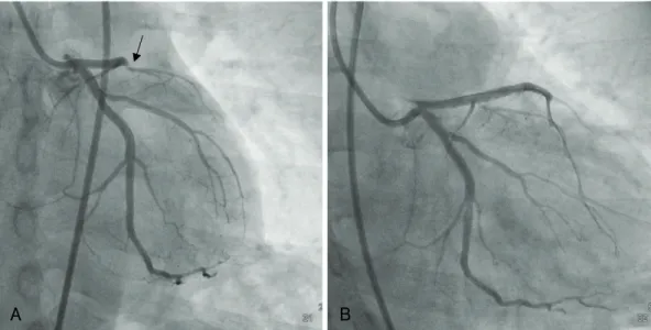
Fig. 2. Left coronary angiogram showed total occlusion of the left anterior descending artery (arrow) (A) and the lesion was treated with two sirolimus-eluting stents (B).
290·Postcardiac Injury Sydrome and Acute Mediastinitis
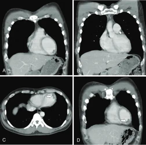
Fig. 3. Axial (A and B) and coronal (C and D) CT images of the chest showed a soft tissue density lesion surrounding the ascending aortic wall and subtle streaky fatty infiltrations with a small gas bubble (arrow), and also showed pericardial thickening and a subtle air density, suggesting acute mediastinitis with aortitis and acute pericarditis. A small bilateral pleural effusion with basal lung atelectasis was also noted.
A B
C D
Fig. 4. A follow-up chest CT (coronal view; A, B and D and axial view; C) showed that the previously noted soft tissue density lesion sur- rounding ascending aortic wall and subtle streaky fatty infiltrations with a small gas bubble had resolved. However, multiple low density nodules (arrow) were found in the apex of the left ventricle, suggesting acute thrombi.
A B
C D
Hong-Kyu Lim, et al.·291
ac procedures, such as pacemaker implantation, PCIS has rarely been reported after coronary angioplasty. Valan- der and colleagues2) described a case of PCIS in which Dressler’s syndrome developed after an extensive MI.2) More recently, Hearne et al.10) reported a case of PCIS 3 weeks after coronary intervention.
Prior to the development of cardiovascular surgery, most cases of mediastinitis arose from esophageal per- foration or contiguous spread of odontogenic or retro- pharyngeal infections. In modern practice, however, most cases of mediastinitis are complications of cardiovascu- lar or thoracic surgical procedures.11-13) The radiologic hallmark of non-postoperative mediastinitis is medias- tinal widening on chest X-ray. Other rare abnormalities include air fluid levels in the mediastinum or subcuta- neous tissue, and mediastinal air on lateral chest radi- ographs. A CT diagnosis of mediastinitis is based on the presence of mediastinal air and fluid collections with or without peristernal abnormalities, such as soft tissue ede- ma or sternal separation.14-16)
The case presented herein showed that PCIS occurr- ed within a few days of an AMI, and that the clinical con- dition was similar in appearance to acute mediastinitis.
Indeed, PCIS rarely occurs several days after an AMI.
Moreover, it has never been reported that PCIS present- ing as mediastinitis occurs after an AMI.
Our case is the first case of PCIS presenting as acute mediastinitis. One should consider this diagnosis after coronary angioplasty when the patient develops sudden chest pain and if the chest CT reveals pericardial thick- ening with small gas bubbles.
REFERENCES
1) Khan AH. The postcardiac injury syndromes. Clin Cardiol 1992;
15:67-72.
2) Velander M, Grip L, Mogensen L. The postcardiac injury syn- drome following percutaneous transluminal coronary angioplasty.
Clin Cardiol 1993;16:353-4.
3) Wood MA, Ellenbogen KA, Hall J, Kay GN. Post-pericardio- tomy syndrome following linear left atrial radiofrequency abla- tion. J Interv Card Electrophysiol 2003;9:55-7.
4) Soloff LA, Zatuchni J, Janton OH, O’Neill TJ, Glover RP. Re- activation of rheumatic fever following mitral commissurotomy.
Circulation 1953;8:481-97.
5) Dressler W. A post-myocardial infarction syndrome: preliminary report of a complication resembling idiopathic, recurrent, benign pericarditis. JAMA 1956;160:1379-83.
6) Miller RH, Horneffer PJ, Gardner TJ, Rykiel MF, Pearson TA.
The epidemiology of the postpericardiotomy syndrome: a com- mon complication of surgery. Am Heart J 1988;116:1323-9.
7) Loughlin V, Murphy A, Russell C. The post-pericardiotomy syn- drome and penetrating injury of the chest. Injury 1987;18:412-4.
8) De Scheerder I, DeBuyzere M, Robbrecht J, et al. Postoperative immunological response against contractile proteins after coro- nary bypass surgery. Br Heart J 1986;56:440-4.
9) Kennedy HL, Das SK. Postmyocardial infarction (Dressler’s) syndrome: report of a case with immunological and viral studies.
Am Heart J 1976;91:233-9.
10) Hearne C, Forjuoh SN. Postcardiac injury syndrome after cor- onary angioplasty and stenting. J Am Board Fam Pract 2003;16:
73-4.
11) Milano CA, Kesler K, Archibald N, Sexton DJ, Jones RH. Me- diastinitis after coronary artery bypass graft surgery: risk factors and long-term survival. Circulation 1995;92:2245-51.
12) Farrington M, Webster M, Fenn A, Phillips I. Study of cardio- thoracic wound infections at St. Thomas’ Hospital. Br J Surg 1985;
72:759-62.
13) Ottino G, De Paulis R, Pansini S, et al. Major sternal wound in- fection after open heart surgery: a multivariate analysis of risk factors in 2,579 consecutive operative procedures. Ann Thorac Surg 1987;44:173-9.
14) Kay HR, Goodman LR, Teplick SK, Mundth ED. Use of com- puted tomography to assess mediastinal complications after me- dian sternotomy. Ann Thorac Surg 1983;36:706-14.
15) Carrol CL, Jeffrey RB Jr, Federle MP, Vernacchia FS. CT eval- uation of mediastinal infections. J Comput Assist Tomogr 1987;
11:449-54.
16) Templeton PA, Fishman EK. CT evaluation of poststernotomy complications. AJR Am J Roentgenol 1992;159:45-50.