Screening for Abdominal Aortic Aneurysm during Transthoracic Echocardiography in Patients
with Significant Coronary Artery Disease
Sung Ho Lee,
1* Sung-A Chang,
2* Shin Yi Jang,
2Sang-Chol Lee,
2Young Bin Song,
2Seung Woo Park,
2Seung-Hyuk Choi,
2Hyeon-Cheol Gwon,
2Jae K. Oh,
2,3and Duk-Kyung Kim
21Division of Cardiology, Department of Medicine, Kangbuk Samsung Hospital, Sungkyunkwan University School of Medicine, Seoul;
2Division of Cardiology, Department of Medicine, Heart Vascular Stroke Institute, Samsung Medical Center, Sungkyunkwan University School of Medicine, Seoul, Korea;
3Division of Cardiovascular Diseases, Mayo Clinic College of Medicine, Rochester, MN, USA.
Received: December 3, 2013 Revised: March 12, 2014 Accepted: March 27, 2014
Corresponding author: Dr. Duk-Kyung Kim, Division of Cardiology, Department of Medicine, Heart Vascular Stroke Institute,
Samsung Medical Center,
Sungkyunkwan University School of Medicine, 81 Irwon-ro, Gangnam-gu,
Seoul 135-710, Korea.
Tel: 82-2-3410-3419, Fax: 82-2-3410-3849 E-mail: [email protected]
*Sung Ho Lee and Sung-A Chang equally contributed to this work.
∙ The authors have no financial conflicts of interest.
© Copyright:
Yonsei University College of Medicine 2015 This is an Open Access article distributed under the terms of the Creative Commons Attribution Non- Commercial License (http://creativecommons.org/
licenses/by-nc/3.0) which permits unrestricted non- commercial use, distribution, and reproduction in any medium, provided the original work is properly cited.
Purpose: Coronary artery disease (CAD) shares several risk factors with abdomi- nal aortic aneurysm (AAA). We evaluated the prevalence during transthoracic echocardiography (TTE) and risk factors of AAA in patients with CAD. Materials and Methods: A total of 1300 CAD patients were screened from August 2009 to May 2010, and measurement of abdominal aorta size was feasible in 920 patients (71%) at the end of routine TTE. An AAA was defined as having a maximal diame- ter of ≥30 mm. Results: Of the 920 patients, 22 (2.4% of the study population) were diagnosed with AAA; of these AAA patients, 86% were male, and 82% were over 65 years-old. Abdominal aortic size was weakly correlated with aortic root di- ameter (r=0.22, p<0.01). Although the proportions of male gender, hypertension, and dyslipidemia were higher in AAA patients, such differences were not statistical- ly significant. Advanced age [odds ratio (OR)=1.07; 95% confidence interval (CI):
1.01‒1.12; p<0.01], smoking (OR=3.44; 95% CI: 1.18‒10.04; p=0.02), and periph- eral arterial disease (OR=5.88; 95% CI: 1.38‒25.05; p=0.01) were found to be as- sociated with AAA. Conclusion: Although prevalence of AAA is very low in the Asian population, the prevalence of AAA in Asian CAD patients is higher than the general population. Therefore, opportunistic examination of the abdominal aorta during routine TTE could be effective, especially for male CAD patients over 65 years with a history of smoking or peripheral arterial disease.
Key Words: Abdominal aortic aneurysm, screening, coronary artery disease, transthoracic echocardiography
INTRODUCTION
An abdominal aortic aneurysm (AAA) is defined as an aorta size of more than 30
mm or regional dilation of the abdominal aorta by more than 50%. AAA usually
remains asymptomatic unless it ruptures, and in cases of rupture, operative mortal-
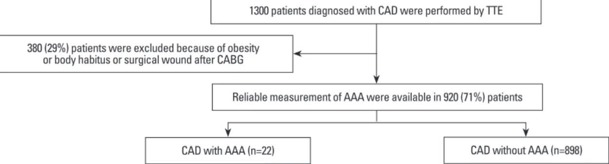
in 1300 consecutive patients who had been diagnosed with significant CAD. Of these patients, reliable measurements of abdominal aorta size were available in 920 patients (71%).
Intestinal gas, obesity or body habitus, and surgical wounds after coronary artery bypass grafting (CABG) were the rea- sons for inadequate visualization of the abdominal aorta in 380 patients (29%). Among these patients, 99 (10.8%) had their TTE done after CABG; echocardiographic windows of the abdominal aorta could not be acquired due to surgi- cal wounds from CABG. The analysis was based on the 920 patients (677 men and 243 women) from whom reli- able aorta size measurements were available (Fig. 1). As a retrospective study, there was no need to obtain informed consent from each patient.
Significant coronary artery stenosis was defined as a nar- rowing of ≥50% in diameter of the epicardial segment of the coronary artery, as observed through coronary angiog- raphy (CAG). All clinical data and laboratory data were collected using an electrical recording system in a prospec- tive manner. This study was approved by the Institutional Review Board of our hospital.
Transthoracic echocardiography
TTE images were acquired by experienced sonographers, interpreted by echocardiographers, and prospectively re- corded in an electrical reporting system. No instructions on food or fluid intake were given prior to the examination.
Routine examinations, which included 2-dimension, M- mode, and Doppler techniques, were performed, and mea- surements were made according to the guidelines of the American Society of Echocardiography. At the end of the examination, the abdominal aorta was visualized with the patient in supine position, as previously described,
6using a Vivid E9 (GE Healthcare, Horten, Norway) or SC2000 ul- trasound (Siemens Medical Solutions, Mountain View, CA, USA) and a 3.7 Hz transducer. First, a longitudinal image ity rate often exceeds 50%.
1However, if patients undergo
elective surgery for AAA, hospital mortality rate is greatly reduced to <5%.
2Therefore, early diagnosis of AAA is cru- cial, and a screening of AAA is recommended especially for a high risk population.
The prevalence of AAA has been reported in Western countries as 1.3‒8.9% in men and 1.0‒2.2% in women.
3-5On the contrary, the prevalence of AAA in the Asian popu- lation has not been studied thoroughly. Previously, we have reported that the prevalence of AAA in the Korean popula- tion is 0.5%, found by the routine screening for AAA dur- ing clinical transthoracic echocardiography (TTE).
6AAA is considered a manifestation of atherosclerosis,
7and coronary artery disease (CAD) may be associated with a higher prevalence of AAA than the normal population
8,9Also, AAA and CAD share several risk factors such as male gender, advanced age, hypertension, smoking, periph- eral arterial disease, and hypercholesterolemia.
10,11A high incidence of adverse cardiovascular events such as cardio- vascular death, myocardial infarction, and stroke were ob- served in patients with AAA and CAD.
12However, there have been few studies regarding the prevalence of AAA in Asians with CAD.
TTE is usually performed on CAD patients for a clinical purpose; thus, echocardiographic protocol can be modified for use as a screening method for AAA. The purpose of this study was to investigate the prevalence of AAA during TTE and the associated risk factors of AAA with CAD patients in Korea.
MATERIALS AND METHODS
Study subjects
From August 2009 to May 2010 at Samsung Medical Center, screening for AAA during TTE was prospectively performed
Fig. 1. Study population. CAD, coronary artery disease; AAA, abdominal aortic aneurysm; TTE, transthoracic echocardiography; CABG, coro- nary artery bypass grafting.
CAD with AAA (n=22) CAD without AAA (n=898)
380 (29%) patients were excluded because of obesity or body habitus or surgical wound after CABG
Reliable measurement of AAA were available in 920 (71%) patients 1300 patients diagnosed with CAD were performed by TTE
index value ≤0.9.
17Statistical analysis
Continuous variables were described as mean±standard de- viation. Categorical variables were expressed as numbers and percentages (%). The chi-square test was used for the comparison of categorical variables, and the independent sample t-test was applied for the comparison of continuous variables. A p-value less than 0.05 was considered statisti- cally significant. Statistical analysis was performed with IBM SPSS Statistics (Version 19.0, IBM SPSS Inc., Chica- go, IL, USA).
RESULTS
The prevalence of AAA in CAD patients
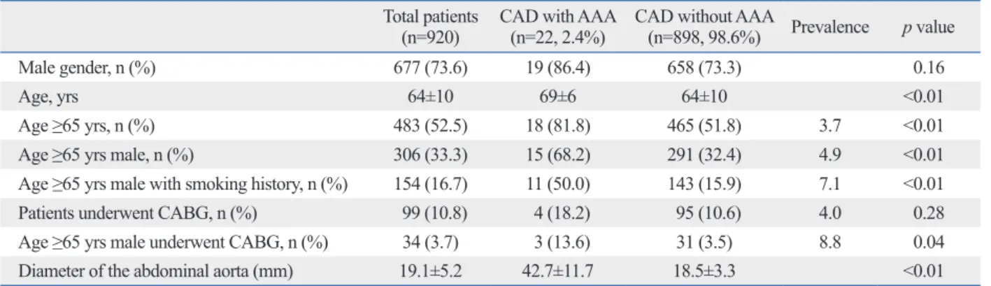
The baseline demographic variables of the study population are shown in Table 1. The mean age of the study population was 64±10 years. The mean abdominal aorta size was 19.1±5.2 mm. AAA was diagnosed in 22 patients of 920 CAD patients (2.4% of the study population), 19 of who were male (86% of AAA patients). Among them, 9 patients had already been diagnosed with AAA by a different imag- ing study and 13 patients (1.4%) were newly diagnosed with AAA from the TTE study.
All 3 women with AAA were over 70 years old. The mean age of AAA patients was 69±6 years, and 82% of AAA patients were older than 65 years. Considering only the male patients aged over 65 years, the 4.9% of patients (15 of 306) were found to have an AAA. The prevalence of AAA in male patients over 65 years with a smoking history was 7.1% (11 of 154).
Patients with AAA were older than those without AAA of the abdominal aorta was visualized with the transducer
marker pointing toward the patient’s feet. The transducer was then rotated 90° counterclockwise and adjusted to scan the orthogonal plane perpendicular to the central line of the abdominal aorta. The infrarenal abdominal aorta was visu- alized below the origin of the renal artery and then traced distally as far as possible. The maximum short-axial diame- ter of the abdominal aorta was measured at the antero-pos- terior plane of the abdominal aorta because that has been shown to be more reproducible than transverse diameter.
13The average examination time required for the evaluation of the abdominal aorta was about 2 minutes. An abdominal aorta greater than 30 mm was recognized as an AAA.
Risk factors
Hypertension was defined as systolic blood pressure of 140 mm Hg or higher, diastolic blood pressure of 90 mm Hg or higher, or use of oral antihypertensive medication.
14Smok- ing history was classified as either current smoker or past smoker. Patients who smoked at least 1 cigarette/day were defined as current smokers, and patients who had quit smok- ing for at least 1 year were defined as past smokers. Dyslip- idemia was defined as total cholesterol level >240 mg/dL, low-density lipoprotein cholesterol level >130 mg/dL, high- density lipoprotein cholesterol level <40 mg/dL for men or
<50 mg/dL for women, triglyceride level >200 mg/dL, or use of antihyperlipidemic medication.
15Diabetes mellitus was defined as fasting plasma glucose level ≥126 mg/dL, random plasma glucose level ≥200 mg/dL in patients with classic hyperglycemic symptoms, use of oral hypoglycemic drugs or insulin, or life style modifications for the treatment of known diabetes.
16Peripheral arterial disease (PAD) was defined as Rutherford claudication stage ≥3, history of treat- ment for chronic limb ischemia, or ankle brachial pressure
Table 1. Demographic Variables of the Study Population
Total patients(n=920) CAD with AAA
(n=22, 2.4%) CAD without AAA
(n=898, 98.6%) Prevalence p value
Male gender, n (%) 677 (73.6) 19 (86.4) 658 (73.3) 0.16
Age, yrs 64±10 69±6 64±10 <0.01
Age ≥65 yrs, n (%) 483 (52.5) 18 (81.8) 465 (51.8) 3.7 <0.01
Age ≥65 yrs male, n (%) 306 (33.3) 15 (68.2) 291 (32.4) 4.9 <0.01
Age ≥65 yrs male with smoking history, n (%) 154 (16.7) 11 (50.0) 143 (15.9) 7.1 <0.01
Patients underwent CABG, n (%) 99 (10.8) 4 (18.2) 95 (10.6) 4.0 0.28
Age ≥65 yrs male underwent CABG, n (%) 34 (3.7) 3 (13.6) 31 (3.5) 8.8 0.04
Diameter of the abdominal aorta (mm) 19.1±5.2 42.7±11.7 18.5±3.3 <0.01
AAA, abdominal aortic aneurysm; CAD, coronary artery disease; CABG, coronary artery bypass grafting.
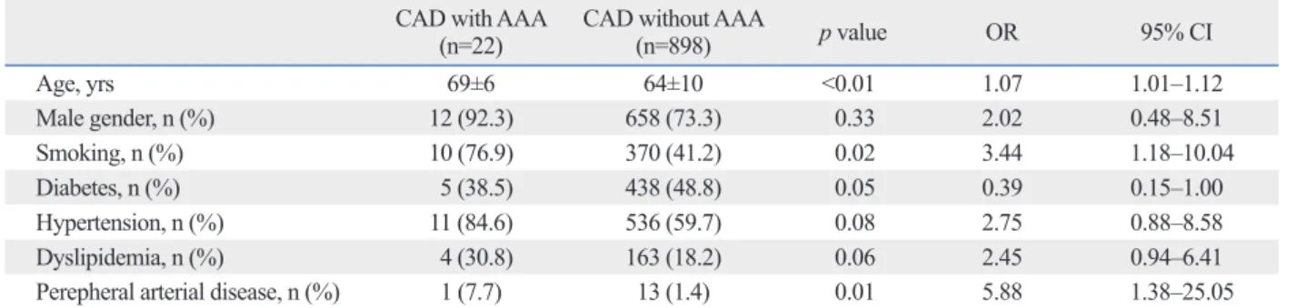
and the prevalence of hypertension and dyslipidemia were higher in AAA, they were not statistically significant. Diabe- tes mellitus was inversely associated with AAA and was sta- tistically insignificant. Multiple logistic regression analysis showed advanced age [odds ratio (OR)=1.07; 95% confi- dence interval (CI): 1.01‒1.12; p<0.01], smoking (OR=
3.44; 95% CI: 1.18‒10.04; p=0.02), and PAD (OR=5.88;
95% CI: 1.38‒25.05; p=0.01) as the independent risk fac- tors for AAA.
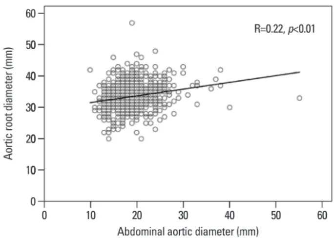
Aortic root (sinuses of valsalva) size measured by TTE was greater in patients with AAA than in those without AAA (AAA: 35 mm; non-AAA: 33 mm; p<0.01). Aortic root size was weakly correlated with abdominal aortic size in Fig. 2 (r=0.22; p<0.01). After adjustment for age (r=0.22;
p<0.01), gender (r=0.24; p<0.01), and smoking status (r=0.23; p<0.01), weak correlation was observed between abdominal aortic size and aortic root size. Other echocardio- (AAA: 69±6; non-AAA: 64±10 years; p<0.01) and more
often male than female, but without statistical significance (male: 86%; female: 73%; p=0.16). The mean AAA size was 42.7±11.7 mm. Among the 13 newly diagnosed pa- tients, AAA was confirmed by abdominal computed to- mography for only 3 patients as the other patients were found to have smaller AAAs. One patient (7.6%) had an aortic diameter greater than 50 mm and underwent an elec- tive surgical open repair of AAA. The other two patients underwent endovascular aneurysm repair in order to treat associated thoracic aortic aneurysms. There were no serious complications after AAA repair.
Risk factors and echocardiographic findings of AAA We compared the cardiovascular risk factors (Table 2) and echocardiographic findings (Table 3) of CAD patients with and without AAA. Although the proportion of male gender
Table 2. Comparison of Cardiovascular Risk Factors in CAD Patients with and without AAA
CAD with AAA(n=22) CAD without AAA
(n=898) p value OR 95% CI
Age, yrs 69±6 64±10 <0.01 1.07 1.01‒1.12
Male gender, n (%) 12 (92.3) 658 (73.3) 0.33 2.02 0.48‒8.51
Smoking, n (%) 10 (76.9) 370 (41.2) 0.02 3.44 1.18‒10.04
Diabetes, n (%) 5 (38.5) 438 (48.8) 0.05 0.39 0.15‒1.00
Hypertension, n (%) 11 (84.6) 536 (59.7) 0.08 2.75 0.88‒8.58
Dyslipidemia, n (%) 4 (30.8) 163 (18.2) 0.06 2.45 0.94‒6.41
Perepheral arterial disease, n (%) 1 (7.7) 13 (1.4) 0.01 5.88 1.38‒25.05
AAA, abdominal aortic aneurysm; CAD, coronary artery disease; OR, odds ratio; CI, confidence interval.
Table 3. Echocardiographic Characteristics of Patients with and without AAA
CAD with AAA (n=22) CAD without AAA (n=898) p value
IVSd (mm) 9.3±1.1 9.1±1.5 0.45
LVPWd (mm) 8.8±1.3 8.9±1.4 0.70
LV EDD (mm) 51±6 51±6 0.81
LV ESD (mm) 32±6 32±7 0.84
LV EF (%) 60±9 59±11 0.58
LA (mm) 39±12 40±6 0.66
E (m/s) 0.63±0.25 0.66±0.21 0.59
A (m/s) 0.73±0.13 0.76±0.20 0.49
E/A 0.85±0.42 0.92±0.52 0.59
DT (msec) 229±44 226±62 0.85
E’ (m/s) 0.05±0.01 0.06±0.04 0.49
A’ (m/s) 0.08±0.01 0.09±0.05 0.59
E/E’ 10.92±2.96 11.55±5.55 0.60
Aortic root (sinuses of valsalva) (mm) 35.9±4.0 33.4±4.0 <0.01
AAA, abdominal aortic aneurysm; CAD, coronary artery disease; IVSd, interventricular septal wall thickness at diastole; LVPWd, left ventricular posterior wall thickness at diastole; LVEDD, left ventricular end-diastolic dimension; LVESD, left ventricular end-systolic dimension; LV EF, left ventricular ejection fraction; LA, left atrium; E, early diastolic mitral inflow velocity; A, late diastolic mitral inflow velocity; DT, deceleration time of E velocity; E’, early diastolic septal mitral annular velocity; A’, late diastolic septal mitral annular velocity.
casians with CAD. In this study, we enrolled patients with any significant CAD diagnosed by CAG. Thus, our study population includes rather mild forms of CAD than other reports. Although the prevalence of AAA in Koreans with CAD is low (2.4% of the study population) when compared with Western reports, this prevalence in CAD patients may be nearly five times higher than the general Korean popula- tion (0.5%) in our previous report.
6To the best of our knowledge, this is the first study to focus on AAA preva- lence in Korean CAD patients.
Several previous studies demonstrated that smoking is strongly associated with the risk of AAA.
10,23-25Smoking has been suggested as indicator for AAA screening even in wom- en of age >65 years.
26Svensjö, et al.
27reported a low preva- lence of AAA among 65-year-old Swedish men and ex- plained that decreasing numbers of smokers compared to previous generations is associated with a change in the epide- miology of the disease. Smoking not only promotes athero- sclerosis but may also block the active site of α1-antitrypsin, which could promote the destruction of the aortic wall by proteolytic enzymes in an additional nonatheroscleritic path- way.
25,28Other studies reported increased plasma levels of matrix metalloproteinases-1, -2, and -9 in patients with AAA,
29-31and this observation may be related to the effects of smoking on elastase activity and elastin degradation in the vessel wall media.
32TTE is almost routinely performed in CAD patients for evaluating left ventricular (LV) systolic function, detecting regional LV wall motion abnormalities and complications, and assessing prognosis. Our study revealed that the feasi- bility of visualization of abdominal aortic size was consid- erably high (71%). Moreover, evaluation of the abdominal aortic size during TTE is not time consuming or laborious, nor is there additional cost. Considering the finding that CAD patients present with a higher prevalence of AAA and are more frequently examined with TTE, patients with CAD could be considered as a selective group for AAA screening, which can take place during TTE to save costs.
That effect would be even more augmented in 65 year old male CAD patients with a history of smoking or peripheral arterial disease.
This study has several limitations. First, the prevalence of AAA in CAD in our study population was low; slightly un- dermining the statistical reliability of the risk factors of AAA. Second, we included the postoperative patients who underwent CABG, and these patients’ echocardiographic windows were frequently poor, which indicates that the fea- graphic variables were not significantly different between
groups (Table 3).
DISCUSSION
We investigated the prevalence of AAA in CAD patients through a modified screening protocol using TTE. We found that the prevalence of those newly diagnosed with AAA by screening using TTE was 2.4% in CAD patients who under- went CAG in Korea. The prevalence tended to increase with age and for males. For example, among males over 65 years old with CAD, the prevalence of AAA reached 4.9%. Sever- al previous studies of Caucasian CAD patients have report- ed varying rates of AAA prevalence ranging from 6.9% to 14% (Supplementary Table 1, only online).
8,9,18-21As most of these studies are composed of males older than 60 years who had severe CAD and underwent CABG, prevalence in these studies may be much higher than those in subjects without CAD. Regarding the disposition of AAA in CAD patients, a recent study with 35418 individuals in Sweden
22showed a higher incidence of AAA in northern Sweden, cor- responding well with reported CAD patterns and suggesting that the presence of CAD is linked to risk of AAA.
However, there is little data about the prevalence of AAA in CAD patients in the Asian population. One previous study composed of Chinese patients with severe CAD and awaiting elective CABG showed a much lower prevalence of 1.8% compared to the prevalence reported with Cauca- sians (Supplementary Table 1, only online).
21The present study shows results similar to the study involving Chinese patients, which supports the claim that the prevalence of AAA is found to be lower in Asians with CAD than in Cau-
Fig. 2. Correlations between the diameter of the abdominal aorta and diam- eters of aortic root.
Abdominal aortic diameter (mm) 0
0 0 0 0 0
10 20 30 40 50 60
Aortic root diameter (mm)
0 10 20 30 40 50 60
R=0.22, p<0.01
abdominal aortic aneurysm and surveillance of small abdominal aortic aneurysms, rationale and recommendations of the French Society for Vascular Medicine. Final document]. J Mal Vasc 2006;31:260-76.
14. Chobanian AV, Bakris GL, Black HR, Cushman WC, Green LA, Izzo JL Jr, et al. The Seventh Report of the Joint National Com- mittee on Prevention, Detection, Evaluation, and Treatment of High Blood Pressure: the JNC 7 report. JAMA 2003;289:2560-72.
15. Expert Panel on Detection, Evaluation, and Treatment of High Blood Cholesterol in Adults. Executive Summary of The Third Report of The National Cholesterol Education Program (NCEP) Expert Panel on Detection, Evaluation, And Treatment of High Blood Cholesterol In Adults (Adult Treatment Panel III). JAMA 2001;285:2486-97.
16. American Diabetes Association. Diagnosis and classification of diabetes mellitus. Diabetes Care 2013;36 Suppl 1:S67-74.
17. Hirsch AT, Haskal ZJ, Hertzer NR, Bakal CW, Creager MA, Hal- perin JL, et al. ACC/AHA Guidelines for the Management of Pa- tients with Peripheral Arterial Disease (lower extremity, renal, mesenteric, and abdominal aortic): a collaborative report from the American Associations for Vascular Surgery/Society for Vascular Surgery, Society for Cardiovascular Angiography and Interven- tions, Society for Vascular Medicine and Biology, Society of In- terventional Radiology, and the ACC/AHA Task Force on Practice Guidelines (writing committee to develop guidelines for the man- agement of patients with peripheral arterial disease)--summary of recommendations. J Vasc Interv Radiol 2006;17:1383-97.
18. Monney P, Hayoz D, Tinguely F, Cornuz J, Haesler E, Mueller XM, et al. High prevalence of unsuspected abdominal aortic aneu- rysms in patients hospitalised for surgical coronary revascularisa- tion. Eur J Cardiothorac Surg 2004;25:65-8.
19. Dupont A, Elkalioubie A, Juthier F, Tagzirt M, Vincentelli A, Le Tourneau T, et al. Frequency of abdominal aortic aneurysm in pa- tients undergoing coronary artery bypass grafting. Am J Cardiol 2010;105:1545-8.
20. Long A, Bui HT, Barbe C, Henni AH, Journet J, Metz D, et al.
Prevalence of abdominal aortic aneurysm and large infrarenal aorta in patients with acute coronary syndrome and proven coronary ste- nosis: a prospective monocenter study. Ann Vasc Surg 2010;24:
602-8.
21. Poon JT, Cheng SW, Wong JS, Ting AC. Prevalence of abdominal aortic aneurysm in Chinese patients with severe coronary artery disease. ANZ J Surg 2010;80:630-3.
22. Hultgren R, Forsberg J, Alfredsson L, Swedenborg J, Leander K.
Regional variation in the incidence of abdominal aortic aneurysm in Sweden. Br J Surg 2012;99:647-53.
23. Singh K, Bønaa KH, Jacobsen BK, Bjørk L, Solberg S. Preva- lence of and risk factors for abdominal aortic aneurysms in a pop- ulation-based study: The Tromsø Study. Am J Epidemiol 2001;
154:236-44.
24. Golledge J, Muller J, Daugherty A, Norman P. Abdominal aortic aneurysm: pathogenesis and implications for management. Arte- rioscler Thromb Vasc Biol 2006;26:2605-13.
25. Lee AJ, Fowkes FG, Carson MN, Leng GC, Allan PL. Smoking, atherosclerosis and risk of abdominal aortic aneurysm. Eur Heart J 1997;18:671-6.
26. Derubertis BG, Trocciola SM, Ryer EJ, Pieracci FM, McKinsey JF, Faries PL, et al. Abdominal aortic aneurysm in women: preva- lence, risk factors, and implications for screening. J Vasc Surg 2007;46:630-5.
sibility of AAA screening using TTE may be low for such patients.
In conclusion, the prevalence of AAA in CAD was low but higher than the general population, as observed in opportu- nistic examinations of abdominal aorta during routine TTE.
The prevalence tended to increase with older males. Of tradi- tional risk factors, advanced age, smoking, and PAD were statistically significant risk factors of AAA in CAD patients.
Therefore, opportunistic examination of the abdominal aorta during routine TTE could be effective, especially for male CAD patients over 65 years with a history of smoking or PAD.
REFERENCES
1. Ingoldby CJ, Wujanto R, Mitchell JE. Impact of vascular surgery on community mortality from ruptured aortic aneurysms. Br J Surg 1986;73:551-3.
2. Graham M, Chan A. Ultrasound screening for clinically occult ab- dominal aortic aneurysm. CMAJ 1988;138:627-9.
3. Sakalihasan N, Limet R, Defawe OD. Abdominal aortic aneu- rysm. Lancet 2005;365:1577-89.
4. Lederle FA, Johnson GR, Wilson SE; Aneurysm Detection and Management Veterans Affairs Cooperative Study. Abdominal aor- tic aneurysm in women. J Vasc Surg 2001;34:122-6.
5. Lederle FA, Johnson GR, Wilson SE, Chute EP, Hye RJ, Ma- karoun MS, et al. The aneurysm detection and management study screening program: validation cohort and final results. Aneurysm Detection and Management Veterans Affairs Cooperative Study Investigators. Arch Intern Med 2000;160:1425-30.
6. Oh SH, Chang SA, Jang SY, Park SJ, Choi JO, Lee SC, et al.
Routine screening for abdominal aortic aneurysm during clinical transthoracic echocardiography in a Korean population. Echocar- diography 2010;27:1182-7.
7. Reed D, Reed C, Stemmermann G, Hayashi T. Are aortic aneu- rysms caused by atherosclerosis? Circulation 1992;85:205-11.
8. Bergersen L, Kiernan MS, McFarlane G, Case TD, Ricci MA.
Prevalence of abdominal aortic aneurysms in patients undergoing coronary artery bypass. Ann Vasc Surg 1998;12:101-5.
9. Madaric J, Vulev I, Bartunek J, Mistrik A, Verhamme K, De Bruyne B, et al. Frequency of abdominal aortic aneurysm in pa- tients >60 years of age with coronary artery disease. Am J Cardiol 2005;96:1214-6.
10. Forsdahl SH, Singh K, Solberg S, Jacobsen BK. Risk factors for abdominal aortic aneurysms: a 7-year prospective study: the Tromsø Study, 1994-2001. Circulation 2009;119:2202-8.
11. Cornuz J, Sidoti Pinto C, Tevaearai H, Egger M. Risk factors for asymptomatic abdominal aortic aneurysm: systematic review and meta-analysis of population-based screening studies. Eur J Public Health 2004;14:343-9.
12. Miura T, Soga Y, Doijiri T, Aihara H, Yokoi H, Iwabuchi M, et al.
Prevalence and clinical outcome of polyvascular atherosclerotic disease in patients undergoing coronary intervention. Circ J 2013;77:89-95.
13. Becker F, Baud JM; Groupe de Travail Ad Hoc. [Screening for
2006;43:95-100.
30. Longo GM, Xiong W, Greiner TC, Zhao Y, Fiotti N, Baxter BT.
Matrix metalloproteinases 2 and 9 work in concert to produce aor- tic aneurysms. J Clin Invest 2002;110:625-32.
31. Kakafika AI, Mikhailidis DP. Smoking and aortic diseases. Circ J 2007;71:1173-80.
32. Murphy EA, Danna-Lopes D, Sarfati I, Rao SK, Cohen JR. Nico- tine-stimulated elastase activity release by neutrophils in patients with abdominal aortic aneurysms. Ann Vasc Surg 1998;12:41-5.
27. Svensjö S, Björck M, Gürtelschmid M, Djavani Gidlund K, Hell- berg A, Wanhainen A. Low prevalence of abdominal aortic aneu- rysm among 65-year-old Swedish men indicates a change in the epidemiology of the disease. Circulation 2011;124:1118-23.
28. Cohen JR, Sarfati I, Ratner L, Tilson D. Alpha 1-antitrypsin phe- notypes in patients with abdominal aortic aneurysms. J Surg Res 1990;49:319-21.
29. Kowalewski R, Sobolewski K, Małkowski A, Wolan´ska M, Gacko M. Evaluation of enzymes involved in proteoglycan degra- dation in the wall of abdominal aortic aneurysms. J Vasc Res
population AAA (%) AAA (mm)
Monney, et al.18 Switzerland M>60 395 CABG 40 (10.1) ≥30
Dupont, et al.19 France 217 (M 189, F 28) CABG 15 (6.9) ≥30
Bergersen, et al.8 USA M>50 192 CABG 24 (13) ≥30
Madaric, et al.9 Slovak Republic M>60 109 Stenosis ≥50% 16 (14) ≥30
Long, et al.20 France 304 (M 234, F 70) ACS with stenosis ≥50% 20 (6.6) ≥30
Poon, et al.21 Hong Kong 624 (M 499, F 125) CABG 11 (1.8) ≥30
AAA, abdominal aortic aneurysm; CAD, coronary artery disease; CABG, coronary artery bypass grafting; ACS, acute coronary syndrome.