INTRODUCTION
Elastin is involved in the maintenance of vascular compli-
ance. A change in the balance between elastin synthesis and degradation may lead to the development of a pathological vascular condition.1 Elastin degradation has been reported to be a determinant of arterial stiffness and has prognostic value for clinical outcomes in high-risk populations.2 In many stud- ies, elastin degradation peptides have been proposed as par- ticipating in the progression of atherosclerosis through the ac- tivation of various biological processes.3
For decades, anti-elastin antibodies have been assayed by immunological techniques. Levels of the antibody and the changes that occur in normal and pathological conditions have also been investigated in prior studies.4-6 Therein, blood levels of elastin peptides and those of the antibodies against elastin peptides did not correlate.7,8 Moreover, elastin degra- dation by diverse enzymes can release various epitopes, and any antibody-disease association may depend on the origin of the epitopes.9 To date, the clinical relevance of antibody levels
Circulating Anti-Elastin Antibody Levels and Arterial Disease Characteristics: Associations with Arterial Stiffness and Atherosclerosis
Seung-Hyun Lee1*, Kihyuk Shin2*, Sungha Park1,3, Seok-Min Kang1,3, Donghoon Choi1,3, Seung-Hyo Lee2, and Sang-Hak Lee1,3
1Division of Cardiology, Department of Internal Medicine, Severance Hospital, Yonsei University College of Medicine, Seoul;
2Graduate School of Medical Science and Engineering, Korea Advanced Institute of Science and Technology, Daejeon;
3Cardiovascular Research Institute, Yonsei University Health System, Seoul, Korea.
Purpose: Elastin is a major arterial structural protein, and elastin-derived peptides are related to arterial change. We previously reported on a novel assay developed using aortic elastin peptides; however, its clinical implications remain unclear. In this study, we assessed whether anti-elastin antibody titers reflect the risk of coronary artery disease (CAD) or its characteristics.
Materials and Methods: We included 174 CAD patients and 171 age- and sex-matched controls. Anti-elastin antibody titers were quantified by enzyme-linked immunosorbent assay. Parameters of arterial stiffness, including the augmentation index (AI) and heart-to-femoral pulse wave velocity (hfPWV), were measured non-invasively. The clinical and angiographic characteristics of CAD patients were also evaluated. Associations between anti-elastin levels and vascular characteristics were examined by linear regression analysis.
Results: The median blood level of anti-elastin was significantly lower in the CAD group than in the controls [197 arbitrary unit (a.u.) vs. 63 a.u., p<0.001]. Levels of anti-elastin were significantly lower in men and in subjects with hypertension, diabetes melli- tus, hyperlipidemia, or high hfPWV. Nevertheless, anti-elastin levels were not dependent on atherothrombotic events or the angi- ographic severity of CAD. In a multivariate analysis, male sex (β=-0.38, p<0.001), diabetes mellitus (β=-0.62, p<0.001), hyperlipid- emia (β=-0.29, p<0.001), and AI (β=-0.006, p=0.02) were ultimately identified as determinants of anti-elastin levels.
Conclusion: Lower levels of anti-elastin are related to CAD. The association between antibody titers and CAD is linked to arterial stiffness rather than the advancement of atherosclerosis.
Key Words: Elastin, antibody formation, vascular stiffness, coronary artery disease, atherosclerosis Yonsei Med J 2015 Nov;56(6):1545-1551
http://dx.doi.org/10.3349/ymj.2015.56.6.1545 pISSN: 0513-5796 · eISSN: 1976-2437
Received: November 17, 2014 Revised: December 26, 2014 Accepted: December 27, 2014
Co-corresponding authors: Dr. Sang-Hak Lee, Division of Cardiology, Depart- ment of Internal Medicine, Severance Hospital, Yonsei University College of Medi- cine, 50-1 Yonsei-ro, Seodaemun-gu, Seoul 03722, Korea.
Tel: 82-2-2228-8461, Fax: 82-2-2227-7732, E-mail: [email protected] and Dr. Seung-Hyo Lee, Graduate School of Medical Science and Engineering, Korea Advanced Institute of Science and Technology, 291 Daehak-ro, Yuseong-gu, Dae- jeon 34141, Korea.
Tel: 82-42-350-4235, Fax: 82-42-350-4240, E-mail: [email protected]
*Seung-Hyun Lee and Kihyuk Shin contributed equally to this work.
•The authors have no financial conflicts of interest.
© Copyright: Yonsei University College of Medicine 2015
This is an Open Access article distributed under the terms of the Creative Com- mons Attribution Non-Commercial License (http://creativecommons.org/ licenses/
by-nc/3.0) which permits unrestricted non-commercial use, distribution, and repro- duction in any medium, provided the original work is properly cited.
is not thoroughly understood and interpretation of antibody levels warrants caution. Although a few studies have investi- gated anti-elastin antibody levels in atherosclerotic cardiovas- cular disease, across these studies, the relationship between antibody levels and atherosclerosis has been inconsistent.5,6,10
The aim of this study was to evaluate the association be- tween anti-elastin antibody levels and coronary artery disease (CAD). We also assessed relationships between antibody lev- els and cardiovascular risk factors using a newly developed enzyme-linked immunosorbent assay (ELISA). We found that lower levels of anti-elastin are related to CAD and that this re- lation is linked to arterial stiffness.
MATERIALS AND METHODS
Study patients
This study included 171 patients with CAD and 174 control subjects without CAD. Participants were drawn from the data- base of the Cardiovascular Genome Center at Yonsei Universi- ty Health System, Seoul, Korea. CAD patients were recruited when undergoing coronary angiography for chest discomfort or chest pain. Subjects aged 30–70 years with stenosis >50% in at least one epicardial coronary artery were included. The ex- clusion criteria included uncontrolled high blood pressure (BP) (systolic BP >180 mm Hg or diastolic BP 110 mm Hg); uncon- trolled diabetes mellitus (fasting blood glucose 180 mg/dL); a history of structural heart disease with or without heart fail- ure; thyroid [thyroid stimulating hormone <lower limit of nor- mal or upper limit of normal (ULN)], liver (serum amino- transferase >2×ULN) or kidney disease (serum creatinine >1.5 mg/dL); acute or chronic inflammatory disease; and malig- nant neoplasm. Control subjects were matched for sex and age (±5 years), and selected from the database of a communi- ty health check-up center in Mapo-gu, Seoul, Korea. Written informed consent was obtained from all participants, and the study protocol was approved by the Institutional Review Board of Severance Hospital, Seoul, Korea.
Collection of clinical and angiographic data
At the time of enrollment, subjects were interviewed regard- ing their individual medical histories and underwent com- plete physical examinations. Hypertension was defined as a BP >140/90 mm Hg on two or more occasions or undergoing antihypertensive treatment. Diabetes mellitus was defined as a fasting blood glucose ≥126 mg/dL, postprandial blood glu- cose ≥200 mg/dL, or current treatment with hypoglycemic medications. Hyperlipidemia was defined as low-density li- poprotein-cholesterol ≥160 mg/dL. CAD characteristics were evaluated by a cardiologist who was blinded to the study’s purpose. The cardiologist first evaluated the most severe clini- cal presentation of CAD in the patient’s history, and then the number of coronary arteries with at least one stenosis of >50%.
After a 12-h fasting period, venous blood samples were col- lected, centrifuged, and stored at -80°C.
Measurement of augmentation index and pulse wave velocity
The augmentation index (AI) was measured in the sitting po- sition after a 5-min rest using a radial artery tonometry device (SphygmoCor, AtCor Medical, Sydney, Australia), as described previously.11 The central BP was calibrated to the brachial BP measured using the OMRON HEM 7080IT (Omron Health- care, Kyoto, Japan). Briefly, using a high-fidelity micromanom- eter (Millar Instruments, Houston, TX, USA), peripheral pres- sure waveforms from the radial artery were recorded at the wrist of the dominant arm. Central systolic BP, diastolic BP, pulse pressure, augmentation pressure, and AI were acquired from the pulse waveform analysis. Augmentation pressure is the difference between the second and first systolic peak pres- sures, and the AI is defined as the ratio of augmentation pres- sure to aortic pulse pressure.
The heart-to-femoral pulse wave velocity (hfPWV) was de- termined by a VP-2000 pulse wave unit (Nippon Co. Ltd., Ko- maki, Japan), as described previously.12 Briefly, after an over- night fast and a 5-min rest, the PWV was measured in a supine position. Carotid and femoral artery pressure waveforms were recorded from multi-element tonometry sensors at the left common carotid and left femoral arteries. The electrocardio- gram was monitored from electrodes placed on both wrists.
Heart sounds were detected by a microphone placed on the left sternal edge in the third intercostal space. The hfPWV, a mark- er of central aortic stiffness, was calculated using the equation Lhf/(Thc+Tcf). Lhf refers to the distance from the heart to the femoral artery, Thc is the interval between S2 and the notch of the carotid pulse wave, and Tcf is the time interval between the carotid and femoral artery pulse wave.
Enzyme-linked immunosorbent assay (ELISA) for an- ti-elastin antibody titer
The anti-human elastin antibody quantification assay was per- formed using a modified ELISA protocol.13 Briefly, human aortic elastin peptides were purchased from EPC (Owensville, MO, USA) and dissolved and used to coat ELISA plates (Greiner, Kremsmunster, Austria). After incubation and wash- ing with phosphate-buffered saline (PBS) containing Tween 20 (BioRad, Bedford, MA, USA) (PBS/Tween), plates were blocked by I-block (Tropix, Bedford, MA, USA) and incubat- ed. After washing with PBS/Tween, human serum samples were diluted and incubated. After washing, biotinylated chicken anti-human IgG antibody (Immunology Consultants Laboratory, Newberg, OR, USA) was added and the samples were incubated. Plates were washed again, then alkaline phosphatase-conjugated streptavidin (BD bioscience, San Di- ego, CA, USA) was added, and the samples were incubated.
After a final wash, alkaline phosphatase substrate (Sigma, St.
Louis, MO, USA) was added and the plates were allowed to develop until the standard curve was readily apparent. The colorimetric reaction was terminated by adding sodium hy- droxide, and the optical density of the individual wells was determined. We selected one sample from a subject with em- physema that showed a very high optical density as a stan- dard for relative quantification in all assays.
Statistical analysis
Differences between cases and controls were evaluated using chi-square tests for categorical variables and Student’s t-tests
for continuous variables. As anti-elastin levels had a log-nor- mal distribution, they were log-transformed before analysis.
The relationship between anti-elastin titers and clinical or vascular characteristics was analyzed by Student’s t-test or an analysis of variance test. To identify independent associations between anti-elastin levels and vascular characteristics, uni- variate and multivariate linear regression analyses were per- formed. Variables that showed p<0.15 in the univariate analy- sis were entered into the multivariate analysis. SPSS software, version 18 (SPSS Inc., Chicago, IL, USA) was used for all statis- tical analyses.
RESULTS
Characteristics of the study subjects
The characteristics of the 345 study subjects (mean age: 51.0
±7.7 years, men: 63%) are presented in Table 1. Hypertension, diabetes mellitus, and hyperlipidemia were observed with greater frequency in the CAD group than in the control group.
CAD patients also had a higher diastolic BP, AI, and hfPWV. The blood anti-elastin antibodies levels were significantly lower in the CAD group than in the control group (p<0.001). In the CAD group, 39% of subjects experienced myocardial infarction and 41% of coronary angiographies revealed multi-vessel involve- ment.
Determinants of anti-elastin
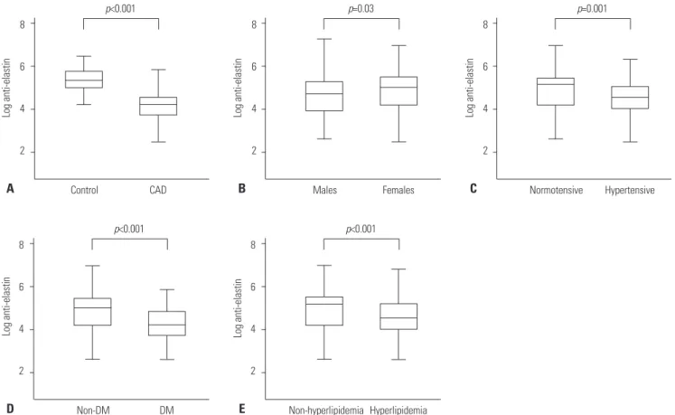
The log anti-elastin levels were significantly lower in CAD pa- tients, men, and individuals with hypertension, diabetes mel- litus, hyperlipidemia, or high hfPWV (Figs. 1 and 2). In CAD patients, anti-elastin levels were not dependent on myocardi- al infarction history or the number of diseased vessels (Fig. 2).
In univariate analysis, male sex, hypertension, diabetes melli- tus, hyperlipidemia, and hfPWV were inversely correlated with log anti-elastin levels. In multivariate analysis, male sex, dia- betes mellitus, hyperlipidemia, and AI were identified as de- terminants of log anti-elastin levels (Figs. 1 and 2, Table 2). In the analysis of potential AI determinants, the relationship be- tween index and log anti-elastin levels was again found to be significant and independent of other clinical risk factors (Sup- plementary Table 1, only online).
DISCUSSION
The present study assessed the relationship between anti- elastin antibody levels and CAD and its characteristics. This study revealed that anti-elastin levels are lower in CAD pa- tients than in matched controls. Anti-elastin levels were asso- ciated with sex, hypertension, diabetes mellitus, hyperlipid- emia, and arterial stiffness, but not with myocardial infarction history or angiographic severity of CAD. AI, a parameter of ar- Table 1. Baseline Characteristics of All Study Subjects
All subjects (n=345)
Control (n=174)
CAD
(n=171) p value
Age, yrs 51.0±7.7 50.9±7.7 51.2±7.7 0.75
Males 219 (63) 111 (64) 108 (63) 0.91
Medical history
Hypertension 147 (43) 55 (32) 92 (53) <0.001 Diabetes mellitus 60 (17) 10 (6) 50 (29) <0.001 Hyperlipidemia 122 (35) 41 (24) 81 (47) <0.001
Current smoker 78 (23) 38 (22) 39 (23) 0.90
Physical findings
Body mass index, kg/m2 25.2±3.2 25.0±3.3 25.4±3.0 0.23 Systolic BP, mm Hg 123±18 121±15 125±20 0.07
Diastolic BP, mm Hg 77±12 76±13 79±11 0.02
Arterial stiffness
Augmentation index 15.2±18.9 12.1±19.4 18.2±17.8 0.003
hfPWV, cm/sec 899±157 874±158 923±153 0.003
CAD characteristics Clinical diagnosis
Angina pectoris 107 (61)
Myocardial infarction 67 (39)
Number of diseased vessels
1 102 (59)
2 40 (23)
3 32 (18)
Anti-elastin, a.u. 113
(59, 211)
197 (147, 270)
63
(40, 96) <0.001 Log anti-elastin 4.70±0.86 5.24±0.60 4.17±0.73 <0.001 Medications
ACE inhibitors or ARBs 139 (40) 31 (18) 108 (63) <0.001 Calcium channel blockers 103 (30) 28 (16) 75 (44) <0.001 Beta blockers 117 (34) 15 (9) 102 (60) <0.001
Diuretics 54 (16) 18 (10) 36 (21) 0.01
Statins 152 (44) 21 (12) 131 (77) <0.001
Biguanides 30 (9) 8 (5) 22 (13) 0.01
Sulfonylurea 20 (6) 4 (2) 16 (9) 0.01
BP, blood pressure; CAD, coronary artery disease; a.u., arbitrary unit; ACE, angiotensin converting enzyme; ARB, angiotensin receptor blocker; hfPWV, heart-to-femoral pulse wave velocity.
Values are presented as the mean±SD, n (%), or median (interquartile range).
terial stiffness, was identified as a determinant of antibody lev- els independent of other confounding risk factors. Our results suggest that lower anti-elastin levels are related to CAD and that this relation is linked to arterial stiffness rather than ad- vancement of atherosclerosis. We quantified this antibody us- ing a novel assay against aortic elastin peptides, and our result may be used as basic data for further validation studies.
One major finding of our study was lower anti-elastin anti- body levels in CAD patients than in controls. This result is consistent with that reported by Baydanoff, et al.,6 which indi- cated that anti-human aortic elastin antibody levels were low- er in patients with atherosclerosis. Although its mechanism was not fully elucidated at that time, the authors suggested an adaptive restriction of the immune response to avoid aggrava- tion of the atherosclerosis as a possible cause. However, the antibody levels and elastin-derived peptide levels did not cor- relate. In the same patients with atherosclerosis, their elastin degradation peptide levels were higher than those in other subjects.8 In another study,7 the antibody and peptide levels did not correlate either. Clarification of the relationship be- tween antibody levels and elastin-derived peptide concentra- tions may help better understand their physiology. Mean- while, a few studies have shown results that differ from ours.
In one study, anti-elastin peptide antibody levels were higher in patients with ischemic heart disease and obliterative arte-
riosclerosis of the legs.5 In another, a significant increase in se- rum alpha-anti-elastin antibody levels was demonstrated in patients with carotid plaque destabilization.10 It was also re- ported that cathepsins K and S at vascular matrix remodeling can degrade elastin, and this supports that elastolytic prod- ucts can be elevated in progressed atherosclerosis.14 Further- more, studies have shown that atherosclerosis-associated cyto- kines augment cysteinyl cathepsin activity, which can promote elastin degradation, in various vascular diseases.15 Taken to- gether, caution should be exercised when interpreting anti- elastin antibody levels in clinical situations.
As mentioned above, the mechanisms and implications of varying anti-elastin antibody levels have not been fully under- stood. However, several possible roles have been proposed.
Anti-elastin antibody binds elastin peptides and may block their chemotactic effect on inflammatory cells.16 Thus, lower antibody levels may be insufficient to block this effect of elas- tin peptides. Secondly, hyperactivity of the immune response to elastin may cause more vascular damage. Therefore, a de- crease in antibody levels may result from the impaired im- mune reaction against elastin to reduce the aggravation of atherosclerosis.6 Nevertheless, uncovering the significance of the lower antibody levels in our study requires further analy- sis. For instance, in the current study, antibody levels were in- dependently associated with sex, diabetes mellitus, hyperlip-
Fig. 1. Associations between anti-elastin levels and clinical characteristics. Levels of log anti-elastin were lower in CAD patients than in control sub- jects (A). Anti-elastin levels were also significantly lower in males (B) and subjects with hypertension (C), DM (D), or hyperlipidemia (E), compared to each subgroup’s respective counterpart. CAD, coronary artery disease; DM, diabetes mellitus.
8 6
4 2
Control p<0.001
CAD
Log anti-elastin
A
Log anti-elastin
8 6
4 2
Non-DM p<0.001
D DM
Log anti-elastin
8 6
4 2
Non-hyperlipidemia p<0.001
Hyperlipidemia E
Log anti-elastin
8 6
4 2
Normotensive p=0.001
Hypertensive C
Log anti-elastin
Males p=0.03
Females B
8 6
4 2
idemia, and an arterial stiffness index. Although antibody level was significantly associated with CAD, other cardiovas- cular risk factors also showed correlations with antibody lev- els. Thus, it is assumed that the relationship between antibody levels and CAD can be influenced in part by the vascular con- ditions developed by other risk factors. We found arterial stiff- ness to be an independent determinant of anti-elastin. Risk factors can coexist in a single patient and influence one anoth- er. Therefore, caution should be taken in interpreting the level of an antibody that is interrelated to other factors. Another point to consider is whether the antibody levels reflect a gen-
eral vascular pathology or CAD specifically. Although the an- tibody levels were correlated with the presence of CAD in our study, they were not associated with disease advancement.
The antibody concentration was linked to the arterial stiffness index, and this concentration may indicate functional change of non-coronary vasculature as well as the risk of CAD.
The relationship between elastin degradation and arterial stiffness has been investigated in many studies. Ruseva, et al.17 showed that the anti-elastin antibody level is lower when elas- tin degradation is experimentally slowed in rats. Maclay, et al.4 measured cutaneous elastin degradation and reported that
Fig. 2. Associations between anti-elastin levels and arterial stiffness or the severity of atherosclerotic CAD. Though the levels of log anti-elastin did not differ significantly among the five groups stratified according to augmentation index values (A), they were lower in subjects with higher hfPWV (B). Log anti-elastin levels were similar in the angina pectoris and myocardial infarction subgroups (C). Anti-elastin levels were also similar among subgroups with different numbers of diseased vessels (D). Subgroups classified by augmentation index or hfPWV were analyzed in total subjects (n=345), while those by clinical or angiographic severity of CAD were analyzed in CAD patients (n=171). CAD, coronary artery disease; hfPWV, heart-to- femoral pulse wave velocity.
8
6
4
2
Augmentation index, quintiles p=0.16
Log anti-elastin
A
8
6
4
2
Angina pectoris Myocardial infarction p=0.74
Log anti-elastin
C
Log anti-elastin
8
6
4
2
hfPWV, quintiles p=0.003
B
Log anti-elastin
8
6
4
2
1 2 3
Number of diseased vessels p=0.78
D
this was positively associated with PWV. Our results are not in agreement with these reports, and therefore, clarification of the association between antibody level and stiffness is need- ed. Notably, whether the blood antibody level correlates with arterial stiffness, whether changes in antibody levels have clinically significant implications, and which clinical factors may influence antibody levels remain to be clarified. In this re- gard, our analysis of the association between antibody levels and clinical risk factors might serve as helpful basic data.
A prior study demonstrated that female mice have higher anti-elastin antibody levels than males, which is consistent with our data.18 The authors suggested that reproductive organ remodeling in female mice may be the cause of this finding, although this does not sound plausible in our subjects. Previ- ous studies on the association between diabetes mellitus and antibody levels did not show consistent results. The fibulin-1 level, an elastin-associated protein, was lower in patients with type 2 diabetes than in controls.19 Conversely, in another study conducted in diabetic children, anti-elastin IgG level was pos- itively correlated with diabetes duration, hemoglobin A1c lev- el, and the level of antibodies to advanced glycation end-prod- ucts. Although low-density lipoproteins interact with arterial elastin during the development of atherosclerosis,20 the data on the blood levels of elastin peptide or the antibody against it in hypercholesterolemia are limited. Interestingly, Fülöp, et al.5 showed that anti-elastin antibody levels decreased in sub- jects with IV hyperlipoproteinemia with hypertriglyceridemia, whereas we found that antibody levels were lower in patients
with a history of hypercholesterolemia. On the other hand, it was shown in the study of Baydanoff, et al.8 that anti-elastin an- tibody levels decrease after the age of 60 years. In their study, no significant change in antibody levels was noticed until that age. In our study, antibody levels did not vary with subjects’
age. However, because the age range in our subjects was rela- tively narrow, it is difficult to say whether age is significantly associated with antibody levels.
Our study was designed with well-matched cases and con- trol subjects, and systemically assessed the effects of CAD or individual cardiovascular risk factors on anti-elastin antibody levels. In particular, the analysis of associations with detailed vascular characteristics is the strength of this study. Several findings were demonstrated for the first time in the current study, although they require further validation. Some limita- tions of our study can be pointed out. The contribution of AI to anti-elastin levels was significant but weak (beta=-0.006) and its clinical impact could be modest. In our study, we could only measure anti-elastin antibody levels. A simultaneous exami- nation of elastin peptide level may have given us more insight into the pathological role and clinical interpretation of antibody levels. In addition, patients with CAD had slightly but signifi- cantly higher diastolic BP. Although it did not show significant relationship with arterial stiffness in our data, it could be one of confounders in the analyses. Lastly, we excluded subjects with uncontrolled hypertension or diabetes mellitus and these fac- tors with medications might have partially affected our results.
To summarize, anti-elastin antibody levels were lower in Table 2. Determinants of Log Anti-Elastin in All Study Subjects
Total subjects (n=345)
Univariate analysis Multivariate analysis
Beta SE p value Beta SE p value
Age 0.002 0.006 0.75 - - NS
Sex (male) -0.210 0.096 0.03 -0.377 0.105 <0.001
Hypertension -0.324 0.093 0.001 - - NS
Diabetes mellitus -0.626 0.119 <0.001 -0.615 0.117 <0.001
Hyperlipidemia -0.343 0.096 <0.001 -0.285 0.092 0.002
Smoking -0.114 0.112 0.31 - - NS
Systolic BP -0.004 0.003 0.11 - - NS
Body mass index -0.025 0.015 0.09 - - NS
Augmentation index -0.004 0.002 0.12 -0.006 0.003 0.02
hfPWV -0.001 0.000 0.01 - - NS
Medications
ACE inhibitors or ARBs -0.111 0.100 0.27 - - NS
Calcium channel blockers -0.126 0.097 0.20 - - NS
Beta blockers -0.163 0.104 0.12 - - NS
Diuretics -0.061 0.122 0.62 - - NS
Statins -0.175 0.109 0.11 - - NS
Biguanides -0.099 0.165 0.55 - - NS
Sulfonylurea -0.180 0.197 0.36 - - NS
BP, blood pressure; hfPWV, heart to femoral pulse wave velocity; ACE, angiotensin-converting enzyme; ARB, angiotensin receptor blocker; SE, standard error;
NS, not significant.
CAD patients and associated with male sex and a history of hypertension, diabetes mellitus, or hyperlipidemia. Antibody levels negatively correlated with arterial stiffness, but not with myocardial infarction history or the severity of CAD. In con- clusion, a lower anti-elastin antibody titer is related to CAD and this relation is linked to arterial stiffness rather than to the ad- vancement of atherosclerosis. Our results may provide the ba- sic data for further validation studies of this novel assay.
ACKNOWLEDGEMENTS
This research was financially supported by the Basic Science Research Program through the National Research Foundation of Korea (NRF) funded by the Ministry of Education, Science, and Technology (2012R1A1A2039828), by MEST through the National Research Foundation of Korea (Grant No. 2012R1A4 A1029061), by a Creative Allied Project (CAP) grant funded by the Korean Research Council of Fundamental Science and Technology (KRCF), and by the KAIST Future Systems Health- care Project of the Ministry of Science, ICT, and Future Plan- ning.
REFERENCES
1. Zieman SJ, Melenovsky V, Kass DA. Mechanisms, pathophysiolo- gy, and therapy of arterial stiffness. Arterioscler Thromb Vasc Biol 2005;25:932-43.
2. Smith ER, Tomlinson LA, Ford ML, McMahon LP, Rajkumar C, Holt SG. Elastin degradation is associated with progressive aortic stiffening and all-cause mortality in predialysis chronic kidney disease. Hypertension 2012;59:973-8.
3. Maurice P, Blaise S, Gayral S, Debelle L, Laffargue M, Hornebeck W, et al. Elastin fragmentation and atherosclerosis progression:
the elastokine concept. Trends Cardiovasc Med 2013;23:211-21.
4. Maclay JD, McAllister DA, Rabinovich R, Haq I, Maxwell S, Hart- land S, et al. Systemic elastin degradation in chronic obstructive pulmonary disease. Thorax 2012;67:606-12.
5. Fülöp T Jr, Wei SM, Robert L, Jacob MP. Determination of elastin peptides in normal and arteriosclerotic human sera by ELISA.
Clin Physiol Biochem 1990;8:273-82.
6. Baydanoff S, Nicoloff G, Alexiev C. Age-related changes in anti- elastin antibodies in serum from normal and atherosclerotic sub- jects. Atherosclerosis 1987;63:267-71.
7. Hong YJ, Kim J, Oh BR, Lee YJ, Lee EY, Lee EB, et al. Serum elastin-
derived peptides and anti-elastin antibody in patients with sys- temic sclerosis. J Korean Med Sci 2012;27:484-8.
8. Baydanoff S, Nicoloff G, Alexiev C. Age-related changes in the lev- el of circulating elastin-derived peptides in serum from normal and atherosclerotic subjects. Atherosclerosis 1987;66:163-8.
9. Skjøt-Arkil H, Clausen RE, Rasmussen LM, Wang W, Wang Y, Zheng Q, et al. Acute Myocardial Infarction and Pulmonary Dis- eases Result in Two Different Degradation Profiles of Elastin as Quantified by Two Novel ELISAs. PLoS One 2013;8:e60936.
10. Tzvetanov P, Hegde V, Al-Hashel JY, Atanasova M, Sohal AP, Rous- seff RT. Abnormal levels of serum anti-elastin antibodies in pa- tients with symptomatic carotid stenosis. Clin Neurol Neurosurg 2014;116:9-12.
11. Wilkinson IB, Fuchs SA, Jansen IM, Spratt JC, Murray GD, Cock- croft JR, et al. Reproducibility of pulse wave velocity and augmen- tation index measured by pulse wave analysis. J Hypertens 1998;
16(12 Pt 2):2079-84.
12. Lee SH, Choi S, Jung JH, Lee N. Effects of atrial fibrillation on arte- rial stiffness in patients with hypertension. Angiology 2008;59:459- 63.
13. Lee SH, Goswami S, Grudo A, Song LZ, Bandi V, Goodnight-White S, et al. Antielastin autoimmunity in tobacco smoking-induced emphysema. Nat Med 2007;13:567-9.
14. Sukhova GK, Shi GP, Simon DI, Chapman HA, Libby P. Expres- sion of the elastolytic cathepsins S and K in human atheroma and regulation of their production in smooth muscle cells. J Clin In- vest 1998;102:576-83.
15. Cheng XW, Huang Z, Kuzuya M, Okumura K, Murohara T. Cyste- ine protease cathepsins in atherosclerosis-based vascular disease and its complications. Hypertension 2011;58:978-86.
16. Senior RM, Griffin GL, Mecham RP, Wrenn DS, Prasad KU, Urry DW. Val-Gly-Val-Ala-Pro-Gly, a repeating peptide in elastin, is che- motactic for fibroblasts and monocytes. J Cell Biol 1984;99:870-4.
17. Ruseva B, Atanasova M, Georgieva M, Shumkov N, Laleva P. Ef- fects of selenium on the vessel walls and anti-elastin antibodies in spontaneously hypertensive rats. Exp Biol Med (Maywood) 2012;
237:160-6.
18. Atanasova M, Konova E, Georgieva M, Dimitrova A, Coquand-Gan- dit M, Faury G, et al. Age-related changes of anti-elastin antibodies in senescence-accelerated mice. Gerontology 2010;56:310-8.
19. Laugesen E, Høyem P, Christiansen JS, Knudsen ST, Hansen KW, Argraves WS, et al. Plasma levels of the arterial wall protein fibu- lin-1 are associated with carotid-femoral pulse wave velocity: a cross-sectional study. Cardiovasc Diabetol 2013;12:107.
20. Podet EJ, Shaffer DR, Gianturco SH, Bradley WA, Yang CY, Guyton JR. Interaction of low density lipoproteins with human aortic elastin. Arterioscler Thromb 1991;11:116-22.