http://dx.doi.org/10.3988/jcn.2013.9.1.36 J Clin Neurol 2013;9:36-42
Clinical Efficacy of Plasmapheresis in Patients with Neuromyelitis Optica Spectrum Disorder and Effects on Circulating Anti-Aquaporin-4 Antibody Levels
Su-Hyun Kim, Woojun Kim, So-Young Huh, Kyue Yim Lee, In Ja Jung, Ho Jin Kim
Department of Neurology, Research Institute and Hospital of National Cancer Center, Goyang, Korea
Received July 15, 2012 Revised August 21, 2012 Accepted August 21, 2012 Correspondence Ho Jin Kim, MD, PhD Department of Neurology, Research Institute and
Hospital of National Cancer Center, 323 Ilsan-ro, Ilsandong-gu, Goyang 410-769, Korea Tel +82-31-920-2438 Fax +82-31-925-5524 E-mail [email protected]
Background and PurposezzAlthough plasmapheresis is becoming standard practice as a res- cue therapy for neuromyelitis optica (NMO), evidence for the therapeutic efficacy of plasma- pheresis is limited, and the effect of plasmapheresis on anti-aquaporin-4 (AQP4) levels in pa- tients with NMO has not been reported. Here, our objective was to evaluate the clinical efficacy of therapeutic plasmapheresis and its effect on anti-AQP4 antibody levels in patients with NMO spectrum disorder (NMOSD).
MethodszzWe retrospectively reviewed the medical records of 15 patients with NMOSD who had 18 acute attacks and received plasmapheresis because they did not respond to high-dose in- travenous methylprednisolone (IVMP) therapy. Anti-AQP4 antibodies were measured before and after plasmapheresis. The primary outcomes were functional improvements immediately and 6 months after plasmapheresis, and the secondary outcome was the change in anti-AQP4 antibody serum levels following plasmapheresis.
ResultszzPlasmapheresis following IVMP therapy led to significant improvement in 50% of the 18 attacks in 15 patients immediately after the procedure was completed, and in 78% (14 attacks) after 6 months. Plasmapheresis was generally well tolerated in all patients. Anti-AQP4 antibody serum levels declined significantly following plasmapheresis, to a mean of 15% of the preplasmapheresis levels. Lower scores on the visual outcome scale recorded before an attack were associated with significant immediate improvement upon the completion of plasmaphere- sis (p=0.03).
ConclusionszzPlasmapheresis following IVMP therapy effectively removed anti-AQP4 anti- bodies and was accompanied by a substantial improvement in the neurological disability of pa- tients with NMOSD. Lower levels of pre-existing neurological damage may be associated with an improved acute response to plasmapheresis. J Clin Neurol 2013;9:36-42 Key Wordszz plasmapheresis, neuromyelitis optica, anti-aquaporin-4 antibody.
Open Access
cc This is an Open Access article distributed under the terms of the Cre- ative Commons Attribution Non-Commercial License (http://creative- commons.org/licenses/by-nc/3.0) which permits unrestricted non-com- mercial use, distribution, and reproduction in any medium, provided the ori- ginal work is properly cited.
Introduction
Neuromyelitis optica (NMO) is an idiopathic inflammatory disease of the central nervous system (CNS) that is character- ized by severe optic neuritis and myelitis. High-dose intrave- nous methylprednisolone (IVMP) is typically administered to
treat acute exacerbations of NMO, but this is ineffective in some patients. Therapeutic plasmapheresis appears to be ef- fective in patients with CNS inflammatory demyelinating dis- eases (IDDs) who do not recover after IVMP treatment. The efficacy of plasmapheresis may be due to the removal of circu- lating autoantibodies and other immunologically active sub- stances (e.g., complement and cytokines) from the blood.1 Since an autoantibody that targets aquaporin-4 (AQP4) was discovered in patients with NMO,2 numerous clinical and ex- perimental studies have implicated anti-AQP4 antibody-medi-
ated autoimmunity in the pathogenesis of NMO.3-8 According- ly, NMO may be particularly amenable to treatment by plas- mapheresis. Previous studies and case series have found that plasmapheresis is effective in suppressing acute attacks in 50- 89% of patients with NMO.9-13 However, many of the previous observational studies on the clinical efficacy of plasmapheresis retrospectively evaluated all cases of CNS IDDs including NMO; thus, the exact efficacy of plasmapheresis for NMO at- tacks may have been underestimated.9-11 Furthermore, recent plasmapheresis case series in patients with NMO involved sm- all numbers of patients.12,13 Plasmapheresis is becoming the preferred standard rescue therapy for NMO when high-dose IVMP treatment elicits only a weak response.14-16 However, ev- idence for the therapeutic efficacy of plasmapheresis for acute attacks of NMO is still limited. Moreover, the extent to which anti-AQP4 antibodies are removed by plasmapheresis and whether removing anti-AQP4 antibodies is critical for the ther- apeutic efficacy of plasmapheresis in patients with NMO re- main unclear.
The above situation prompted this study to assess the clinical efficacy and safety of plasmapheresis in patients with NMO spectrum disorder (NMOSD) and also to evaluate the changes in anti-AQP4 antibody levels following plasmapheresis.
Methods
Patients
We retrospectively reviewed the medical records of 15 patients who had NMO or who were seropositive for a limited form of NMO17,18 and who had received plasmapheresis for acute at- tacks between March 2010 and September 2011. Plasmapher- esis was performed when severe disability was sustained or worsened after high-dose IVMP therapy (1 g for 5 days) as in- dicated by Expanded Disability Status Scale (EDSS) scores
≥7.0 or a visual acuity worse than 20/200. Three patients had two series of plasmapheresis sessions with intervals of more than 6 months. Therefore, 18 plasmapheresis series performed in 15 individual patients were evaluated. This study was ap- proved by the Institutional Review Board of the National Can- cer Center (Protocol #NCCCTS-11-546), and informed con- sent was obtained from all patients.
Plasmapheresis
Patients were treated using double-filtration plasmapheresis (Plasauto EZ, Asahi Kasei Medical, Tokyo, Japan). Between 1 and 1.5 plasma volumes were treated in each session every other day. Vascular access was established with a double-lu- men catheter in the central vein. All but one of the patients re- ceived tapered oral corticosteroids during plasmapheresis. Five patients were maintained on prior immunosuppressive treat-
ments (mycophenolate mofetil or azathioprine) during the plas- mapheresis. During double-filtration plasmapheresis, 5% albu- min or normal saline was used as the replacement fluid. Each patient underwent six sessions of plasmapheresis.
Outcome assessments
Functional improvements immediately and 6 months after pl- asmapheresis were the primary outcomes, and changes in an- ti-AQP4 antibody levels following plasmapheresis were the secondary outcome measured. Because the EDSS focuses on ambulation-related disability, the targeted neurological deficit may not be fully reflected in the EDSS score (e.g., upper ex- tremity paresis). Therefore, the outcome of plasmapheresis was evaluated according to the criteria of Keegan et al.9 as follows:
“no improvement” was defined as no improvement in neuro- logical function, “mild improvement” presented as definite im- provement in neurological status without impact on function,
“moderate improvement” appeared as definite improvement in function (e.g., walking or the use of an upper extremity), and
“marked improvement” was a major improvement in function.
Either a “moderate improvement” or “marked improvement”
was defined as a significant improvement. In parallel, the EDSS score was also evaluated in attacks that included myelitis or brain involvement, and visual acuity was assessed separately for each eye using the following Visual Outcome Scale (VOS):
0=20/20; 1=scotoma, but better than 20/30; 2=20/30-20/59;
3=20/60-20/199; 4=20/200-20/800; 5=able to count fingers only; 6=light perception; and 7=no light perception.19 Adverse events were recorded throughout the study.
Blood collection and measurement of serum anti-AQP4 antibodies and cytokines
Serum samples were obtained before and after plasmapheresis for all attacks. Serum samples were also obtained pre- and poststeroid therapy and serially after each session in eight at- tacks. All samples were immediately centrifuged, and sera were stored at -80°C until analysis. The quantitative change in anti-AQP4 antibody levels was measured using an enzyme- linked immunosorbent assay.20 The levels of anti-AQP4 anti- body were quantified using a standard curve.
Statistical methods
Wilcoxon’s signed-rank test for paired data was used to ana- lyze the changes in the EDSS and VOS scores and the anti- AQP4 antibody level after plasmapheresis. Clinical variables were compared between patients with and without significant responses using the χ2 for frequency comparisons and the Mann-Whitney test for mean comparisons. All statistical anal- yses were performed using GraphPad PRISM (San Diego, CA, USA), and probability values of p<0.05 were considered statis-
tically significant.
Results
Eighteen plasmapheresis treatments performed for acute at- tacks of NMOSD were evaluated in 15 patients (8 with NMO and 7 with a limited form of NMO). Thirteen patients (87%) were women. The median age at the time of plasmapheresis was 40 years (range, 12-53 years), and the median disease du- ration was 4 years (range, 0.1-13 years). Plasmapheresis was administered because of 11 optic neuritis attacks in 8 patients, 5 myelitis attacks in 5 patients, and 2 attacks involving the brain in 2 patients. Patients had a median of 5 relapses (range, 1-12) prior to treating an attack with plasmapheresis. The baseline VOS score for patients who had optic neuritis attacks was 1.1±1.2 (mean±SD), and the baseline EDSS score for patients who had attacks of myelitis or brain involvement was 3.5±2.5 (mean±SD). The immunotherapies that each patient received
before plasmapheresis are listed in Table 1 and 2.
Plasmapheresis was associated with significant improve- ments immediately in 9 attacks (50%; 2 with moderate im- provement and 7 with marked improvement) and with signifi- cant improvements at 6 months in 14 attacks (78%; 5 with mo- derate improvement and 9 with marked improvement). Neuro- logical disability at relapse was severe (EDSS scores of 7.4±
1.0 in patients with attacks of myelitis or brain involvement and VOS scores of 4.9±1.6 in patients with attacks of optic neu- ritis), but was sustained or worsened despite high-dose IVMP therapy (EDSS score, 7.6±0.7; VOS score, 4.8±0.9; p=0.180 and 0.527, respectively). Plasmapheresis was initiated at 16±8 days after the onset of symptoms. The neurological disability improved significantly upon the completion of plasmapheresis (EDSS score, 6.1±1.6; VOS score, 3.6±1.5; p=0.041 and 0.024, respectively) and further improved at 6 months following plas- mapheresis (EDSS score, 4.6±1.8; VOS score, 2.6±1.6; p=
0.016 and 0.009, respectively). Of the nine attacks showing
Table 1. Clinical characteristics, treatments, and outcomes of 11 optic neuritis attacks in 8 patients Attack no./sex
/age at plasmapheresis,
years
Days from symptom onset to plasmapheresis
Type of lesion
VOS score before attack
VOS score at attack
VOS score after IVMP
VOS score upon completing
plasmapheresis
VOS score at 6 months
after plasmapheresis
Immuno- therapy before plasmapheresis
1/F/53 24 O 0 5.0 6.0 2.0 2.0
2/F/49 18 O 0 1.0 4.0 2.0 0
3/F/24 15 O 3.0 6.0 5.0 5.0 5.0 RTX
4/F/12‡ 14 O 0 7.0 6.0 5.0 3.0
5/F/37§ 20 O 1.0 6.0 5.0 3.0 2.0 MMF
6/M/36 10 O* 0.0/2.0 3.0/4.0 3.0/4.0 3.0/4.0 3.0/4.0
7/F/48† 11 O 0 5.0 4.0 2.0 1.0
8/F/38§ 12 O 2.0 4.0 4.0 4.0 3.0 RTX
9/F/12‡ 16 O 3.0 6.0 6.0 6.0 4.0 MMF
10/F/35 10 O 0 5.0 4.0 2.0 1.0 MMF
11/F/48† 9 O 1.0 5.0 5.0 5.0 4.0 MMF
*Bilateral optic neuritis, †,‡,§Each patient had two separate attacks that were treated with plasmapheresis.
F: female, IVMP: intravenous methylprednisolone, M: male, MMF: mycophenolate mofetil, O: optic nerve, RTX: rituximab, VOS: Visual Outcome Scale.
Table 2. Clinical characteristics, treatments, and outcomes of 7 attacks involving the spinal cord or brain in 7 patients Attack no./sex
/age at plasmapheresis,
years
Days from symptom onset to
plasmapheresis
Type of lesion
EDSS score before attack
EDSS score at attack
EDSS score after IVMP
EDSS score upon completing plasmapheresis
EDSS score at 6 months after plasmapheresis
Immuno- therapy before plasmapheresis
1/F/51 18 B 0 8.5 8.5 5.5 3.0
2/F/46 16 S 4.0 7.0 7.0 5.0 3.5
3/F/30 45 S 4.5 6.5 7.0 6.5 5.0
4/F/53 12 S 4.0 6.0 7.5 5.5 4.5 RTX
5/M/51 14 S 6.0 8.0 8.0 8.0 7.0 AZT
6/F/25 13 B 0.0 7.0 7.0 4.0 2.5
7/F/43 9 S 6.0 8.5 8.5 8.5 7.0
AZT: azathioprine, B: brain, RTX: rituximab, EDSS: Expanded Disability Status Scale, IVMP: intravenous methylprednisolone, S: spinal cord.
significant improvement upon the completion of plasmaphere- sis, the clinical response typically commenced after the third session (median value; range, sessions 1-4). Meanwhile, 5 of the 18 attacks (28%) showed no functional improvement upon the completion of plasmapheresis, but there was an improve- ment 6 months after plasmapheresis.
All patients were seropositive for anti-AQP4 antibodies.
Plasmapheresis consistently resulted in a marked reduction in serum anti-AQP4 antibody levels (by 84.5±14.8% of the pre-
plasmapheresis serum levels). We also investigated serial chan- ges in anti-AQP4 antibody levels before and after steroid ther- apy and following every session of plasmapheresis in eight attacks (Fig. 1). The anti-AQP4 antibody levels decreased slightly after steroid therapy (by 14.3±15.2% of the pre-ste- roid-therapy level), but the decrease was significantly larger af- ter plasmapheresis following all attacks. The reduction rate in anti-AQP4 antibodies of each sessions plateaued after the five sessions.
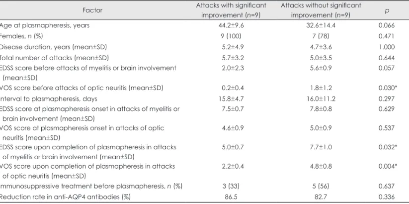
Table 3. Comparison of attacks with and without significant improvement upon the completion of plasmapheresis
Factor Attacks with significant
improvement (n=9)
Attacks without significant
improvement (n=9) p
Age at plasmapheresis, years 44.2±9.6 32.6±14.4 0.066
Females, n (%) 9 (100) 7 (78) 0.471
Disease duration, years (mean±SD) 5.2±4.9 4.7±3.6 1.000
Total number of attacks (mean±SD) 5.7±3.2 5.0±3.5 0.644
EDSS score before attacks of myelitis or brain involvement (mean±SD)
2.0±2.3 5.6±0.9 0.057
VOS score before attacks of optic neuritis (mean±SD) 0.2±0.4 1.8±1.2 0.030*
Interval to plasmapheresis, days 15.8±4.7 16.0±11.2 0.297
EDSS score at plasmapheresis onset in attacks of myelitis or brain involvement (mean±SD)
7.5±0.7 7.8±0.8 0.629
VOS score at plasmapheresis onset in attacks of optic neuritis (mean±SD)
4.6±0.9 5.0±0.9 0.537
EDSS score upon completion of plasmapheresis in attacks of myelitis or brain involvement (mean±SD)
5.0±0.7 7.7±1.0 0.032*
VOS score upon completion of plasmapheresis in attacks of optic neuritis (mean±SD)
2.2±0.4 4.8±0.8 0.004*
Immunosuppressive treatment before plasmapheresis, n (%) 3 (33) 5 (56) 0.637
Reduction rate in anti-AQP4 antibodies (%) 86.5 82.7 0.336
*Level of significance <0.05.
AQP4: aquaporin-4, EDSS: Expanded Disability Status Scale, VOS: Visual Outcome Scale.
600 550 500 450 400 350 300 250 200 150 100 50 0
AQP4 antibody titers (SU)
Acute attack After
steroid 1st 2nd 3rd 4th 5th 6th
A
Fig. 1. Longitudinal titer (A) and cumu- lative reduction rate (B) of anti-aquapo- rin-4 (AQP4) antibodies before and af- ter steroid therapy at each plasmaphe- resis session.
100 90 80 70 60 50 40 30 20 10 0 -10
Cumulative reduction rate of AQP4 antibody
(%)
Acute attack After
steroid 1st 2nd 3rd 4th 5th 6th
B
No significant differences in sex ratio, age at the time of pl- asmapheresis, previous numbers of attacks, disease duration, attack severity, EDSS score before an attack, delay in plasma- pheresis after symptom onset, or rate of reduction in anti-AQP4 antibodies were identified as an explanation for the variable acute response after the completion of plasmapheresis. Howev- er, lower VOS scores before an attack were associated with a significant immediate improvement upon the completion of plasmapheresis (p=0.03) (Table 3).
One patient developed anemia and one experienced hypo- kalemia, but no serious complications occurred during plasma- pheresis. Significant transient leukocytosis (12000-22000/μL) was common immediately after plasmapheresis, but in all cas- es this reversed 1 day later. No clinical symptoms associated with transient leukocytosis were observed. Direct contact of blood mononuclear cells with the filter membrane may induce leukocytosis.21
Discussion
We analyzed the clinical effectiveness of therapeutic plasma- pheresis and its effect on anti-AQP4 antibody levels in patients with acute attacks of NMOSD who responded insufficiently to high-dose IVMP therapy. Plasmapheresis following IVMP therapy was well tolerated. A significant functional improve- ment was seen in 50% of attacks immediately after the comple- tion of plasmapheresis, and in 78% of attacks at 6 months after plasmapheresis. The response rate to plasmapheresis in our se- ries is comparable to the response rates of 50-89% observed in NMO patients in previous studies.9-13 Plasmapheresis was ef- fective at decreasing anti-AQP4 antibody levels to 15% of the preplasmapheresis serum levels.
Considering the absence of controls, the observed effects in our study may be at least partly attributable to the natural cour- se following attacks or to a delayed effect of IVMP therapy.
However, immediate functional improvement following plas- mapheresis was observed in half of the attacks that had been unresponsive to IVMP therapy or where the condition wors- ened despite the therapy. These results are consistent with a previous study finding significantly better outcomes in the ste- roid-and-plasmapheresis-treated group than in the steroid-on- ly-treated group.22 The clear temporal association between plasmapheresis and the clinical response over a short period of time in the present study suggests that this therapy is pivotal in facilitating recovery from neuronal injury. Although previous plasmapheresis studies involving CNS IDDs (including NMO) found rapid improvement in 33-37% of patients,9,10 the hetero- geneity of CNS IDD pathology may have influenced the re- sponse rate. Moreover, recent studies on CNS IDDs have con- centrated on long-term efficacy, with the primary outcome
being improvement at 6 months after plasmapheresis.11 Thus, the beneficial effects of the acute response may have been un- derestimated.11 Considering humoral immunity-mediated im- munopathogenesis in NMO, the acute response following plas- mapheresis may be more profound than that in other CNS IDDs. Indeed, the observation of the rate of acute improvement being higher (50%) in this study than in previous studies in- cluding all CNS IDDs9,10 may indicate that the acute therapeu- tic efficacy of plasmapheresis in NMOSD is better than previ- ously thought. Similarly, a recent case series found rapid clinical improvement following plasmapheresis in 50-67% of patients with NMO.12,13
The immediate response to plasmapheresis in acute attacks of NMO may be related to a rapid reduction in anti-AQP4 an- tibody levels. The anti-AQP4 antibody levels declined by an average of 14% of the initial levels following IVMP therapy (with little variation), whereas an average of six sessions of plasmapheresis consistently induced marked reductions (85%) relative to the preplasmapheresis levels (Fig. 1). This observa- tion agrees with previous findings that the exchange molecules decreased to less than 20% of their initial level after five ses- sions.23,24 The rate of reduction of anti-AQP4 antibodies was greatest at the beginning of plasmapheresis and reached a pla- teau after five sessions (Fig. 1). Therefore, at least five sessions of plasmapheresis might be required to effectively remove an- ti-AQP4 antibodies. Similarly, previous studies have suggest- ed that five or six standard plasmapheresis sessions are requir- ed to substantially reduce blood levels of IgG.25 However, the optimal minimum number of sessions for clinical recovery still needs to be determined.
Meanwhile, the reduction rate of anti-AQP4 antibodies did not differ significantly between attacks with and without sig- nificant improvement upon the completion of plasmapheresis.
This finding suggests that a reduction in anti-AQP4 antibody is not the only factor affecting the clinical efficacy of plasma- pheresis. In the present study, a significant improvement upon the completion of plasmapheresis was associated with a low baseline level of neurological disability. The baseline VOS score was much higher in attacks that showed no significant improvement than in those with significant improvement (p=
0.03) (Table 3). This observation indicates that the extent of pre-existing neuronal damage and the “neurologic reserve” to promote restoration may be important factors for clinical re- covery following plasmapheresis. This hypothesis was sup- ported by the clinical results seen in three patients in whom two series of plasmapheresis were applied on the same lesion for two separate attacks. All three patients achieved significant improvements following the first plasmapheresis series; how- ever, the clinical response was much slower after the second plasmapheresis series than after the first series in all three pa-
tients, and one patient showed no functional improvement fol- lowing the second series. Bonnan and colleagues suggested that a low basal impairment is associated with a better out- come.22 A recent study also found that a shorter disease dura- tion was associated with a beneficial response to plasmapher- esis, which is roughly consistent with our findings.11 Additio- nally, previous studies involving patients with CNS IDDs found that starting plasmapheresis early (within 15 to 20 days) is the most important predictor of a favorable response to the proce- dure.9-11 However, plasmapheresis was initiated within 20 days in 89% of our patients, which might explain why the time in- terval to plasmapheresis appeared to have no significant asso- ciation with clinical efficacy upon the completion of plasma- pheresis in the present study.
Plasmapheresis did not initially result in significant im- provement for five attacks (28%), although there was a marked improvement at 6 months after plasmapheresis. This result agrees with previous observations of delayed responses in 4-48% of patients.9-11 Actually, most of the attacks in our series were further improved at 6 months after plasmapheresis. Whe- ther the late improvement is a delayed effect of plasmapheresis or the natural course of the disease remains uncertain. Never- theless, the effective removal of anti-AQP4 antibodies by plasmapheresis may be beneficial in preventing further attacks for a period of time and thus providing patients with an ex- tended recovery time without further relapse, which may pos- itively influence the clinical course of the disease.
Our analysis was limited by the study having a retrospective design, being based in a single center, examining only a small number of patients, and not including a control group of pa- tients with similar symptoms who did not receive plasmapher- esis. However, considering the rarity of the disease, the limit- ed evidence for the efficacy of plasmapheresis in NMO, and the ethical difficulties in conducting randomized controlled tri- als, our data provide clinical and laboratory support for the value of plasmapheresis in steroid-resistant acute attacks of NMO.
In conclusion, plasmapheresis should be considered for NMOSD patients with severe attacks who do not respond to IVMP therapy. The extent of the underlying neuronal damage may influence the acute response to plasmapheresis. Further clinical trials are needed to confirm the reported findings and identify the optimal protocol for delivering plasmapheresis.
Acknowledgements
This work was supported by Grant No. A110587 from the Korean Min- istry for Health and Welfare (S.H. Kim).
REFERENCES
1. Lehmann HC, Hartung HP, Hetzel GR, Stüve O, Kieseier BC. Plasma exchange in neuroimmunological disorders: Part 1: Rationale and treat-
ment of inflammatory central nervous system disorders. Arch Neurol 2006;63:930-935.
2. Lennon VA, Wingerchuk DM, Kryzer TJ, Pittock SJ, Lucchinetti CF, Fujihara K, et al. A serum autoantibody marker of neuromyelitis opti- ca: distinction from multiple sclerosis. Lancet 2004;364:2106-2112.
3. Saadoun S, Waters P, Bell BA, Vincent A, Verkman AS, Papadopoulos MC. Intra-cerebral injection of neuromyelitis optica immunoglobulin G and human complement produces neuromyelitis optica lesions in mice.
Brain 2010;133:349-361.
4. Weinshenker BG, Wingerchuk DM, Vukusic S, Linbo L, Pittock SJ, Lucchinetti CF, et al. Neuromyelitis optica IgG predicts relapse after longitudinally extensive transverse myelitis. Ann Neurol 2006;59:566- 5. Matiello M, Lennon VA, Jacob A, Pittock SJ, Lucchinetti CF, Winger-569.
chuk DM, et al. NMO-IgG predicts the outcome of recurrent optic neu- ritis. Neurology 2008;70:2197-2200.
6. Bennett JL, Lam C, Kalluri SR, Saikali P, Bautista K, Dupree C, et al.
Intrathecal pathogenic anti-aquaporin-4 antibodies in early neuromye- litis optica. Ann Neurol 2009;66:617-629.
7. Bradl M, Misu T, Takahashi T, Watanabe M, Mader S, Reindl M, et al.
Neuromyelitis optica: pathogenicity of patient immunoglobulin in vivo. Ann Neurol 2009;66:630-643.
8. Kim W, Kim SH, Kim HJ. New insights into neuromyelitis optica. J Clin Neurol 2011;7:115-127.
9. Keegan M, Pineda AA, McClelland RL, Darby CH, Rodriguez M, Weinshenker BG. Plasma exchange for severe attacks of CNS demye- lination: predictors of response. Neurology 2002;58:143-146.
10. Llufriu S, Castillo J, Blanco Y, Ramió-Torrentà L, Río J, Vallès M, et al. Plasma exchange for acute attacks of CNS demyelination: Predic- tors of improvement at 6 months. Neurology 2009;73:949-953.
11. Magaña SM, Keegan BM, Weinshenker BG, Erickson BJ, Pittock SJ, Lennon VA, et al. Beneficial plasma exchange response in central ner- vous system inflammatory demyelination. Arch Neurol 2011;68:870- 12. Watanabe S, Nakashima I, Misu T, Miyazawa I, Shiga Y, Fujihara K, 878.
et al. Therapeutic efficacy of plasma exchange in NMO-IgG-positive patients with neuromyelitis optica. Mult Scler 2007;13:128-132.
13. Wang KC, Wang SJ, Lee CL, Chen SY, Tsai CP. The rescue effect of plasma exchange for neuromyelitis optica. J Clin Neurosci 2011;18:43- 14. Collongues N, de Seze J. Current and future treatment approaches for 46.
neuromyelitis optica. Ther Adv Neurol Disord 2011;4:111-121.
15. Sellner J, Boggild M, Clanet M, Hintzen RQ, Illes Z, Montalban X, et al. EFNS guidelines on diagnosis and management of neuromyelitis optica. Eur J Neurol 2010;17:1019-1032.
16. Szczepiorkowski ZM, Winters JL, Bandarenko N, Kim HC, Linen- berger ML, Marques MB, et al. Guidelines on the use of therapeutic apheresis in clinical practice--evidence-based approach from the Apheresis Applications Committee of the American Society for Apher- esis. J Clin Apher 2010;25:83-177.
17. Wingerchuk DM, Lennon VA, Pittock SJ, Lucchinetti CF, Weinshenk- er BG. Revised diagnostic criteria for neuromyelitis optica. Neurology 2006;66:1485-1489.
18. Wingerchuk DM, Lennon VA, Lucchinetti CF, Pittock SJ, Weinshenk- er BG. The spectrum of neuromyelitis optica. Lancet Neurol 2007;6:
805-815.
19. Wingerchuk DM, Hogancamp WF, O’Brien PC, Weinshenker BG.
The clinical course of neuromyelitis optica (Devic’s syndrome). Neu- rology 1999;53:1107-1114.
20. Kim W, Lee JE, Li XF, Kim SH, Han BG, Lee BI, et al. Quantitative measurement of anti-aquaporin-4 antibodies by enzyme-linked immu- nosorbent assay using purified recombinant human aquaporin-4. Mult Scler 2012;18:578-586.
21. Narita YM, Hirahara K, Mizukawa Y, Kano Y, Shiohara T. Efficacy of plasmapheresis for the treatment of severe toxic epidermal necrolysis:
Is cytokine expression analysis useful in predicting its therapeutic effi- cacy? J Dermatol 2011;38:236-245.
22. Bonnan M, Valentino R, Olindo S, Mehdaoui H, Smadja D, Cabre P.
Plasma exchange in severe spinal attacks associated with neuromyeli- tis optica spectrum disorder. Mult Scler 2009;15:487-492.
23. Brecher ME. Plasma exchange: why we do what we do. J Clin Apher
2002;17:207-211.
24. Bonnan M, Cabre P. Plasma exchange in severe attacks of neuromye- litis optica. Mult Scler Int 2012;2012:787630.
25. Okafor C, Ward DM, Mokrzycki MH, Weinstein R, Clark P, Balogun RA. Introduction and overview of therapeutic apheresis. J Clin Apher 2010;25:240-249.