Laser epithelial keratomileusis (LASEK) is a surgery that corrects refractive errors to a desired level by creating a corneal epithelial flap, irradiating the exposed Bowman's layer with excimer laser, and then re-placing the flap,
1,2with the use of therapeutic contact lenses after surgery. With regards to recovery after LASEK, the scar healing process and epithelium recovery may differ depending on whether the epithelial flap survives or not. In the cases wherein the epithelial flap adheres and survives, the shedding of the epithelium's monolayer structure occurs gradually for 3-4 days after LASEK surgery, and the epithelial cells are shed to the underside of therapeutic lenses gradually.
3Generally, therapeutic contact lenses perform the role of accelerating scar healing and protecting the corneal surface mechanically as well as reducing the discomfort caused by the abnormality on the corneal surface and maintaining
appropriate humidity on the corneal surface.
4,5In addition, therapeutic contact lenses have to be selected based on their appropriateness for therapy, taking into consideration the oxygen permeability, water content, raw materials, diameter, thickness, the base curve, the peripheral curve, the rim, and other factors.
4-7After LASEK surgery, for the corneal epithelium to recover, selection of appropriate therapeutic lenses is required. Among other factors, it is required to select the appropriate base curve radius since the keratometry value changes after LASEK surgery. Therefore, in our study, the effect of the base curve radius (BCR) on the corneal healing period after LASEK surgery was examined.
The Subject and Methods
The subjects were 47 patients (92 eyes) who wore therapeutic lenses after LASEK surgery performed by a single surgeon from October 2004 to December 2005 at Seoul national university hospital. Pre-operative examinations performed including a slit-lamp examination, manifest refractive errors, cycloplegic refractive errors, tonometry, fundus examination, pupil size, Schirmer test, corneal topography, wavefront analysis, corneal pachymetry, and
Effect of Base Curve Radius of Therapeutic Lenses on Epithelial Healing after Laser-Assisted Subepithelial Keratectomy
Je Hyun Seo, MD,
1,2Won Ryang Wee, MD,
1,2Jin Hak Lee, MD,
1,3Mee Kum Kim, MD,
1,2Department of Ophthalmology, Seoul National University College of Medicine1, Seoul, Korea
Korea Seoul Artificial Eye Center, Seoul National University Hospital Clinical Research Institute
2, Seoul, Korea Department of Ophthalmology, Seoul National University Bundang Hospital
3, Seoul, Korea
Purpose: To determine the effect of the base curve radius (BCR) of therapeutic soft contact lens (T-lens) on epithelial healing after laser-assisted subepithelial keratectomy (LASEK).
Methods: Ninety-two eyes in 47 patients with myopia were prospectively evaluated after LASEK. All the patients wore T-lenses with the BCR (R1) randomly chosen after LASEK. The T-lenses were removed after complete healing of the epithelial wounds. We calculated an estimated BCR (R2) from postoperative topography using a diopter conversion table. The patients were divided into two groups according to the differences between the BCR (R1) and the estimated BCR (R2). The flat fitting group was R1 > R2 (Group A), and the steep fitting group was R1<R2 (Group B). Patient's age, epithelial healing time, ablation amount, and BCR were compared between these two groups.
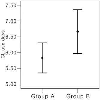
Results: Group A (R1>R2) had 53 eyes, and Group B (R1<R2) had 39 eyes. Group A showed a shorter epithelial healing time than Group B (5.8±1.7 days vs. 6.7±2.1 days, p=0.04).
Conclusions: The flat fitting group showed a shorter epithelial healing time than the steep fitting group after LASEK.
Korean Journal of Ophthalmology 21(2):85-89, 2007
Key Words: Base curve radius (BCR), LASEK, Therapeutic contact lens
Received: July 29, 2006 Accepted: February 27, 2007
Reprint requests to Mee Kum Kim, MD. Department of Ophthal- mology, Seoul National University College of Medicine, 28 Yongon- dong, Chongno-gu, Seoul 110-744, Korea. Tel: 82-2-2072-2438, Fax:
82-2-741-3187, E-mail: [email protected]
* This study was presented in part at the 95nd annual metting of
the Korean Ophthalmological Society, April 2006.
keratometry. Patients who underwent refractive surgery previously, with dry eye syndrome, cataract, keratoconus, glaucoma, retinal disease, optic neuropathy, connective tissue disease, or systemic diseases were excluded.
LASEK surgery was performed by spreading the palpebral fissure with a speculum, administering 0.5 % proparacaine hydrochloride (Alcaine
Ⓡ, Alcon, USA), placing an alcohol solution cone 8.5 mm in diameter (J2905, Janach, UK) on the cornea, diluting with distilled water, placing 20 % alcohol solution on the cone, waiting for 30 seconds, adding balanced salt solution, and washing sufficiently. Subsequently, using an epithelial microhoe (J2915A, Janach, UK), the corneal epithelium was resected smoothly. And at later time, the corneal epithelial flap was gathered at 12 o'clock direction using a spatula. At the time of resection, the basement membrane was resected carefully from the Bowman's layer.
Cases whose corneal epithelial fragment was not created as a complete flap were excluded from the subject group. After the irradiation with excimer laser (Star S4, VISX, Santa Clara, USA), the corneal epithelial flap and the corneal stroma were washed sufficiently with buffer solution and carefully placed in the original site with a spatula. After LASEK surgery, therapeutic lenses, of which the base curve radius (R1) was within 8.6 - 9.0 mm range, were selected randomly and worn. The characteristics of therapeutic lens used in our study are summarized in Table 1. Patients were examined 1 day, 4 days, 6 days, and 8 days after surgery.
Also, eye drops of 0.1 % Fluorometholone (Fluorometholone
Ⓡ, Santen, Japan), Levofloxacin (Cravit
Ⓡ, Santen, Japan) were administered 4 times per day, and until the defect of the corneal epithelium recovered completely, the eyes were examined without removing the corneal epithelial flap.
We calculated the estimated BCR (R2) by calculating keratometry value on corneal topography after surgery, using the diopter conversion table. According to the differences of BCR (R1) and the estimated BCR (R2), they were divided into group A and group B. Group A was defined as the group with lenses that had a BCR larger than the BCR calculated by postsurgical keratometry (flat group: R1>R2); group B was the group with lenses that had a BCR smaller than the BCR calculated by postsurgical keratometry (Steep group:
R1<R2). The age, gender, corneal curve radius, and the corneal epithelial healing period of the two groups were compared.
Statistical analysis was performed using the SPSS v12.0 (SPSS Inc., Chicago, USA). An independent T-test was used
for comparing the two groups and for the cases showing significant results in univariant regression analysis, multivariant regression analysis was performed. Cases with a p value less than 0.05 were considered significant.
Results
The subject group had a total of 47 patients (92 eyes);
group A had 53 eyes and group B had 39 eyes. In group A, the healing period after LASEK surgery was on average 5.8±1.7 days; in group B, the corneal epithelial healing period after LASEK surgery was on average 6.7±2.1 days, a statistically significant difference (p=0.04, independent T-test, Figure 1).
Table 1. Descriptive data and characteristics of the therapeutic contact lens used in this study
Series BCR (mm) Diameter (mm) Water content (%) Material Producer
Biomedics 55 8.6 14.2 55 Ocufilcon D Hanaoptical
Anyday 8.7 14.2 55 Hema/MAA copolymer Hanaoptical
Focus Vistinct 8.8 14.0 55 Vifilcon A CIBA vision
Anyday 8.9 14.2 55 Hema/MAA copolymer Hanaoptical
Acuve 9.0 14.2 58 Etafilcon A Johnson & Johnson
Fig. 1. Estimated mean and standard errors for number of days wearing therapeutic contract lens in Group A and Group B.
Group A showed a shorter epithelial healing time than Group B (5.8±1.7 days vs. 6.7±2.1 days, p=0.04). This implies selecting a flat fitting BCR lens is more beneficial on wound healing than a steep fitting lens after LASEK.
Group A=Patients with flat fitting (R1>R2), Group B=Patients with steep fitting (R1<R2).
R 1: Base curve radius (BCR) of therapeutic contact lens (T-lens) which was randomly selected for patients who underwent LASEK.
R 2: BCR of T-lens which were converted from post operative diopters of patients who underwent LASEK by using a diopter conversion table.
Group A Group B 5.00
5.50 6.00 6.50 7.00 7.50
CL u se d a y s
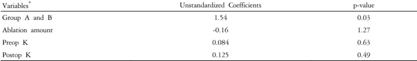
In addition, the difference in the mean value between the BCR of therapeutic lenses and the BCR anticipated after surgery (R1-R2) in group A was 0.5 mm. In group B, the difference was -0.4 mm. Even though age, gender ratio, and the BCR of the contact lens used were not different, the preoperative keratometry, postoperative keratometry, and the ablation depth between the two groups were significantly different (Table 2 and 3). To examine the effect of various factors, for variables with p values lower than 0.2 in univariant analysis, multivariance regression analysis was performed. The result showed that it was not associated with other variables and a significant difference was observed only in the BCR (Table 3). In other words, when therapeutic lenses that are flatter than the BCR according to the keratometry were worn after LASEK, the healing of the epithelium was faster.
Discussion
LASEK is a refractive surgery that has been used frequently together with LASIK and PRK. Previous reports
have suggested that complications associated with the corneal flap in LASIK could be avoided by use of LASEK, that it is applicable to patients whose corneal thicknesses are thin,
1and that the frequent pain or subepithelial haze after PRK could be reduced.
8It is considered that reasons causing the less subepithelial haze are that after LASEK surgery, the scar healing process is similar to PRK, nonetheless, it allows the faster migration of epithelial cells, and thus ultimately, less cellular reaction in the stroma is induced. The cytokine that is involved in the subepithelial haze, TGF-β1 was detected to be less in the tear than PRK. The epithelial flap plays a physical barrier function and thus reduces the secretion of reflex tearing, consequently, the contact with Fas ligand and other cytokines inducing stromal reactions present in the tear was reduced.
9-11Nevertheless, some researchers claimed that the subepithelial haze after PRK and LASEK was not different, and the postsurgical visual acuity immediately after LASEK was rather poor, because of edema in the epithelial cell flap itself. It did not adhere well to the stroma, and over time it was destroyed naturally, replaced with the new epithelium growing from the vicinity. Hence, the regeneration
Table 3. Multiple regression analysis on days of CL use, R1=0.27, R2=0.19
Variables
*Unstandardized Coefficients p-value
Group A and B 1.54 0.03
Ablation amount -0.16 1.27
Preop K 0.084 0.63
Postop K 0.125 0.49
*
Variables of which p-value were significant on single variable analysis.
We constructed a multiple variable model to predict therapeutic contact lens use days contending group A or group B variable and other covariates, but the group A or group B variable was the only significant predictor.
Group A=Patients with flat fitting (R1>R2), Group B=Patients with steep fitting (R1<R2).
R 1: Base curve radius (BCR) of therapeutic contact lens (T-lens) which was randomly selected for patients who underwent LASEK.
R 2: BCR of T-lens which were converted from post operative diopters of patients who underwent LASEK by using diopter conversion table.
Table 2. Demographics and descriptive statistics for ophthalmic measurements of enrolled patients
Group A (n=53) Group B (n=39) p-value
Age, years 23.5±4.04 23.8±4.4 0.66
*Male, n (%) 15 (28.3%) 13 (30%) 0.87
†Ablation amount, μm 51.6±20.2 70.3±14.8 0.02
*Preop K, Diopter 43.7±1.48 42.4±1.34 0.03
*Postop K, Diopter 40.2±1.46 36.7±1.19 0.02
*Lens wearing, days 5.83±1.74 6.66±2.14 0.04
*Mean ∆ (R1-R2) mm 0.51±0.26 -0.41±0.27 0.00
*BCR of used lens, mm 8.79±0.13 8.79±0.15 0.91
*Data are given as mean±standard deviation (range) of variables unless otherwise indicated.
Group A=Patients with flat fitting (R1>R2), Group B=Patients with steep fitting (R1<R2).
R 1: Base curve radius (BCR) of therapeutic contact lens (T-lens) which was randomly selected for patients who underwent LASEK.
R 2: BCR of T-lens which were converted from postoperative diopters of patients who underwent LASEK by using diopter conversion table.
*
Independent T-test.
†