Original Article
Practical patterns for stereotactic body radiotherapy to hepatocellular carcinoma in Korea: a survey of the Korean Stereotactic Radiosurgery Group
Sun Hyun Bae 1 , Mi-Sook Kim 2, *, Won Il Jang 2 , Chul-Seung Kay 3 , Woochul Kim 4 , Eun Seog Kim 5 , Jin Ho Kim 6 , Jin Hee Kim 7 ,
Kwang Mo Yang 8 , Kyu Chan Lee 9 , A Ram Chang 10 , and Sunmi Jo 11
1
Department of Radiation Oncology, Soonchunhyang University College of Medicine, Bucheon,
2Korea Institute of Radio- logical & Medical Sciences, Seoul,
3Incheon St Mary ’s Hospital, The Catholic University of Korea, Inchon,
4Inha University Hospital, Inha University School of Medicine, Incheon,
5Soonchunhyang University College of Medicine, Cheonan,
6
Seoul National University, College of Medicine, Seoul,
7Keimyung University Dongsan Medical Center, Keimyung University School of Medicine, Daegu,
8Dongnam Institute of Radiological & Medical Sciences, Busan,
9Gachon Uni- versity Gil Medical Center, Gachon University of Medicine and Science, Incheon,
10Soonchunhyang University Col- lege of Medicine, Seoul, and
11Haeundae Paik Hospital, Inje University School of Medicine, Busan, Republic of Korea
*For reprints and all correspondence: Mi-Sook Kim, Department of Radiation Oncology, Korea Institute of Radiological &
Medical Sciences, 75, Nowon-ro, Nowon-gu, Seoul 01812, Republic of Korea. E-mail: [email protected] Received 5 October 2015; Accepted 17 December 2015
Abstract
Objective: To investigate practical patterns for stereotactic body radiotherapy to hepatocellular carcinoma in Korea.
Methods: In June 2013, the Korean Stereotactic Radiosurgery Group of the Korean Society for Radiation Oncology conducted a national patterns-of-care survey about stereotactic body radiother- apy to the liver lesion in hepatocellular carcinoma, consisting of 19 questions and 2 clinical scenarios.
Results: All 208 radiation oncologists (100%), who are regular members of Korean Society for Radiation Oncology, responded to this survey. Among these, 95 radiation oncologists were specialists for hepatol- ogy; 64 physicians did not use stereotactic body radiotherapy for hepatocellular carcinoma, and 31 physicians used stereotactic body radiotherapy. Most physicians (52%) performed stereotactic body radiotherapy to hepatocellular carcinoma in ≤5 cases per year. Physicians applied stereotactic body radiotherapy according to tumour size and baseline Child –Pugh class. All physicians agreed the use of stereotactic body radiotherapy to 2.8-cm hepatocellular carcinoma with Child –Pugh class of A, while 23 physicians (74%) selected stereotactic body radiotherapy for Child –Pugh class of B. Nineteen physicians (61%) selected stereotactic body radiotherapy to 5-cm hepatocellular carcinoma with Child –Pugh class of A, and only 14 physicians (45%) selected stereotactic body radiotherapy for Child –Pugh class of B. On the other hand, the preferred dose scheme was same as 60 Gy in three fractions.
Conclusions: Among radiation oncologists in Korea, there was diversity in the practice for stereotac- tic body radiotherapy to the liver lesion in hepatocellular carcinoma. Additional prospective studies are necessary to standardize the practice and establish Korea-specific practice guidelines for hepatocellular carcinoma stereotactic body radiotherapy.
Key words: hepatocellular carcinoma, Korea, stereotactic body radiotherapy, survey
Advance Access Publication Date: 29 January 2016 Original Article
© The Author 2016. Published by Oxford University Press. All rights reserved. For Permissions, please email: [email protected] 363
at KEIMYUNG UNIV MEDICAL LIBRARY on January 5, 2017 http://jjco.oxfordjournals.org/ Downloaded from
Introduction
Since stereotactic radiosurgery (SRS) was developed for the treatment of intracranial malignancies, stereotactic body radiotherapy (SBRT) was derived from SRS for the treatment of extracranial malignancies.
Blomgren et al. (1) reported the first clinical use of SBRT to liver le- sions in patients with hepatocellular carcinoma (HCC) at the Karolins- ka Institute in Stockholm in 1995. Since then, several prospective and retrospective studies on liver SBRT in HCC patients have reported a promising local control rate (LCR) and low risk of severe toxicity (2 – 5). Now, SBRT is considered as the alternative treatment option for HCC that is inoperable or unsuitable for other local treatments, and the National Comprehensive Cancer Network guidelines and the practice guidelines from Korean Liver Cancer Study Group and National Cancer Center recommend SBRT as a local treatment modal- ity for the management of HCC (6,7).
Despite increased use of SBRT for HCC, current patterns of prac- tice are unknown. Considering the vast heterogeneity in the manage- ment of HCC and some differences in equipment availability for SBRT, a wide variation in patterns of practice is expected (8). Recently, we conducted a nationwide survey on the use of SBRT in Korea and reported a continuous increase in its use, as well as a variety of prac- tices surrounding its use (9). Therefore, the Korean Stereotactic Radio- surgery Group of the Korean Society for Radiation Oncology (KOSRO) conducted a national patterns-of-care survey to better understand practical patterns of SBRT for HCC in Korea.
Patients and methods
In Korea, SBRT for the treatment of extracranial malignancies is cov- ered by the National Health Insurance Service when the number of fractions is 4 or fewer. Therefore, for this survey, we defined SBRT as radiotherapy with delivery of a high dose of radiation using ≤4 frac- tions to liver lesions in HCC patients. We sent the survey by e-mail to all 228 radiation oncologists, who are regular members of KOSRO, at 85 institutions in Korea in June 2013. A 19-questionnaire survey was designed to identify a specialist for hepatology and to determine prac- tices surrounding the use of SBRT, including prescribed dose, moving organ control system, treatment machine and planning system. The full contents of the survey are available in Supplementary material 1.
If the respondents were not a specialist for hepatology, they ended the survey on the first question and returned it by e-mail. If the respondents were specialist for hepatology, they were asked to complete the rest of the survey. Because they were able to select multiple answers to certain questions, the total percentage was >100% for selected questions. After 3 months, at which time we had collected the completed surveys by e-mail, we sent an additional survey including two clinical scenarios to only SBRT users: 2.8-cm HCC and 5-cm HCC. In this clinical setting, they selected SBRT or other fractionation schemes on their own judge- ment. The full contents of the survey are available in Supplementary material 2.The completed survey was returned by e-mail within 1 month. In the event of non-response, we contacted by telephone and sent e-mails in order to achieve a 100% response rate. This study was conducted under the authorization and cooperation of the Korea Radiation Oncology Group (KROG 13-14).
Results
The use of SBRT
In June 2013, 228 radiation oncologists were registered as regular members of KOSRO. Among these, 20 radiation oncologists were
not involved in clinical practice owing to active service in the military, a temporary layoff or work as a general practitioner in primary health care. The remaining 208 KOSRO members actively participated in clinical practice, and all (100%) responded to the survey by August 2013. Among these, 95 KOSRO members responded that they acted as a specialist for hepatology. Of these, 31 physicians (33%) from 27 institutions used SBRT to treat liver lesions in HCC patients. The most common reason for the use of SBRT was the delivery of a higher dose than that possible with conventional radiotherapy (68%), followed by the shortening of treatment duration in order to start another treat- ment as early as possible (23%). Additional reasons were the shorten- ing of treatment duration to improve patients ’ convenience and to reduce the mechanical load of the treatment machine (6%), and par- ticipation in a clinical trial (3%). When the respondents were allowed up to two answers, the most common reason for not using SBRT was the lack of appropriate patients for SBRT (56%), followed by the lack of special equipment (34%), and the use of other fractions such as hy- pofractionation or conventional fractionation (28%). Additional rea- sons were adoption of other treatment modalities (8%), the lack of experience with use of SBRT (6%), the existence of another SBRT ex- pert (5%) and preparation for use of SBRT (3%).
In Korea, the number of physicians using SBRT has shown a grad- ual increase, since one physician applied SBRT to the liver lesion in HCC patients in 2003 (Fig. 1A). In 2013, the number of SBRT cases per physician per year was ≥50 in 2 physicians (7%), 40–50 in 1 (3%), 10 –30 in 6 (19%), 6–10 in 6 (19%) and ≤5 in 16 (52%). When HCC patients were consulted to receive radiotherapy for the liver lesion, the application rate of SBRT was mostly <50%
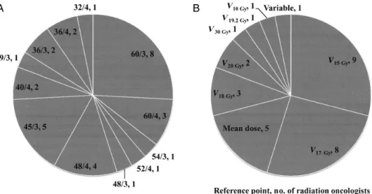
(Fig. 1B). Only two physicians who work at the same institution re- sponded with an application rate of ≥90%. The most commonly pre- scribed doses for SBRT varied among physicians, as shown in Fig. 2A.
The most common scheme was 60 Gy in three fractions (26%), fol- lowed by 45 Gy in three fractions (16%). When physicians adopted liver dose constraints, the normal liver volume [the total liver volume minus the planning target volume (PTV)] was considered as the signifi- cant liver volume by 28 physicians (90%), and the total liver volume by 3 physicians (10%). Reference points of liver dose constraints var- ied, as shown in Fig. 2B; V
15 Gy(at least 700 ml of the normal liver volume had to receive a total dose of <15 Gy, 29%), V
17 Gy(at least 700 ml of the normal liver volume had to receive a total dose of
<17 Gy, 26%) and the mean dose (16%) were the most common liver dose constraints.
The preferred method of planning computed tomography (CT) for SBRT was four-dimensional CT (20 physicians, 65%), followed by in- halation and exhalation-breath-hold CT (19%), free-breathing CT combined with fluoroscopy (10%) and free-breathing CT (6%). The preferred method of immobilization during planning CT was alpha cradle/vacuum-lock, followed by a combination of stereotactic body frame plus alpha cradle/vacuum-lock plus wingboard. For control of liver motion, respiratory-gated radiotherapy with Real-Time Position Management (Varian Medical Systems, Palo Alto, CA) was preferred.
Details of the preferred methods are summarized in Table 1. Various treatment machines were available for liver SBRT: nine physicians (29%) applied two or more specially equipped treatment machines and could select the most suitable treatment machine on a case-by-case basis. The most commonly used treatment machines were RapidArc (Varian Medical Systems) and CyberKnife (Accuray Inc., Sunnyvale, CA). Details of the treatment machines are summarized in Table 2.
For SBRT planning, nine physicians (29%) used two planning sys- tems. The majority (65%) used Eclipse (Varian Medical Systems). In addition, 29% of the physicians used the CyberKnife planning system, 364 A survey of practice for SBRT to HCC in Korea
at KEIMYUNG UNIV MEDICAL LIBRARY on January 5, 2017 http://jjco.oxfordjournals.org/ Downloaded from
Figure 1. (A) Cumulative adoption of stereotactic body radiotherapy (SBRT) to the liver lesion in hepatocellular carcinoma (HCC) after its introduction in 2003.
(B) Application rate of liver SBRT per physician per year in HCC patients.
Figure 2. (A) The most commonly prescribed doses for SBRT to the liver lesion in HCC. (B) Reference points of liver dose constraints for liver SBRT in HCC: V
x Gymeans the normal liver volume receiving <X Gy; variable means that the radiation oncologist selected the reference points case by case.
at KEIMYUNG UNIV MEDICAL LIBRARY on January 5, 2017 http://jjco.oxfordjournals.org/ Downloaded from
16% used the iPlan (BrainLAB AG, Feldkirchen, Germany), 7% used the Pinnacle system (Philips, Milpitas, CA) and 7% used MONACO (Electa, Crawley, UK). For optimal dose distribution for the tumour and the liver, 18 physicians (58%) used multiple planning techniques.
The most commonly applied planning techniques were static inten- sity-modulated radiotherapy (IMRT) (61%), followed by dynamic conformal arc radiotherapy (55%), three-dimensional conformal radiotherapy (3DCRT) with multiple beam arrangements (29%), robotic SBRT (29%) and rotational IMRT (10%).
To assist in target localization, fiducial insertion was always per- formed by three physicians (10%) and sometimes by eight physicians (26%). For target localization before each treatment, the preferred verification method was conebeam CT (74%), followed by orthogonal kilovoltage radiography (13%), orthogonal megavoltage radiography (10%) and fluoroscopy (3%). The application rate of gating treatment was 0% in 13 physicians (43%), <10% in 4 (13%), 10 –40% in 2 (6%), 40 –60% in 2 (6%), 60–90% in 2 (6%), ≥90% in 2 (6%) and 100% in 6 (20%). When patients received SBRT, 11 physicians (35%) delivered it on consecutive days; 18 physicians (58%), with 48-h intervals between fractions and 2 physicians (7%), with 72-h
intervals between fractions. The overall treatment was ended within 1 week (55%) or 2 weeks (45%). At the time, 13 respondents (42%) were participating in clinical trials.
Clinical cases
The first case was a 49-year-old male patient with 2.8-cm HCC at the liver dome. The lesion was inoperable owing to an underlying medical problem and was unsuitable for radiofrequency ablation. He received one cycle of transarterial chemoembolization (TACE). The follow-up CT after TACE showed a viable tumour with incomplete lipiodol up- take (Fig. 3A), and he was consulted for radiotherapy. The normal liver volume was measured as 1359 ml on planning CT. If the baseline liver function was Child –Pugh (CP) class of A, all 31 physicians agreed on the use of SBRT (Fig. 3B). However, in case of CP class of B, 23 physicians (74%) selected SBRT (Fig. 3C), while 8 physicians (26%) selected altered fractionation schedules (6 physicians adopted hypo- fractionation and 2 physicians adopted conventional fractionation).
The second case was a 57-year-old male patient with 5-cm HCC at the liver dome. He received three cycles of TACE. The follow-up CT after TACE showed a viable tumour with incomplete lipiodol uptake (Fig. 3D), and he was consulted for radiotherapy. The normal liver volume was measured as 1435 ml. In case of CP class of A, SBRT was chosen by 19 physicians (61%, Fig. 3E), hypofractionation by 10 (32%) and conventional fractionation by 2 (7%). In case of CP class of B, SBRT was chosen by only 14 physicians (45%, Fig. 3F), hypofractionation by 13 (42%) and conventional fractionation by 4 (13%). Although physicians selected SBRT according to tumour size and baseline liver function, the preferred dose scheme was 60 Gy in three fractions for all cases.
During the SBRT planning process, magnetic resonance imaging (MRI) to assist target delineation was always used by only three phy- sicians and sometimes used by six physicians. The majority (71%) did not use MRI. The most commonly applied interval from completion of SBRT to the first follow-up imaging study was 4 weeks (65%), fol- lowed by 8 weeks (32%), and 12 weeks (3%). The preferred imaging techniques for surveillance were CT alone (42%) or a combination of CT and MRI (39%). Two physicians selected the type of follow-up image (CT or MRI) based on the pretreatment image. Some physicians preferred a combination of PET-CT and other techniques: CT (one physician) or MRI (one physician), or CT and MRI (two physicians).
Discussion
In Korea, practical patterns of radiotherapy to HCC have changed over time. The Korean Liver Cancer Study Group conducted the first national survey on radiotherapy for HCC patients in 2006 (10).
Among 53 institutions that were running in Korea at that time, only 10 institutions (19%) treated at least 5 HCC patients with external beam radiotherapy between 2004 and 2005. Applied planning techniques were 3DCRT (82%), IMRT (1%), two-dimensional conventional radiotherapy (8%) and CyberKnife (9%). On the other hand, 27 in- stitutions (32%) used SBRT to the liver lesion in HCC in 2013; 15 in- stitutions (18%) treated at least 6 HCC patients with SBRT per year.
However, the application rate of SBRT for HCC is still lower than that of other cancers, especially primary or metastatic lung cancers, consid- ering that a previous national survey conducted by our group on the use of SBRT in Korea for the treatment of various tumours reported that 38 institutions (45%) have used SBRT (9). Our current survey that the application rate of SBRT to HCC was mostly <50%, and that Table 1. Details of the preferred methods for immobilization and
control of liver motion for stereotactic body radiotherapy to hepatocellular carcinoma in Korea
Methods of immobilization No. of radiation oncologists (%)
Alpha cradle/vacuum-lock 9 (29)
Alpha cradle/vacuum-lock + wingboard 5 (16)
Stereotactic body frame 2 (7)
Stereotactic body frame + alpha cradle/
vacuum-lock
1 (3) Stereotactic body frame + alpha cradle/
vacuum-lock + wingboard
6 (19)
Wingboard 4 (13)
No use 4 (13)
Methods of liver motion control No. of radiation oncologists (%) Respiratory gating method: Varian RPM 15 (48) Respiratory gating method: ExacTrac
gating-Novalis gating
2 (7) Forced shallow breathing with abdominal
compression
10 (32) Real-time tumour-tracking methods 2 (7)
No use 2 (6)
Table 2. Details of the treatment machines for stereotactic body radiotherapy to hepatocellular carcinoma in Korea
Types of treatment machines No. of radiation oncologists
a(%)
RapidArc_Varian 10 (32)
CyberKnife 9 (29)
Clinac iX_Varian 6 (19)
Novalis_Varian 6 (19)
TrueBeam_Varian 4 (13)
Tomotherapy 3 (10)
VMAT_Elekta 2 (7)
Novalis Tx_Varian 1 (3)
a