Physiological and Biochemical Responses to Ozone Toxicity in Five Species of genus Quercus S eedlings
Du-Hyun Kim
1*, Sim-Hee Han
1, Ja-Jung Ku
1, Kab-Yeon Lee
1and Pan-Gi Kim
21Divison of Forest Genetic Resources, Korea Forest Research Institute, Suwon, 441-350, Korea
2Division of Forest Environment and Resources, Kyungpook National University, Sangju, 742-711, Korea
(Received May 7, 2008; Revised June 13, 2008; Accepted June 25, 2008)
참나무속 5종의 오존 독성에 대한 생리생화학적 반응
김두현1
*
·한심희1·구자정1·이갑연1·김판기21국립산림과학원 산림유전자원부, 2경북대학교 산림환경자원학부 (2008년 5월 7일 접수; 2008년 6월 13일 수정; 2008년 6월 25일 수락)
ABSTRACT
Physiological and biochemical changes of five species of genus
Quercusexposed to ozone fumigation were investigated to assess their tolerance against ozone toxicity. At the end of 150 ppb ozone fumigation, chlorophyll contents, photosynthetic characteristics, malondialdehyde (MDA) and antioxidative enzyme activities were measured in the leaves of five
Quercusspecies (
Quercus acutissima, Q. aliena, Q. palustris, Q. serrata,and
Q. variabilis). Chlorophyll and carotenoid contents, net photosynthesis and carboxylation efficiency decreased after ozone treatment, indicating that O
3-exposed plants underwent physiological inhibition. The reduction rate of total chlorophyll contents and carboxylation efficiency were respectively 15% and 34% for
Q. alienaand 38% and 62% for
Q. variabilis. The amount of MDA increased with the highest increase rate of 140% in
Q. acutissimawhich also showed the highest increase rate (60%) of superoxide dismutase (SOD). Ascorbate peroxidase (APX) activity increased in
Q. variabilis,
Q. serrataand
Q. acutissimaby ozone treatment. Based on our results, ozone tolerance of the five
Quercusspecies was ranked as
Q. aliena>
Q. palustris>
Q. serrata>
Q. variabilis>
Q. acutissima. We concluded that chlorophyll contents, photosynthesis, MDA content and antioxidative enzymes were the important physiological markers for tolerance against ozone stress, which were closely related with one another.
Key words
: Oak, Chlorophyll content, Photosynthesis, MDA, Antioxidative enzyme
I. INTRODUCTION
For more than 50 years, the phytotoxicity of ozone (O
3) has been demonstrated for forest tree species (Kar- nosky
et al., 2007). Studies concerning the dynamics of ozone (O
3) between atmosphere and plants have been carried out under the United Nations Economic Com- mission for Europe-Convention on Long Range Trans- boundary Air Pollution (UNECE CLRTAP) (Mills
et al., 2000). It is now clearly established that O
3can
cause a range of effects including visible leaf injury and reduction of growth and yield, and can alter the sensitivity of plants to biotic and abiotic stresses (Fuhrer and Achermann, 1994). Despite the increasing environmental awareness and regulations designed to limit industrial and vehicular emissions, ozone levels potentially harmful to human health and vegetation have increased every year in Korea (Ministry of Envi- ronment, 2005).
Oak trees,
Quercusspecies, which are one of the
Corresponding Author: Du-Hyun Kim ([email protected])most abundant tree species in Korean, are important economically and ecologically as the ornamental or roadside tree. They are known as a tolerance species to air pollutants, but the information on the biochemical and physiological responses and tolerant mechanisms for
Quercusspecies under environmental stress was few
.It appears that distinct O
3induced effects are largely confined to fast-growing tree species (Skärby
et al., 1998). Investigations on the effects of O
3on the slow- grown species, such as
Quercus, are scarce and did not consider O
3to be a significant factor in the complex of damage to the oak (Thomas
et al., 2002, Samuelson
et al., 1996; Hanson
et al., 2005). On the other hand, Hanson
et al. (1994) and Kelting
et al. (1995) reported reductions in foliar assimilation to O
3exposures in red oak (
Q. rubra). No information has been provided to compare the sensitivity among
Quercusspecies to O
3that this study focuses on. However, difficulties in ranking the O
3sensitivity of different species may arise since intra-specific variations have been reported in several plant species (Inclán
et al., 1999; Minnocci
et al., 1999; Ranieri
et al., 2001). Inclán
et al. (1999) indi- cated the differential sensitivity to O
3of five Mediter- ranean woody species, and Elvira
et al. (2004) reported intra-specific variations in two population of
Q. coc- ciferato O
3.
During the past three decades, occurrence of oak declines has been recorded in many European countries (Thomas
et al., 2002). In the Mediterranean region, mainly
Q. ilexhas been affected (Müller-Edzards
et al., 1997), and a decline has been also reported for decid- uous oak species (
Q. cerris, Q. frainetto, Q. pubescens and Q. robur) in Italy (Ragazzi
et al., 1998), and for
Q.suber
on the Iberian Peninsula (Sousa Santos and Moura Martins, 1993). On the contrary, studies of red oak, however, showed an enhanced sensitivity to O
3in 30-year-old mature trees than two year-old seedlings due to higher stomatal conductances in the older trees (Samuelson
et al., 1996; Weinstein
et al., 1998;
Wullschleger
etal., 1996).
The factors determining plant sensitivity or tolerance are not clearly understood but it is thought to be related to many underlying physiological, anatomical, bio- chemical and environmental factors (Alonso
et al., 2001; Pääkkönen
et al., 1998, Ribas
et al., 2005).
Ozone effects on tree biomass are the result of several processes occurring at the cellular and physiological levels. Acceleration of leaf senescence has been widely
reported as one of the most characteristic processes derived form ozone exposure. These processes involve chlorophyll degradation and reductions in CO
2assimi- lation (Elvira
etal., 1998; Zheng
et al., 2002). Physiolog- ical effects of ozone exposure include photosynthesis reduction and an increase of antioxidant systems. Pho- tosynthesis reduction results in a decreased growth rate in either volume or biomass.
The biochemical markers have been used to select the best tolerant species, cultivar and variety to O
3or other pollutants (Han
et al., 2006). Paoletti
et al. (2003) also adopted antioxidant levels as biomarkers of ozone sensitivity that could be used for screening of great diversity of tree species’ potential sensitivity to ozone.
However, there was not an exact answer about the differ- ences of biochemical and physiological responses among species, cultivar or varieties as well as among biochemical makers.
Therefore, this study was undertaken to investigate cellular responses to ozone as mechanism for oxidant impact on photosynthesis in seedlings of oak trees. The aims of the present study were to (1) determine the effects of ozone exposure on photosynthetic pigments, photosynthesis, antioxidant enzyme activities and lipid peroxidation, (2) evaluate the potential occurrence of interspecific variations in O
3sensitivity in
Quercus, and (3) evaluate a differential sensitivity to ozone expo- sure in
Quercus.
II. MATERIALS AND METHODS
Seeds of five species of genus
Quercus(
Q. acutis- sima, Q. aliena, Q. palustris, Q. serrata,and
Q. vari- abilis) were germinated in sand soil on March 12,
2007. One-year-old seedlings were transplanted into
plastic pots (H 20 × W 15 cm) containing artificial
soil which consisted of 1:1:1 sand: peat moss: ver-
miculite (volume basis). Five seedlings per treatment
were transferred into O
3chamber and were arranged
in two blocks. O
3treatment was divided into two
chambers: control chamber was circulated with the
clean air and the other one was fumigated with 150
ppb O
3. O
3fumigation time was 8 hrs a day. O
3con-
centration in chamber was registered 5
±1 ppb in
control and 150
±10 ppb in treatment chamber dur-
ing fumigation period. The fumigation system has
been described in detail by Lee
et al. (2003). The
experiment was started on June 22, 2007 and it was
conducted for four weeks.
2.1. Photosynthetic pigments
At the end of O
3fumigation, the leaves of control and O
3-treated seedling of oak trees were excised and soaked in dimethyl sulfoxide (DMSO) in a glass vial.
The vial was tightly capped and incubated at 70
oC for 2 hrs in the dark. The concentration of the extracted pigments (total chlorophyll, chlorophyll a, chlorophyll b and carotenoid) was calculated on the basis of their absorbance values at 664, 645 and 470 nm according to Lichtenthaler (1987).
2.2. Photosynthesis and biomass
Net photosynthesis of fully expanded leaves was measured with an infrared gas analyzer (Li-6400, Li- COR, USA). Environmental parameters were main- tained stably for measuring (mean temperature: 20.0
±0.1
oC; relative humidity: 68.2
±3.2%; leaf-to-air vapour pressure deficit: 1.2
±0.2 kPa). All determinations were performed at 1100
μmol m
−2s
−1photon flux den- sity (PFD). Net photosynthesis (A,
μmol CO
2m
−2s
−1) was determined at light saturation level between 10 a.m. and 3 p.m.
To calculate carboxylation efficiency,
ACi-curve was made (Farquhar
et al., 1980; Kim and Lee, 2001). The carboxylation efficiency was determined from the ini- tial slope of a linear regression using the linear portion of the
ACi-curve (0-150 ppb intercellular CO
2).
For biomass measurements, shoots and roots were carefully removed, and then thoroughly rinsed twice with distilled water. Shoot and root dry weights were recorded after drying the tissues at 70
oC.
2.3. Lipid peroxidation
Lipid peroxidation was determined by measuring the amount of malondialdehyde (MDA) produced by thiobar- bituric acid reaction as described by Heath and Parker (1968). The crude extract was mixed with the same vol- ume of 0.5% (w/v) thiobarbituric acid solution containing 20% (w/v) trichloroacetic acid. The mixture was heated at 95
oC for 30 min. and then quickly cooled in an ice-bath.
The mixture was centrifuged at 3000 × g for 10 min. and the absorbance of the supernatant was monitored at 532 and 600 nm. After subtracting the non-specific absorbance (600 nm), MDA concentration was determined by its molar extinction coefficient (155 mM
−1cm
−1) and the results were expressed as
μmol MDA g
−1FW.
2.4. Antioxidative enzyme activities
Fresh leaves (0.1 g) were homogenized under ice-
cold condition with 5 mL of 50 mM phosphate buffer (pH 7.0), 10 mM ascorbic acid (AsA) and 1.0% (w/v) polyvinylpyrrolidone. The homogenate was centrifuged at 20,000
×g for 30 min. and the supernatant was col- lected for enzyme assays.
Superoxide dismutase (SOD) was assayed based on the inhibition of reduction of nitro-blue tetrazolium in the presence of xanthine at 530 nm according to the method of Beauchamp and Fridovich (1971). Ascor- bate peroxidase (APX) activity was determined by the method of Nakano and Asada (1981). The assay was carried out in a reaction mixture containing 50 mM phosphate buffer (pH 7.0), 0.5 mM AsA, 0.1 mM EDTA, 0.1 mM H
2O
2, and 0.1 mL enzyme extract. The change in A
290was recorded for 1 min. after the addi- tion of H
2O
2. Activity of glutathione reductase (GR) was assayed as described in Carlberg and Mannervik (1985). The assay was carried out in a reaction mixture containing 50 mM phosphate buffer (pH 7.8), 0.1 mM NADPH, 0.5 mM GSSH and 0.1 mL enzyme extract.
The change in A
340was recorded for 5 min. after the addition of enzyme extract. Catalase (CAT) activity was determined by following a two-step procedure (Fossati
et al., 1980). The rate of dismutation of H
2O
2to water and molecular oxygen is proportional to the concentration of catalase. Therefore, the sample con- taining catalase was incubated in the presence of a known concentration of H
2O
2. After incubation for exactly one minute, the reaction was quenched with sodium azide. The amount of H
2O
2remaining in the reaction mixture was then determined by the oxidative coupling reaction of 4-aminophenazone (4-aminoan- tipyrene) and 3,5-dichloro-2-hydroxybenzenesulfonic acid (DHBS) in the presence of H
2O
2and catalyzed by horseradish peroxidase (HRP). The resulting quinone- imine dye was measured at 520 nm. All enzyme activ- ities were measured using UV-120 (SHIMADZU, Japan).
2.6. Tolerance and injury index
Based on physiological and biochemical responses,
ozone tolerance ability of five
Quercusspecies was
ranked. Injury index was determined by the effect of
ozone on MDA, total chlorophyll content, carboxyla-
tion efficiency and biomass. Tolerance index was cal-
culated using increase rate of SOD, APX, CAT and GR
activities. Standard index on each parameter was calcu-
lated as (X-Xavg)/SD, where X is the average differ-
ence rate between control and ozone treatment for each
species, Xavg is the average difference rate between
control and ozone treatment for all five species and SD is the standard deviation of difference rate for five spe- cies. Standard index calculated by multiplied with weight value 12.5 to make even impact for all 8 param- eters. The total sum of tolerance index that is calculated from 4 antioxidative enzymes and injury index includ- ing sum of MDA, chlorophyll content, carboxylation efficiency and biomass were aggregated to evaluate the tolerance against ozone stress.
2.6. Statistical analysis
To compare the effect on control and O
3treatment, ANOVA was performed on experimental data (statisti- cal significance,
p ≤0.05). Statistical analyses were performed using the statistical package SAS System for Windows, Version 8.01 (SAS Institute, USA).
III. RESULTS
3.1. Photosynthetic pigments
After 28 day of ozone exposure, the contents of pho- tosynthetic pigments showed significant (
p< 0.05) dif-
ferences among five species, between control and ozone treatment and species × ozone treatment interac- tion. Chlorophyll and carotenoid contents were decreased by ozone treatment, indicating that O
3-exposed plants underwent physiological inhibition (Table 1). Ozone treatment negatively affected on the photosynthetic pigments in the leaves of five
Quercusspecies. The content of chlorophyll
ain the seedlings of five species was 44.1% (
Q. variabilis) to 15.1% (
Q. palustris) lower in O
3-exposed plant when compared with the control, respectively. For the content of chlorophyll
b,
Q. palus-tris
and
Q. serratashowed reduction rate ranged from 27.2% to 18.6%, whereas
Q. alienashowed an increase of 17.8% under ozone exposure. The reduction rate of total chlorophyll contents in five species ranged from 14.9% (
Q. aliena) to 38.2% (
Q. variabilis) in O
3- exposed plant comparing control, respectively. Caro- tenoid content showed highest reduction in
Q. variabi- lis(27.7%) and lowest reduction in
Q. serrata(9.5%) under ozone fumigation. The highest reduction rate on the ratio of chlorophyll
ato chlorophyll
bwas observed in the leaves of
Q. aliena(38.9%), whereas
Q. palustris Table 1. Effects of ozone fumigation on photosynthetic pigments in the leaves of five species of genus QuercusSpecies Ozone Chl a Chl b Chla+b Car Chl a/b Chl/Car
mg g−1
Q. acutissima Control 3.77±0.02 1.81±0.02 5.58±0.03 0.86±0.01 2.08±0.02 6.50±0.05 150 ppb 2.49±0.01 1.46±0.02 3.95±0.03 0.68±0.10 1.70±0.02 5.79±0.07
%* -33.9 -19.3 -29.2 -20.9 -18.3 -10.9
Q. aliena Control 2.96±0.01 1.02±0.01 3.98±0.02 0.83±0.01 2.90±0.01 4.77±0.02 150 ppb 2.20±0.01 1.20±0.01 3.40±0.01 0.68±0.01 1.83±0.01 5.02±0.03
% -25.7 17.7 -14.9 -18.1 -36.9 5.2
Q. palustris Control 2.70±0.01 0.92±0.01 3.62±0.01 0.70±0.01 2.93±0.01 5.15±0.02 150 ppb 2.29±0.02 0.67±0.01 2.96±0.02 0.60±0.01 3.44±0.02 4.89±0.01
% -15.2 -27.2 -18.2 -14.3 17.4 -5.1
Q. serrata Control 3.73±0.06 1.72±0.03 5.45±0.05 0.83±0.01 2.17±0.06 6.53±0.04 150 ppb 3.10±0.02 1.40±0.01 4.50±0.02 0.75±0.01 2.21±0.02 5.96±0.05
% -16.9 -18.6 -17.4 -9.6 -1.8 -8.7
Q. variabilis Control 3.40±0.05 1.67±0.01 5.10±0.06 0.83±0.06 2.05±0.01 6.18±0.03 150 ppb 1.90±0.02 1.25±0.01 3.15±0.03 0.60±0.01 1.51±0.01 5.28±0.02
% -44.1 -25.2 -38.2 -27.7 -26.3 -14.6
Species (S) *** *** *** *** *** ***
Ozone (O3) *** *** *** *** *** ***
S× O3 *** *** *** *** *** ***
All the values are means of five replicates ± SD; mixed effects linear model: *** p < 0.001.
*Reduction rate (%) was calculated as: % = [(control-treatment)/control] × 100.
increased by 17.4% under ozone exposure
,respec-
tively. For the ratio of total chlorophyll to carotenoid,
Q. variabilisshowed highest reduction (14.6%).
3.2. Net photosynthesis, carboxylation efficiency and biomass
The net photosynthesis showed significant (
p< 0.05) differences among species and between control and ozone treatment at the end of ozone exposure (Fig. 1).
However, there was no significant interaction between species and ozone treatment for net photosynthesis.
The reduction rate of net photosynthesis comparing with control in the seedlings of five species varied from 23.8% (
Q. variabilis) to 58.6% (
Q. acutissima). Car- boxylation efficiency differed significantly (
p< 0.05) among species and between control and ozone treat- ment at the end of ozone exposure (Fig. 1). Species × ozone treatment interaction was not significant for car- boxylation efficiency in the seedlings of five species.
The reduction rate of carboxylation efficiency was the highest in the leaves of
Q. acutissima(62.1%), and
Q.aliena
that showed lowest reduction rate was also decreased 33.6% for carboxylation efficiency in com- parison with control. Substantial progression of photo- synthetic-pigment destruction by ozone fumigation suggested the hinder of photosynthesis and decrees of photosynthesis resulted in reduction of biomass. Biom- ass showed the same result with net photosynthesis and carboxylation efficiency. There were significant (
p<
0.05) differences among species and between control and ozone treatment while there was no significant dif- ference with species × ozone treatment interaction.
Among five species,
Q. acutissima(30.9%) and
Q. ser-Fig. 1. Effects of ozone fumigation on net photosynthesis, car- boxylation efficiency and biomass in the leaves of five species of genus Quercus. Qa: Q. acutissima, Qal: Q. aliena, Qp: Q.
palustris, Qs: Q. serrata, and Qv: Q. variabilis. All the values are means of five replicates ± SD; mixed effects linear model.
Net photosynthesis: species p=0.0003***, ozone p= 0.0001***, species × ozone p=0.2213; carboxylation effi- ciency: species p=0.0001***, ozone p= 0.0006***, species × ozone p=0.7530; dry weight: species p<0.0001***, ozone p<0.0001***, species × ozone p=0.2626. *, ** and *** signif- icant at p≤0.05, 0.01 and 0.001.
Fig. 2. Effect of ozone fumigation on MDA content in the leaves of five species of genus Quercus. Qa: Q. acutissima, Qal: Q. aliena, Qp: Q. palustris, Qs Q. serrata, and Qv: Q.
variabilis. All the values are means of five replicates ± SD;
mixed effects linear model. Species p=0.0001***, ozone p=0.0001***; species × ozone p= 0.0001*** significant at p
≤0.001.
rata
(33.3%) showed high reduction and
Q. variabilis(14.0%) showed low reduction in biomass.
3.3. Lipid peroxidation and antioxidative enzyme activity
There was a significant difference in leaf MDA con- tent among five species and between control and ozone treatment (Fig. 2). Especially, the amount of MDA was increased under ozone fumigation and showed the highest increase rate of 140% in
Q. acutissimaamong five species, whereas it was reduced in
Q. alienaand
Q. palustrisunder ozone fumigation.
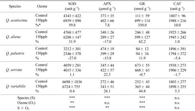
The responses of antioxidative enzymes varied each other under ozone fumigation (Table 2). For SOD, there was a significant (
p< 0.05) difference among five species. There was a significant difference between control and ozone treatment, and species × ozone treat- ment interaction showed a significant difference for SOD activity.
Q. acutissimashowed the highest increase rate of SOD activity (59.8%), however,
Q. palustrisshowed highest reduction of SOD activity (-27%).
APX activity differed significantly (
p< 0.05) between control and ozone treatment as well as among five spe- cies, and species × ozone treatment interaction was also
significant for APX activity. APX activity increased in the leaves of
Q. acutissima(7.8%),
Q. serrata(22.3%) and
Q. variabilis(26.1%) under ozone exposure, whereas
Table 2. Effects of ozone fumigation on antioxidative enzyme activities in the leaves of five species of genus Quercus
Species Ozone SOD
(unit g−1) APX
(μmol g−1) GR
(nmol g−1) CAT
(unit g−1) Q. acutissima Control
150ppb
%*
4343±422 6939±890
59.8
373±35 402±66
7.8
111± 59 499±114 350.0
1807± 96 1988±216
10.0 Q. aliena Control
150ppb
%
4768±477 6288±147
31.9
348±26 289±27 -17.0
246± 48 399±127
62.2
1925±266 1945±242
1.0 Q. palustris Control
150ppb
%
3212±301 2346±378
-27.0
474±19 399±29 -15.8
84± 12 94± 34 11.9
1896±391 1794±372
-5.4 Q. serrata Control
150ppb
%
4059±281 4015±336
1.1
345±44 422±57 22.3
673± 55 668± 63
-0.7
1938±273 1906±229
-1.7 Q. variabilis Control
150ppb
%
6698±1836 6724±7550
0.4
272±46 343±51 26.1
252± 43 365± 44
44.8
1803±277 1898±255
5.3 Species (S)
Ozone (O3) S× O3
*****
***
***n.s.
**
******
***
n.s.n.s.
n.s.
All the values are means of five replicates ± SD; mixed effects linear model: ** p< 0.05, *** p<0.001, and n.s.: not significant.
*Increasing rate (%) was calculated as: % = [(control-treatment)/control] × 100
Fig. 3. Correlation of tolerance and injury indices. Data for this figure were obtained from the same measurements as in Tables 1, 2 and Figs. 1, 2. Quercus Qa: Q. acutissima, Qal: Q. aliena, Qp: Q. palustris, Qs: Q. serrata, and Qv: Q.
variabilis. The differences between the values of exposed ozone and unexposed ozone on photosynthetic pigment con- tent, photosynthesis, biomass and MDA content were eval- uated for injury index, and four antioxidative enzyme activities were used for tolerance index.
Q. aliena
(-17.0%) and
Q. palustris(-15.8%) decreased in comparison with control plants. GR activity differed significantly (
p< 0.05) between control and ozone treatment as well as among five species, and species × ozone treatment interaction was also significant for GR activity. GR activity remarkably increased in the leaves of
Q. acutissima(350%) and
Q. aliena(62.2%) under ozone exposure. However,
Q. serratashowed no sig- nificant difference in comparison with control plants.
For CAT activity, the differences among species, treat- ments and species × ozone interaction were not signif- icant.
3.4. Rank of tolerance ability using tolerance and injury index
Based on physiological and biochemical responses mentioned above, ozone tolerance ability of five
Quer- cusspecies was ranked as
Q. aliena>
Q. palustris>
Q.serrata
>
Q. variabilis>
Q. acutissima(Fig. 3).
IV. DISCUSSION
In our research, the degree of ozone-injury was quan- titatively estimated and compared among five oak spe- cies, based on four injury and four tolerance indices. A gradient in O
3sensitivity was found for the species involved in this experiment. The results indicate that the differences between control and treatment in the sensitivity to ozone were distinct in aspects of low lev- els of chlorophyll and carotenoid content of O
3-fumi- gated leaves (Table 1). Reduction in the chlorophyll content of leaves, following exposure of plants to ozone, has been reported in many species such as
Pinus ponderosa(Anderson
et al., 2003),
Acerspecies (Han
et al., 2007), Citrus (Iglesias
et al., 2006) and
Quercus ilex(Ribas
et al., 2005). Knudson
et al. (1977) and Sakaki
et al. (1983) found higher reduction in chloro- phyll
athan chlorophyll
bin O
3-exposed plants of
Phaseolus vulgarisL. and
Spinacia oleraceaL. and our results also support this founding (Table 1). Ozone exposure induces the activation of leaf senescence- related process that was linked to chlorophyll degrada- tion, photosynthetic decline and lipid peroxidation (Ribas
et al., 2005; Bielenberg
et al., 2002).
The most apparent effect of oxidative stress induced by ozone described in numerous species, is leaf dam- age. In our experiments, visible injury symptoms (i.e., dark pigmented stipple in
Q. serrata, brown pigmented stipple in
Q. palustrisand necrosis in
Q. acutissima)
were observed in O
3-treated plants. It has been reported that a main detrimental effect of ozone at the subcellu- lar level is photosystem damage and reductions in pho- tosynthesis (Anderson
et al., 2003), and collapse of mesophyll cells (Long and Naidu, 2002)
Adult holm oaks (
Q. ilex) may be considered to be O
3tolerant in terms of photosynthesis, as only 9% reduc- tion was recorded in the O
3-treated leaves compared to the controls (Paoletti
et al., 2007) and also, Felzer
et al. (2007) reported higher sensitivity to ozone in 30-year- old mature trees than two year-old seedlings due to greater photosynthetic rates leading to higher stomatal conductances in the older trees. However, we observed severe reduction rate of net photosynthesis ranging from 23.8% to 58.6%, and carboxylation efficiency ranging from 33.6% to 62.1% by ozone treatment for all five species in spite of one-year-old seedlings. Pho- tosynthesis reduction leads to growth and biomass reduction, and reduction rate ranged from 14% to 33.3% (Fig. 1A, 1B, and 1C). Vitale
et al. (2008) showed severely limited photosynthesis in three-year-old seed- ling of
Q. ilexby 250 ppb of O
3fumigation like our results. Therefore, the reasons of differential results about photosynthesis and biomass to O
3tolerance could be variance among species or difference of O
3concentration.
We analyzed the level of lipid peroxidation and anti- oxidant activity, including SOD, APX, GR, CAT, because each of these traits have been previously asso- ciated with oxidative stress induced by O
3(Puckette
et al., 2007; Iglesias
et al., 2006). MDA concentration, which estimates the state and integrity of membrane through the degree of lipid peroxidation, has been shown to correlate with the level of ozone exposure. In addition, O
3-treated plants showed an increase in MDA content, indicating the state of membrane lipid peroxi- dation has a correlation with the degree of O
3exposure to plants (Prince
et al., 1990; Yoshida
et al., 1994;
Ranieri
et al., 1996; Iglesias
et al., 2006). In our results,
there was the increase of MDA content after exposure
of O
3in
Q. acutissima,
Q. serrataand
Q. variabilisspecies that showed severe reduction of chlorophyll
content and net photosynthetic rate. However, the other
two species,
Q. alienaand
Q. palustris, which showed
no reduction rate in the ratio of
chlorophyll ato
b, did
not increase in MDA content (Fig. 2). Therefore, the
reduction of chlorophyll content and net photosynthesis
had a tendency of the increase of MDA content in sen-
sitive species.
Lipid peroxidation is the ultimate effect of O
3attack to membranes, once the antioxidant defenses of mem- branes fail to cope with O
3and oxygen radicals (Calat- ayud
et al., 2002; Iglesias
et al., 2006). Plant responds to O
3-induced oxidative stress by activation of a num- ber of antioxidative stress-related defense mechanisms.
Carotenoid is also protective agents against oxidative stress, and has been associated with photoprotective mechanisms and ascorbate pool that plays a crucial role to the defense of hydrogen peroxides reaction (Tauz
et al., 2002; Iglesias
et al., 2006). However, in our study, we did not find a concomitant decrease in both caro- tenoid content and antioxidative enzyme activities.
In the present study, there were significant differ- ences in the activities of SOD and GR between treat- ments. On the contrary, APX and CAT had no significant differences between treatments. SOD and APX activi- ties were higher in O
3-treated
Q. palustristhan those of control plants, whereas GR activities were not signifi- cantly increased (Table 2). Additionally,
Q. acutissimathat showed high reduction rate in photosynthetic pig- ments content, photosynthesis and highest increase rate of MDA content had highest increase rate in SOD and GR activities (see above). Although cellular responses to ozone are dependent on multiple factors, in general higher activities of scavenger antioxidant metabolite usually increase protection against stress (Sharma and Davis, 1997; Calatayud
et al., 2002; Iglesias
et al., 2006). In our experiment, sensitive species induced increase of APX activity, on the other hand, it was decreased in tolerance
Quercusspecies (
Q. alienaand
Q. palustris). APX is a component of ascorbate-glu- tathione pathway and both chloroplast and cytosolic isozymes of APX. Elevated APX activities in O
3- treated plants contribute to tolerance against O
3through the removal of H
2O
2by APX. SOD represents another major class of antioxidant enzymes that play an impor- tant role in eliminating O
2during oxidative stress. No difference in total SOD and GR mRNA amount and enzyme activity levels were observed in which plants were treated with 150 ppb O
3. However, different ecotype plants of
Arabidopsis thalianaexposed to 200 ppb O
3had higher 83% SOD activity and 98% GR activity than controls (Sharma and Davis, 1997). No detectable changes in CAT activity were observed in O
3-treated plants (Kubo
et al., 1995; Rao
et al., 1996).
Sharma and Davis (1997) concluded that differences in these results were due to the dissimilar ecotypes used and the O
3treatments. As indicated in the previous
studies, we may assume that APX, SOD and GR activ- ities were highly connected with O
3induced stress in sensitive species as much as their activities highly increased. However, the 150 ppb O
3treatment for tol- erance species did not alleviate the oxidative stress or those enzymes already used detoxification as defensive response. We also may not observe changes in CAT activities in O
3-treated plants. Our data indicate that the exposure of five
Quercusspecies to high ozone levels (150 ppb) altered several physiological parameters, and antioxidant mechanisms were triggered especially in sensitive species.
O
3-induced physiological and biochemical responses can be used as a valuable criterion to rank the sensitiv- ity of these species to the ozone pollutant (Fig. 3).
Among five species,
Q. acutissimawas the most sensi- tive species that has detrimental effects on its highest reduction rate of chlorophyll, net photosynthesis and carboxylation efficiency, and highest increase rate of lipid peroxidation by ozone fumigation in our research.
Therefore, in conclusion, the results of this experiment support three-tested hypotheses. Ozone exposure enhanced leaf senescence-related processes and induced reduc- tion of chlorophyll content, photosynthetic decline and depletion of biomass accumulation. Our present study suggests that 1) physiological markers such as chloro- phyll contents, photosynthesis, MDA content and anti- oxidative enzymes were considered as the very important indicators in order to evaluate the tolerance against ozone stress, and 2) parameters were closely related with each other and thus they cannot be used as a single marker. The results of this study highlight the species specificity of ozone responses.
적 요
오존에 노출된 참나무속 5종의 오존에 대한 내성 능 력을 평가하기 위하여 생리생화학적 변화를 조사하였 다. 150ppb 오존에 노출된 참나무속 5종(상수리나무, 갈참나무, 대왕참나무, 졸참나무, 굴참나무)의 잎에서 엽록소 함량, 광합성 특성, MDA 함량 및 항산화효소 활성이 측정되었다. 엽록소, 카로테노이드 함량, 순광합 성 속도 및 탄소고정효율은 오존 처리 후에 감소하였 다. 오존에 노출된 수목의 총 엽록소 함량과 탄소고정 효율의 감소율은 갈참나무의 경우 15%와 34% 였으 며, 굴참나무의 경우 38.3%와 62.1%였다. MDA 함 량은 오존 처리 하에서 증가하였으며, 상수리나무에서 140%까지 증가를 보였다. 상수리나무의 SOD 활성 증
가율(60%)은 가장 높았으며, APX 활성은 굴참나무, 졸참나무, 상수리나무에서 증가를 보였다. 생리생화학 적 반응을 기초로 한 참나무속 5종의 내성 능력은 갈 참나무, 대왕참나무, 졸참나무, 굴참나무, 상수리나무 순이었다. 결론적으로 엽록소 함량, 광합성 특성, MDA 함량, 항산화효소와 같은 생리학적 지표들은 오 존 스트레스에 대한 내성을 평가하기 위한 매우 중요 한 지표들로 생각되며, 이러한 모수들은 서로 밀접한 관계를 가진다.
REFERENCES
Alonso, R., S. Elvira, F. J. Castillo, and B. S. Gimeno, 2001:
Interactive effects of ozone and drought stress on pig- ments and activities of antioxidative enzymes in Pinus halepensis.Plant, Cell & Environment24, 905-916.
Anderson, P. D., B. Palmer, J. L. J. Houpis, M. K. Smith, and J.C. Pushnik, 2003: Chloroplastic responses of pon- derrosa pine (Pinus ponderosa) seedlings to ozone expo- sure. Environment International29, 407-413.
Beauchamp, C. and I. Fridovich, 1971: Superoxide dismu- tase: Improved assays and an assay applicable to acryla- mide gels. Analytical Biochemistry44, 276-297.
Bielenberg, D. G., J. P. Lynch, and E. J. Pell, 2002: Nitro- gen dynamics during O3-dincuded accelerated senes- cence in hybrid polar. Plant, Cell & Environment 25, 501-512.
Calatayud, Á., J. W. Ramirez, H. D. Iglesias, and E. Bar- reno, 2002: Effects of ozone on photosynthetic CO2
exchange, chlorophyll a fluorescence and antioxidant sys- tems in lettuce leaves. Physiologia Plantarum, 116, 308- Carlberg, I. and B. Mannervik, 1985: Glutathione reduc-316.
tase. Methods in Enzymology113, 485-490.
Elvira, S., R. Alonso, F. J. Castillo, and B. S. Gimeno, 1998: On the response of pigments and antioxidants of Pinus halepensis seedlings to Mediterranean climatic factors and long-term ozone exposure. New Phytologist 138, 419-432.
Elvira, S, V. Bermejo, E. Manrique, and B. S. Gimeno, 2004: On the response of two populations of Quercus coccifera to ozone and its relationship with ozone uptake. Atmospheric Environment. 38, 2305-2311.
Farquhar, G.D., von S. Caemmerer, and J.A. Berry, 1980:
A biochemical model of photosynthetic CO2 assimila- tion in leaves of C3 species. Planta149, 78-90.
Fossati, P., L. Prencipe, and G. Berti, 1980: Use of 3,5- dichloro-2-hydroxy benzenesulfonic acid /4-aminophena- zone chromogenic system in direct enzyme assay of uric acid in serum and urine. The Clinical Chemistry Meth- odology26, 227-231.
Felzer, B. S., T. Cronin, J. M., Reilly, J. M. Melillo, and X.
Wang, 2007: Impacts of ozone on trees and crops.
Comptes Rendus Geoscience339, 784-798.
Fuhrer, J. and B. Achermann, 1994: Critical levels for ozone; AUN-ECE Workshop report. FAC Report No. 16 Swiss federal research station for Agricultural Chemis- try and Environmental Hygience, Liebefeld-Bern.
Han, S. H., D. H., Kim, K. Y. Lee, J .J. Ku, and P. G. Kim, 2007: Physiological damages and biochemical allevia- tion to ozone toxicity in five species of genus Acer. Journal of Korean Forest Society96, 551-560.
Han, S. H., J. C. Lee, W. Y. Lee, Y. Park, and C. Y. Oh, 2006: Antioxidant characteristics and phytoremediation potential of 27 texa of roadside trees at industrial com- plex area. Korean Horticultural of Agricultural and For- est Meteorology8, 159-168.
Hanson, P. J., S. D. Wullschleger, R. J. Norby, T. J. Tschap- linski, and C. A. Gunderson, 2005: Importance of chang- ing CO2, temperature, precipitation, and ozone on carbon and water cycles of an upland-oak forest: incorporating experimental results into model simulations. Global Change Biology11, 1402-1423.
Hanson, P. J., L. J. Samuelson, and S. D. Wullschleger, 1994: Seasonal patterns of light-saturated photosynthe- sis and leaf conductance for mature and seedling Quer- cus rubra L. Foliage: differential sensitivity to ozone.
Tree Physiology14, 1351-1366.
Heath, R. L. and L. Parker, 1968: Photoperoxidation in iso- lated chloroplasts. I. Kinetics and stoichiometry of fatty acid peroxidation. Archives of Biochemistry and Bio- physics125, 189-198.
Iglesias, D. J., Á. Calatayud, Barreno, E. Primi-Millo, and M. Talon, 2006. Responses of citrus plants to ozone: leaf biochemistry, antioxidant mechanisms and lipid peroxi- dation. Plant Physiology and Biochemistry44, 125-131.
Inclán, R., A. Ribas, M. Pujadas, J. Teres, and B. S. Gimeno, 1999: The relative sensitivity of different Mediterranean plant species to ozone exposure. Water, Air, and Soil Pol- lution116, 273-277.
Karnosky, D. F., J. M. Skelly, K. E. Percy, and A. H. Chap- pelka, 2007: Perspectives regarding 50 years of research on effects of tropospheric ozone air pollution on US for- ests. Environmental Pollution147, 489-506.
Kelting, D. L., J. A. Burger, and G. S. Edwards, 1995: The effects of ozone on the root dynamics of seedlings and mature red oak (Quercus rubra L.). Forest Ecology and Management769, 197-206.
Kim, P. G., and E. J. Lee, 2001: Ecophysiology of photo- synthesis 1: Effects of light intensity and intercellular CO2 pressure on photosynthesis. Korean Journal of Agricultural and Forest Meteorology3, 126-133.
Knudson, L. L., T. W. Tibbitts, and G. E., Edwards, 1977:
Measurement of ozone injury by determination of leaf chlorophyll concentration. Plant Physiology 60, 606-
Kubo, A., H., Saji, K. Tanaka, and N. Kondo, 1995:608.
Expression of Arabidopsis cytosolic ascorbate peroxi- dase gene in response to ozone or sulfur dioxide. Plant Molecular Biology29, 479-489.
Long, S. P., and S. P. Naidu, 2002: Effects of oxidants at the biochemical, cell and physiological levels, with par- ticular reference to ozone. Air pollution and plant Life, J.
N. B. Bell, and M. Treshow (Eds.),, Wiley, London, 69- Lichtenthaler, H. K., 1987: Chlorophylls and carotenoids:88.
pigments of photosynthetic biomembranes. Methods in Enzymology148, 350-382.
Lee, J. C., S. H. Han, P. G. Kim, S. S. Jang, and S. Y. Woo, 2003: Growth, physiological responses and ozone uptake of five Betula species exposed to ozone. Korean Jour- nal of Ecology26, 165-172.
Mills, G., G. Ball, F. Hayes, J. Fuhrer, L., Skarby, L., B.
Gimeno. L. De Temmerman, and A. Heagle, 2000:
Development of a multi-factor model for predicting the effects of ambient ozone on the biomass of white clove.
Environmental Pollution109, 533-542.
Ministry of Environment, 2005: Annual report of air qual- ity in Korea 2004, 145pp.
Minnocci, A., A. Pannicucci, L. Sebastiani, G. Lorenzini, and C. Vitagliano, 1999: Physiological and morphologi- cal responses of olive plants to ozone exposure during a growing season. Tree Physiology19, 391-397.
Müller-Edzards, C., W. De Nries, and J. W. Erisman, 1997:
Ten years of monitoring forest condition in Europe. UN/
ECE-EC Technical Background Report. Brussels, Geneva:
EC-UN/ECE.
Nakano, Y., and K. Asada, 1981: Hydrogen peroxide is scavenged by ascorbate-specific peroxidase in spinach chloroplasts. Plant Cell Physiology22, 867-880.
Pääkkönen, E., J. Vahala, M. Pohjola, T. Holopainen, and L. Kärenlampi, 1998: Physiological, stomatal and ultra- structural ozone responses in birch (Betula pendula Roth) are modified by water stress, Plant, Cell & Envi- ronment21, 671-684.
Paoletti, E., C. Nali, R. Marabottini, G. Della Rocca, G.
Lorenzini, A. R. Paolacci, M. Ciaffi, and M. Badiani, 2003: Strategies of response to ozone in Mediterranean evergreen species. Establishing Ozone Critical Levels II.
UNECE Workshop Report. IVL report B 1523, IVL Swedish Environmental Research Institute, Göteborg, Sweden, 336-343.
Paoletti, E., G. Seufert, G. Della Rocca, and H. Thomsen, 2007: Photosynthetic responses to elevated CO2 and O3
in Quercus ilex leaves at a natural CO2 spring. Environ- mental Pollution147, 516-524.
Prince, A., P. W. Lucas, and P. J. Lea, 1990: Age depen- dent damage and glutathione metabolism in ozone fumi- gated barley: a leaf section approach. Journal of
Experimental Botany41, 1309-1317.
Puckette, M. C., H. Weng, and R. Mahalingam, 2007:
Physiological and biochemical responses to acute ozone- induced oxidative stress in Medicago truncatula. Plant Physiology and Biochemistry45, 70-79.
Ranieri, A., G. D’Urso, C., Nali, G. Lorenzini, and G. F.
Soldatini, 1996: Ozone stimulates apoplastic antioxidant systems in pumpkin leaves. Physiologia Plantarum97, 381-387.
Ranieri, A., G. Ginuntini, F. Ferraro, C., Nali, B., Baldan, G. Lorensini, and G.R. Soldatini, 2001: Chronic ozone fumigation induces alterations in thylakoid functionality and composition in two poplar clones. Plant Physiology and Biochemistry39, 999-1008.
Ragazzi, A., S. Moricca, and I. Dellavalle, 1998: Status of oak decline studies in Italy and some view of the Euro- pean situation. Proceedings of Workshop on Disease/
Environment Interactions in Forest Decline, Vienna, Austria, 202.
Rao, M. V., C. Paliyath, and D. P. Ormrod, 1996: Ultravio- let-B and ozone-induced biochemical changes in antiox- idant enzymes of Arabidopsis thaliana. Plant Physiology 110, 125-136.
Ribas, À., J. Peñuelas, S. Elvira, and B. S. Gimeno, 2005:
Ozone exposure induces the activation of leaf senes- cence-related processes and morphological and growth changes in seedlings of Mediterranean tree species.
Environmental Pollution134, 291-300.
Sakaki, T., N. Kondo, and K. Sugahara, 1983: Breakdown of photosynthetic pigments and lipids in spinach leaves with ozone fumigation: role of active oxygens. Physiolo- gia Plantarum59, 28–34.
Samuelson, L. J., J. M. Kelly, and P. A. Mays, 1996:
Growth and nutrition of Quercus rubra L. seedlings and mature trees after three seasons of ozone exposure. Envi- ronmental Pollution91, 317-323.
Sharma, Y. K. and K. R. Davis, 1997: The effects of ozone on antioxidant responses in plants. Free Radical Biol- ogy & Medicine23, 480-488.
Skärby, L., H. Ro-Poulsen, F. A. M. Wellburn, and L. J.
Sheppard, 1998: Impacts of ozone on forests: a Euro- pean perspective. New Phytologist139, 109-122.
Sousa Santos, M. N. D, and A. M. Moura Martins, 1993:
Cork oak decline. Notes regarding damage and inci- dence of Hypexylon mediterraneum. Recent advances in Studies on Oak Decline. N.P. Luisi, A. Lerario, and B.
Nannini (Eds), Italy: Universita degli Studi, Dipartmento di by Patologia vegetale, 115-121.
Tauz, M., K. Herbinger, S. Posch, and N. E. Grulke, 2002:
Antioxidant status of Pinus jeffreyi needles from mesic and xeric microsites in early and late summer. Phyton- Annales Rei Botanicae42, 201-207.
Thomas, F. M., R. Blank, and G. Hartmann, 2002: Abiotic and biotic factors and their interactions as causes of oak
decline in Central Europe. Forest Pathology 32, 277- Vitale, M., E. Calvatori, F. Voreto, S. Fares, and F. Manes,307.
2008: Physiological responses of Quercus ilex leaves to water stress and acute ozone exposure under controlled conditions. Water, Air, and Soil Pollution189, 113-125.
Weinstein, A., L. J. Samuelson, and M. A. Arthur, 1998:
Comparison of the response of red oak (Quercus rubra) seedlings and mature trees to ozone exposure using sim- ulation modeling. Environmental Pollution 102, 307- 320.
Wullschleger, S. D., P. J. Hanson, and G. S. Edwards, 1996:
Growth and maintenance respiration in leaves of north- ern red oak seedlings and mature trees after three years of ozone exposure. Plant Cell and Environment19, 577- Yoshida, M., Y. Nouchi, and S. Toyama, 1994: Studies on584.
the role of active oxygen in ozone in injury to plant cells. I. Generation of active oxygen in rice protoplast exposed to ozone. Plant Science95, 197-205.
Zheng, Y., H. Shimizu, and J. D. Barnes, 2002: Limita- tions to CO2 assimilation in ozone-exposed leaves of Plantago major. New Phytologist155, 67-78.