• 검색 결과가 없습니다.

Podosphaera pannosa Causes Powdery Mildew and Rusty Spot on Peach Fruits from Korea

N/A
N/A
Protected

Academic year: 2021

Share "Podosphaera pannosa Causes Powdery Mildew and Rusty Spot on Peach Fruits from Korea"

Copied!
7
0
0

로드 중.... (전체 텍스트 보기)

전체 글

(1)

Received: May 9, 2018 Revised: May 20, 2018 Accepted: May 21, 2018

Ⓒ The Korean Society of Mycology

This is an Open Access article distributed under the terms of the Creative Commons Attrib- ution Non-Commercial License (http://creative- commons.org/licenses/by-nc/4.0/) which permits unrestricted non-commercial use, distribution, and reproduction in any medium, provided the original work is properly cited.

Kor. J. Mycol. 2018 June, 46(2): 193-199 https://doi.org/10.4489/KJM.20180022

pISSN : 0253-651X eISSN : 2383-5249

OPEN ACCESS

RESEARCH NOTE

복숭아 과실에서 흰가루 증상 및 녹얼룩점 증상을 일으키는 Podosphaera pannosa

신현동1*, 조성은1, 최인영2, 서경원2

1

고려대학교 환경생태공학부,

2

전라북도농업기술원 기후변화대응과

Podosphaera pannosa Causes Powdery Mildew and Rusty Spot on Peach Fruits from Korea

Hyeon-Dong Shin

1*

, Sung-Eun Cho

1

, In-Young Choi

2

, Kyoung-Won Seo

2

1

Division of Environmental Science and Ecological Engineering, Korea University, Seoul 02841, Korea

2

Climate Change Response Division, Jeollabuk-do Agricultural Research and Extension Services, Iksan 54591, Korea

*Corresponding author: [email protected]

ABSTRACT

The fungus, Podosphaera pannosa, was identified in 1991 as the cause of powdery mildew symptoms on peach (Prunus persica var. persica) fruit from Korea based on the morphological characteristics of the conidial state. Recently, however, in Serbia and France, the cause of ‘rusty spot’ found on peach fruit was identified as P. leucotricha, and the cause of ‘powdery mildew’

on nectarine (Prunus persica var. nucipersica) fruit was identified as P. pannosa. To confirm the identity of the Korean pathogen, we collected four samples of powdery mildew from Korean peach fruit: three with the ‘powdery mildew’ symptom and one with the ‘rusty spot’ symptom.

Morphological examination of the four samples confirmed P. pannosa as the pathogen. Internal transcribed spacer sequences of rDNA were analyzed for molecular characterization. A phylogenetic tree showed that the Korean isolates were clustered into a clade containing P.

pannosa from Rosa species, with high sequence similarities of more than 99%. Thus, we showed that the powdery mildew and rusty spot symptoms on peach fruits from Korea are associated with P. pannosa.

Keywords: Erysiphaceae, Morphology, Phylogeny, Prunus persica var. nucipersica

우리나라에서 복숭아나무(복사나무, Prunus persica var. persica)의 흰가루병균으로

Phyllactinia corylea P. Karst., Podosphaera pannosa (Wallr.) de Bary, Podosphaera

tridactyla (Wallr.) de Bary의 3종이 기록되어 있다[1]. P. corylea는 1928년에 기록된[2] 이

후에 한국에서 추가로 확인된 적이 없다[3, 4]. P. pannosa는 1991년에 Dong 등[5]에 의해 복

숭아 과실에서 처음으로 보고되었으며, 농촌진흥청 농업기술연구소에 의해 추가로 기록되었

다[3]. P. tridactyla는 1958년에 Park [6]에 의해 복숭아나무에서 처음으로 보고되었으며, 농

(2)

Hyeon-Dong Shin et al.

촌진흥청 농업기술연구소에 의해 잎에 발생되는 것으로 추가 기록되었다[3].

그 중, 복숭아 과실을 침해하는 흰가루병균 P. pannosa의 동정은 무성세대의 형태적 특징 에 의거하였으며[5], 실질적으로 P. tridactyla의 무성세대와 형태적으로 구분하기 어려우므 로[7] 재확인이 필요하다는 의견도 있었다[4]. 그러나, 유럽[8], 미국[9], 일본[10]에서 복숭아 나무 흰가루병균으로 P. pannosa를 보고하면서 발병 부위에 과실을 포함시키거나 병징이 나 타난 복숭아 과실 사진을 제시하기도 하였다. 따라서 복숭아 과실의 흰가루병균은 P.

pannosa로 알려져 왔다.

한편, 유럽에서는 Jankovics 등[11]이 복숭아 과실의 흰가루병균으로 P. leucotricha를 발 표함으로써 복숭아 과실의 흰가루병균에 대한 정체성에 의문점이 제기되었다. 따라서 본 연 구는 한국에서 기록된 복숭아 과실의 흰가루병균의 정체성을 확실하게 밝힘으로써 Prunus 기주에서의 Podosphaera 흰가루병균과의 관계를 이해하는 데 일조하고자 수행하였다.

시료 채집

복숭아 과실에 발생한 흰가루병균을 총 4회 채집하였다. 그 중 3점을 고려대학교 진균표본실 (KUS) 에 보존하였다. 소장된 시료의 내역은 다음과 같다. KUS-F28653 (19 Jun 2015, 강원 도 춘천시 신북읍 발산리, 품종 ‘황도’), F29798 (14 Jun 2017, 강원도 춘천시 신북읍 유포리, 품종 ‘백향’), F29842 (4 Jul 2017, 세종특별자치시 조치원읍 번암리, 품종 ‘미홍’) 등 3점이었 다. 그 외에, 2011년 8월 5일 경상북도 김천시의 복숭아 과수원에서 종이 봉지를 씌워 키운

‘ 황도’에서 수확 당시에 흰가루병 병반이 뚜렷한 과실이 발견되었다. 이 병반은 현미경 관찰 을 통해 흰가루병균의 분생포자경과 분생포자를 관찰하여 P. pannosa로 동정하였으나 표본 으로 제작하기 전에 과실이 부패함으로써 표본으로 보존하지는 못했다.

병징학적 특징

복숭아 과실의 흰가루병균은 과실 표면에 흰색의 집락으로 발견되었다. 유과기에 흰가루병 균의 집락은 뚜렷하면서 전형적인 흰가루(powdery mildew) 증상을 나타냈다(Fig. 1A). 그러 나, 일찍 감염되거나 집락이 노화될수록 과실 표면이 변색되어 전형적인 녹얼룩점(rusty spot) 증상을 나타냈다(Fig. 1D, 1E). 이러한 병징은 대부분 어린 과실에서 나타났으나, 수확 기의 성숙한 과실에서도 새로운 흰가루병균 집락이 나타나기도 하였다(Fig. 1B, 1C).

흰가루 증상에서는 병반의 안쪽이나 가장자리에서 모두 분생포자경과 분생포자가 뚜렷하

였다. 그러나 녹얼룩점 증상에서는 병반의 안쪽에서는 분생포자경과 분생포자가 노화되어

형태적 특징의 검경에는 적합하지 않았으며, 병반의 가장자리에서는 일부 분생포자경과 분

생포자를 확인할 수 있었다. 따라서 흰가루 증상과 녹얼룩점 증상이 감염시기나 품종에 따른

특성으로 보여졌으며 흰가루병균은 형태적으로 차이가 없었다.

(3)

Fig. 1. Symptoms on peach (Prunus persica var. persica) fruits associated with a powdery mildew fungus, Podosphaera pannosa. A, Powdery mildew symptom on a young fruit

‘Hwangdo’; B, Powdery mildew symptoms on a mature fruit ‘Hwangdo’; C, Close-up of powdery mildew symptoms on a mature fruit ‘Hwangdo’; D, Rusty spot symptoms on young fruits ‘Mihong’; E, Close-up of rusty spot symptoms on a young fruit ‘Mihong’.

형태학적 특징

복숭아 과실에 발생한 흰가루병균의 무성세대를 검경하였다. 분류학적 특성을 파악하고 크 기를 측정하기 위해서 명시야광학현미경(BX51; Olympus, Tokyo, Japan)을 사용하였으며, 현미경 사진은 미분간섭현미경(Axio Imager; Carl Zeiss, Oberkochen, Germany)을 이용하 였다. 무성세대의 분류학적 특징 파악 및 현미경 사진 촬영은 신선 시료를 사용하여 수행하였 으며, 증류수를 검경액으로 사용하여 수행하였다.

균사는 과실의 표면에 존재하며, 대부분 파상이며, 때로는 결절을 형성하였다. 균사 부착

기는 발달이 미약하며, 유두상이며, 단생하였다(Fig. 2A, 2B). 분생포자경은 표생균사의 윗

부분으로부터 발달하며, 110~170 × 8~10 µm이며, 3~8개의 세포로 구성되며, 분생포자를 연

쇄상으로 형성하였다(Fig. 1E). 분생포자경의 기부세포는 아래쪽이 곧고, 길이는 40~70 µm

였다(Fig. 2C~2E). 분생포자는 무색의 단세포이며, 뚜렷한 피브로신체를 가지며, 타원형 내

지 달걀형이며, 크기는 25~38 × 12~16 µm였다(Fig. 1F, 1G). 유성세대의 흔적은 발견되지

않았다. 이러한 균학적 특징은 앞선 연구에서 보고된 Podosphaera pannosa와 일치하였다

[12].

(4)

Hyeon-Dong Shin et al.

Fig. 2. Conidial stage of a powdery mildew fungus, Podosphaera pannosa, examined on peach (Prunus persica var. persica) fruits. A, B, Nipple-shaped appressoria on hyphae; C~E, Conidiophores; F, G, Conidia. Arrows indicate fibrosin bodies.

분자계통학적 분석

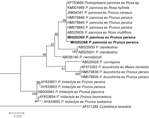
복숭아 과실을 감염하는 흰가루병균 보존시료 중에서 KUS-F28653 및 F29842를 선정하여 internal transcribed spacer (ITS) 영역의 염기서열을 분석하였다. KUS-F29798 시료에서는 rDNA 추출에 실패하여 본 분자계통학적 분석에는 포함시키지 못했다. Takamatsu 등[13]의 방법에 따라 시료에서 채취한 균사와 분생포자로부터 rDNA를 추출하였고, ITS5 [14]와 P3 [15] 프라이머를 사용하여 polymerase chain reaction (PCR)으로 증폭시켰다. 증폭된 PCR 산물을 전기영동을 통하여 확인한 후에 QIAquick PCR Purification kit (Qiagen, Germantown, MD, USA) 로 정제하였다. 이 염기서열을 DNASTAR Lasergene computer package 5.05 (DNASTAR, Madison, WI, USA) 를 이용하여 정리한 후, GenBank에 등록하여 기탁번호를 부여 받았다(accession nos. MH252368, MH252369). GenBank BLAST를 이용하여 ITS 영 역의 염기서열을 비교한 결과, 한국에서 채집된 복숭아 과실의 흰가루병균은 장미류(Rosa spp.) 흰가루병균인 P. pannosa와 99% 이상 일치하였다. 이 결과를 바탕으로 MEGA7 프로 그램[16]을 이용하여 maximum likelihood 방법으로 계통수를 작성하였다. 그 결과 복숭아 과실의 흰가루병균은 P. pannosa의 계통군에 속하였다(Fig. 2). 따라서 한국의 복숭아 과실 을 감염하는 병원균은 P. pannosa임을 분자적으로 처음 확인하였으며, 흰가루 증상 및 녹얼 룩점 증상 모두 P. pannosa에 의해 발생됨을 확인하였다.

고 찰

지금까지 유럽에서는 과수원에서 재배하는 복숭아나무의 잎을 감염하는 대표적인 흰가루병

균으로 Podosphaera pannosa와 P. tridactyla가 알려져 있다[10]. 반면, 미국에서는 복숭아

나무의 과실 흰가루병균으로 P. pannosa [17]와 P. leucotricha [9]가 알려졌다. 최근에 세르

비아와 프랑스의 과수원에서 채집한 10점의 시료를 바탕으로 연구한 Jankovics 등[11]의 결

(5)

과에 따르면, 복숭아 과실에서는 녹얼룩점 증상이 나타나며 P. leucotricha가 관여하고, 천도 과실에서는 흰가루 증상이 나타나며 P. pannosa가 관여하는 것으로 보고했다.

한편, 장미과(Rosaceae) 식물에 발생하는 Podosphaera속 흰가루병균에 대한 균학적 연구 를 통하여 Prunus속 식물의 흰가루병균은 대부분 P. tridactyla이지만 일부는 P. clandestina (Wallr.) Lév. 로 동정하였다[18]. 더구나 P. tridactyla는 단계통군이 아니라 유전적 다양성이 상당히 넓은 복합종으로 여겨진다고 보고하였다[18]. 이들 연구에서는 흰가루병균이 잎에서 유래한 것인지 과실에서 유래한 것인지를 밝히지 않았으나, 특별한 언급이 없는 것으로 보아 모두 잎에서 유래한 것으로 여겨진다.

따라서, 전 세계적으로 지금까지 복숭아나무에서 기록된 흰가루병균은 P. clandestina, P.

leucotricha, P. pannosa, P. tridactyla 등 4종이다. 이 중 P. clandestina가 복숭아 과실에서 기록되었는지 여부는 불확실하지만, 나머지 3종의 흰가루병균이 복숭아 과실에서 기록된 것 은 확실하다. 이 중에서 복숭아 과실에서의 존재를 분자계통학적으로 확인한 것은 P.

leuchotricha가 유일하다. 우리나라를 비롯한 일부 국가에서 복숭아 과실 흰가루병균으로 기 록된 P. pannosa를 본 연구를 통하여 분자계통학적으로 처음 확인하였다.

Fig. 3. Phylogenetic relationship between two Podosphaera pannosa isolates ex Prunus persica

var. persica and some reference isolates retrieved from GenBank, inferred by maximum

likelihood method using the internal transcribed spacer regions. Bootstrap values based on 1,000

replications are indicated above the branches. The scale bar represents 0.02 nucleotide

substitutions per site. The Korean isolates presented in this study are indicated in bold.

(6)

Hyeon-Dong Shin et al.

적 요

1991 년에 우리나라에서 복숭아나무(Prunus persica var. persica) 과실에 흰가루 증상을 일 으키는 병원균은 형태적 특징에 의거하여 Podosphaera pannosa로 동정되었다. 복숭아 과실 이 P. pannosa에 감염된다는 사실은 일본을 비롯한 여러 나라에서 알려져 왔다. 2011년에 Jankovics 등[11]은 세르비아 및 프랑스에서 채집한 10점의 시료를 연구하여 복숭아 과실의

‘ 녹얼룩점(rusty spot)’ 증상은 P. leucotricha에 의한 것이며, 천도(Prunus persica var.

nucipersica) 과실의 ‘흰가루(powdery mildew)’ 증상은 P. pannosa에 의한 것이라고 보고하 였다. 이에 따라, 한국에서 복숭아 과실에 발생한 흰가루병을 4점 채집하여 형태적 검경 및 분 자적 분석을 통해 병원균의 정체를 밝혔다. 3점의 시료는 흰가루 증상, 1점의 시료는 녹얼룩 점 증상을 나타냈다. 형태적으로는 4점 모두 P. pannosa로 동정되었다. 분자적으로는 2점의 시료에서 ITS 염기서열을 분석하여 Rosa spp. 유래의 P. pannosa와 99% 이상 상동성을 확인 하였다. 그리고 maximum likelihood 방법으로 계통수를 작성한 결과 Rosa spp. 유래의 P.

pannosa와 분자계통학적으로 동일한 클러스터에 소속됨을 확인하였다. 이상의 결과를 바탕 으로 한국에서 복숭아 과실의 흰가루 증상과 녹얼룩점 증상은 모두 P. pannosa에 의해 발생 됨을 처음으로 보고한다.

ACKNOWLEDGEMENTS

This work was supported by the BK21 Plus program in 2016-2020 funded by the National Research Foundation of Korea (NRF). This study was also carried out with the support of the “Cooperative Research Program for Agricultural Science & Technology Development (Project No. PJ0124192017)” Rural Development Administration, Republic of Korea.

REFERENCES

1. The Korean Society of Plant Pathology. List of plant diseases in Korea. 5th ed. Seoul:

Korean Society of Plant Pathology; 2009.

2. Nakata K, Takimoto S. List of diseases of cultivated plants in Korea. J Agric Exp Stn Gov Gen Chosen 1928;15:1-146.

3. Rural Development Administration. National Institute of Agricultural Science and Technology. Compendium of fruit tree diseases with color plates. Suwon: National Institute of Agricultural Science and Technology; 1993.

4. Shin HD. Erysiphaceae of Korea. Suwon: National Institute of Agricultural Science and Technology; 2000.

5. Dong CO, Kim SB, Jang HI, Cho MD, Lee EK. Studies on the ecological characteristics and control methods of peach powdery mildew. J Kor Soc Hortic Sci 1991;32:191-8.

6. Park JS. Fungous diseases of plants in Korea (1). Daejeon: Chungnam National University;

1958.

7. Cook RT, Inman AJ, Billings C. Identification and classification of powdery mildew

anamorphs using light and scanning electron microscopy and host range data. Mycol Res

(7)

1997;101:975-1002.

8. Braun U. The powdery mildews (Erysiphales) of Europe. Jena: G. Fischer; 1995.

9. Ogawa JM, Zehr EI, Bird GW, Ritchie DF, Uriu K, Uyemoto JK. Compendium of stone fruit diseases. St. Paul: APS Press; 1995.

10. Farr DF, Rossman AY. Fungal databases, U.S. National Fungus Collections, ARS, USDA [Internet]. Beltsville: Systematic Mycology and Microbiology Laboratory; 2018 [cited 2018 Apr 23]. Available from: http://nt.ars-grin.gov/fungaldatabases/.

11. Jankovics T, Dolovac N, Bulajić A, Krstić B, Pascal T, Bardin M, Nicot PC, Kiss L.

Peach rusty spot is caused by the apple powdery mildew fungus, Podosphaera leucotricha. Plant Dis 2011;95:719-24.

12. Braun U, Cook RT. Taxonomic manual of the Erysiphales (powdery mildews). Utrecht:

CBS-KNAW Fungal Biodiversity Centre; 2012.

13. Takamatsu S, Heluta V, Havrylenko M, Divarangkoon R. Four powdery mildew species with catenate conidia infect Galium: molecular and morphological evidence. Mycol Res 2009;113:117-29.

14. White TJ, Bruns T, Lee S, Taylor J. Amplification and direct sequencing of fungal ribosomal RNA genes for phylogenetics. In: Innis MA, Gelfand DH, Sninsky JJ, White TJ, editors. PCR protocols: a guide to methods and applications. San Diego: Academic Press; 1990. p. 315-22.

15. Kusaba M, Tsuge T. Phylogeny of Alternaria fungi known to produce host-specific toxins on the basis of variation in internal transcribed spacers of ribosomal DNA. Curr Genet 1995;28:491-8.

16. Kumar S, Stecher G, Tamura K. MEGA7: Molecular Evolutionary Genetics Analysis Version 7.0 for Bigger Datasets. Mol Biol Evol 2016;33:1870-4.

17. Yarwood CE. Powdery mildews of peach and rose. Phytopathology 1939;29:282-4.

18. Takamatsu S, Niinomi S, Harada M, Havrynlenko M. Molecular phylogenetic analyses

reveal a close evolutionary relationship between Podosphaera (Erysiphales; Erysiphaceae)

and its rosaceous hosts. Persoonia 2010;24:38-48.

수치

Fig. 1.  Symptoms on peach (Prunus persica var. persica) fruits associated with a powdery  mildew fungus, Podosphaera pannosa
Fig. 2.  Conidial stage of a powdery mildew fungus, Podosphaera pannosa, examined on peach  (Prunus persica var
Fig. 3.  Phylogenetic relationship between two Podosphaera pannosa isolates ex Prunus persica  var

참조

관련 문서

(2013) identified naturally occurring powdery mildew on watermelon in a greenhouse in South Korea as race 1 of P. xanthii using melon differentials during a study of inheritance

Identification of Podosphaera xanthii as the causal agent of powdery mildew disease affecting Echinacea purpurea in Korea.. In-Young Choi 1,2 , Sun-Hee Hong 3 , Yong-Ho Lee 3 ,