Res. Plant Dis. 18(2) : 133−138 (2012) © The Korean Society of Plant Pathology http://dx.doi.org/10.5423/RPD.2012.18.2.133
Erysiphe abeliicola에 의한 꽃댕강나무 흰가루병 발생
조성은·박지현·이승규1·이상현1·신현동*
고려대학교 환경생태공학부, 1국립산림과학원 산림병해충연구과
Occurrence of Powdery Mildew Caused by Erysiphe abeliicola on Glossy Abelia in Korea
Sung-Eun Cho, Ji-Hyun Park, Seung-Kyu Lee1, Sang-Hyun Lee1 and Hyeon-Dong Shin* Division of Environmental Science and Ecological Engineering, Korea University, Seoul 136-701, Korea
1Division of Forest Diseases and Insect Pests, Korea Forest Research Institute, Seoul 130-712, Korea (Received on March 1, 2012; Revised on May 16, 2012; Accepted on May 17, 2012)
In November 2009, a powdery mildew on glossy abelia (Abelia x grandiflora) was found in Seogwipo, Jeju Island, Korea. Further survey in the southern part of Korea, e.g., Jeju, Busan, and Tongyeong confirmed occurrence of the disease. White colonies were present on leaves, young stems, and flowers, detracting from their beauty in landscape plantings. Severely infected lesions were discolored to red-purplish. Based on the morphological characteristics and analysis of rDNA, the fungus associated with the symptoms was identified as Erysiphe abeliicola U. Braun & S. Takam. This work provides the morphological feature of its anamorph for the first time, which is characterized by having multi-lobed hyphal appressoria and short foot-cells of conidiophores. Morphological characteristics of mature chasmothecia were consistent with the previous Japanese record of this species. The sequence of internal transcribed spacer region of ribosomal DNA obtained from a Korean sample showed that this species places in the section Microsphaera of the genus Erysiphe in phylogenetic position, corresponding with the classical taxonomy. This is the first report of E.
abeliicola and its host plant in Korea. The host plant A. x grandiflora is newly listed in the host range of E.
abeliicola.
Keywords : Abelia x grandiflora, Anamorph, Erysiphe abeliicola, Glossy Abelia
꽃댕강나무[Abelia x grandiflora (André) Rehd.]는 인동 과(Caprifoliaceae)에 속하는 반상록성 관목으로 Abelia chinensis와 A. uniflora 사이에서 육성된 종간 잡종이다.
우리나라에는 1930년경 일본으로부터 도입되었는데, 도 입 초기에는 내한성이 약하여 중부 이북 지역에서는 식 재하지 않았지만 최근 들어 전국적으로 식재되고 있다.
특히 대기오염에 잘 견디고 맹아력이 강하여 도로변의 생 울타리와 건물의 진입로 유도식재로 많이 사용되고 있다 (Choi, 2008).
일본에서 기록된 댕강나무류(Abelia spp.)의 병해 중에 서 곰팡이에 의한 것은 Cercospora abeliae에 의한 점무
늬병, Helicobasidium mompa에 의한 자주날개무늬병, Erysiphe abeliicola에 의한 흰가루병 등 3가지가 있다(The Phytopathological Society of Japan, 2000). 그러나 우리나 라에서는 꽃댕강나무는 물론 댕강나무류에서도 식물병이 기록된 바 없다(The Korean Society of Plant Pathology, 2009). 한편, 지금까지 전 세계적으로 댕강나무류에서 기 록된 흰가루병균은 Erysiphe abeliae R.Y. Zheng & G.Q.
Chen, E. abeliicola U. Braun & S. Takam., E. chifengensis T.Z. Liu & U. Braun 등 3종이 있다(Table 1). 이 중, E.
abeliae는 중국(쓰촨성)에서만 알려진 토착종이며(Zheng과 Chen, 1980), E. chifengensis는 중국(내몽고)에서만 기록 되어 있다(Liu와 Braun, 2006). 한편, E. abeliicola는 지금 까지 일본에서만 기록되어 있다(Tanda와 Nomura, 1978).
저자들은 한국에서 2009년부터 꽃댕강나무에 흰가루병이 발생함을 관찰하였으며, 이를 채집하여 병원균을 동정하
*Corresponding author
Phone)+82-2-3290-3063, Fax) +82-2-921-1715 Email) [email protected]
Note Open Access
134 조성은·박지현·이승규·이상현·신현동
였으므로 이에 보고한다.
발생상황 및 표본보존. 꽃댕강나무 흰가루병은 2009년 11월에 서귀포시와 제주시에서 발견되었다. 그 후 2010년 에는 부산시 및 통영시에서 채집됨으로써 우리나라의 남 부지방에서 관상용으로 식재된 꽃댕강나무에 흰가루병이 흔히 발생함을 알 수 있었다. 이상의 모든 시료는 표본으 로 제작하여 고려대학교 식물표본관(KUS)에 영구보존하 였는데, KUS-F24850(무성세대+유성세대, 1 Nov. 2009, 서귀포시 외돌개), F24863(무성세대+유성세대, 2 Nov.
2009, 제주시 노형동), F25628(무성세대+유성세대, 8 Nov.
2010, 부산시 동삼동), F25653(무성세대+유성세대, 15 Nov.
2010, 통영시 무전동) 등 총 4점이다.
병징. 꽃댕강나무는 생울타리용으로 밀식하는 경우가 대부분이므로 흰가루병이 발병된 곳은 모두 집단적으로 감염되어 밀가루를 뒤집어쓴 것처럼 흉한 모습으로 관상 적 가치가 크게 떨어졌다(Fig. 1A). 잎의 앞뒷면에 모두 발생하였지만 대부분 잎 앞면에서 발병이 더 심하였고, 어린 줄기와 꽃에서도 흔히 발생하였다(Fig. 1B). 초기에 하얀 균사로 덮인 잎 앞면의 병환부는 불규칙하게 확대 되었으며(Fig. 1B), 발병이 심해지면서 병환부가 적자색으 로 변하였는데(Fig. 1C), 이에 상응하는 잎 뒷면에서는 변 색이 관찰되지 않았다. 한편, 이 흰가루병균의 유성세대 는 남부지방에서 11월 초부터 형성되었는데, 11월 중순에 는 성숙한 자낭구도 관찰되었지만 대부분 미성숙 상태에 머물렀다. 이 시기까지도 분생포자경에서 분생포자가 활 발히 형성되었으므로 무성세대의 생장도 지속됨을 알 수 있었다. 자낭구의 형성은 잎과 줄기에서 모두 관찰되었으
나, 특히 어린 줄기에 많이 형성되었다(Fig. 1K). 전반적 으로 어린 줄기와 어린잎에도 발병이 심했지만 다른 식 물의 흰가루병에서 흔히 나타나는 줄기마름(shoot blight) 증상은 관찰되지 않았다.
병원균의 형태적 특징. 꽃댕강나무 잎과 줄기에 발생 한 흰가루병균의 무성세대와 유성세대를 검경하였다. 이 균의 분류학적 특성을 파악하고 크기를 측정하기 위해서 명시야광학현미경(BX51, Olympus, Tokyo, Japan)을 사용 하였고, 현미경사진은 미분간섭현미경(Axio Imager, Carl Zeiss, Göttingen, Germany)을 이용하여 촬영하였다. 무성 세대와 유성세대의 검경에서 모두 신선시료를 사용하였 으며, 물을 이용하여 관찰하였다.
균사는 잎 양면에 모두 존재하였으나 대부분 앞면에 분 포하였다. 균사는 직선상 내지 파상이며, 때로는 결절을 형성하였다. 균사 부착기는 굴곡상이며, 단생하거나 쌍을 이루며, 잘 발달하였다(Fig. 1D−E). 분생포자경은 표생균 사의 윗부분으로부터 발달하며, 크기는 60−100×7−9 µm 이며, 3−4개의 세포로 구성되며, 기부세포(foot-cell)의 길 이는 18−26 µm에 불과하여 비교적 짧은 편이며, 기부세 포의 아래쪽은 다소 불룩한 경우도 있었지만 뚜렷하지는 않았고, 기부격벽은 균사의 분지점에 위치하며, 분생포자 를 단생하였다(Fig. 1F−H). 분생포자는 무색의 단세포이 며, 피브로신체(fibrosin body)를 갖지 않으며, 타원형 내 지 장타원형이며, 크기는 26−38×15−20 µm(장폭비=1.5−
2.3)이며, 대부분 분생포자의 배꼽테두리(perihilar position) 바로 옆에서 발아하였다(Fig. 1I). 또한, 분생포자의 표면 구조는 각진 모양으로 주름진 모습(angular/rectangular Table 1. Comparison of morphological characteristics of three species of powdery mildew recorded on Abelia spp. and the present isolate
Characteristics Erysiphe abeliae Erysiphe abeliicola Erysiphe chifengensis Present isolate Chasmothecia
diameter (µm) (88−)94−119 72−108 80−140(−164) 85−110
wall-cells (µm) 6.3−17.5 19.2−26.4×14.4−16.8 6−24 12.5−22.5
no. appendages 12−37 5−7 (5−)10−20(−23) 8−10
shape of appendages uncinate dichotomous dichotomous dichotomous length of appendages (µm) (35−)88−250(−280) 182−308 (55−)70−140(−190) 126−250
no. asci 4−8 2−5 5−10(−13) 4−8
Asci
length×width (µm) 43.2−58.4×30.5−43.2(−50.8) 48−58.8×31.2−46.8 (50−)55−70(−77)×30−45.5 60−70×35−46
no. ascospores 6−8 6 (4−)6−7(−8) 5−6
Ascospores
length×width (µm) 17.5−20(−25)×(10−)11.3−13.8 18−21.6×9.6−12 16.5−23×10−14 17.5−22.5×12.5−16.3 Host plant Abelia sp. Abelia spathulata Abelia biflora Abelia×grandiflora Reference Zheng & Chen (1980) Homma (1937) Liu & Braun (2006)
Fig. 1. Powdery mildew disease of Abelia x grandiflora associated with Erysiphe abeliicola. (A) Severe infections observed in Jeju in November 2009. (B) Close-up view of symptoms. Note the infection on young stems and flowers. Colony of powdery mildew is white without causing leaf discoloration. (C) Close-up view of powdery mildew colony causing red-purplish discoloration. (D−E) Hyphal appressoria. (F−H) Conidiophores. (I) Conidia. (J) Surface view of a conidium showing angular/rectangular wrinkling pattern. (K) Formation of chasmothecia on an infected stem. (L) Chasmothecium accommodating eight asci. (M) Asci containing six ascospores.
Scale bar in I = 20 µm (D−J are in the same magnification). Scale bar in L = 100 µm. Scale bar in M = 50 µm.
136 조성은·박지현·이승규·이상현·신현동 wrinkling pattern)이었다(Fig. 1J).
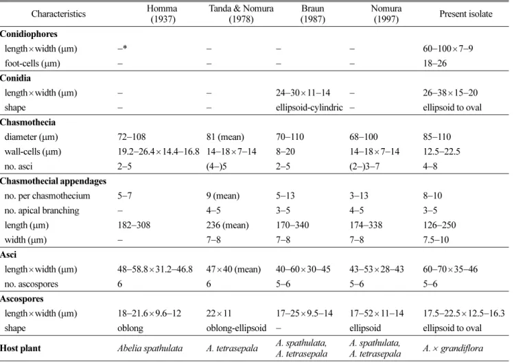
자낭구는 주로 줄기와 잎에 흩어져 형성되며, 미성숙 상태의 자낭구가 많았다(Fig. 1K). 직경은 85−110 µm이 며, 벽세포는 직경 12.5−22.5 µm 정도의 불규칙한 세포 로 구성되며, 4−8개의 자낭을 가졌다(Fig. 1L). 자낭구 부 속사는 8−10개에 이르며, 자낭구의 적도면에 분포하며, 길이는 126−250 µm이고 폭은 7.5−10 µm이며, 끝은 3−5 번 이차 분지되어 있으며, 무색이며 평활하였다. 자낭은 근원형으로서 아랫부분에 짧은 꼭지가 있으며(short stalked), 크기는 60−70×35−46 µm이며, 5−6개의 자낭포자를 지녔 다(Fig. 1M). 자낭포자는 타원형 내지 달걀형이며, 크기 는 17.5−22.5×12.5−16.3 µm이었다. 이와 같은 균학적 특 징은 앞선 연구(Homma, 1937; Tanda와 Nomura, 1978;
Braun, 1987; Nomura, 1997)에서 보고된 Erysiphe abeliicola U. Braun & S. Takam.와 일치하였다(Table 2).
염기서열 분석. 고려대학교 식물표본관에 보존되어 있 는 꽃댕강나무의 흰가루병균 E. abeliicola 시료 중에서
KUS-F25628을 선정하여 ribosomal DNA(rDNA)의 complete internal transcribed spacer(ITS) 영역의 염기서열을 분석하 였다. Takamatsu 등(2009)의 방법에 따라 시료에서 채취 한 자낭구로부터 rDNA를 추출하였고, ITS5(White 등, 1990) 와 P3(Kusaba와 Tsuge, 1995) 프라이머를 사용하여 PCR 로 증폭시켰다. 증폭된 PCR 산물을 전기영동을 통하여 확인한 후에 LaboPassTM PCR kit(Cosmo Genetech, Seoul, Korea)로 정제하였다. 이 염기서열을 DNASTAR computer package 5.05(Lasergene, Madison, WI, USA)를 이용하여 정리한 후, NCBI(National Center for Biotechnology Information)의 GenBank에 등록하여 기탁번호를 부여받았 다(JQ710724). 계통수는 MEGA4 프로그램(Tamura 등, 2007)을 이용하여 neighbor-joining 방법으로 작성하였다 (Fig. 2). GenBank BLAST를 이용하여 ITS 영역의 염기 서열을 비교한 결과, 우리나라 꽃댕강나무의 흰가루병균 E. abeliicola와 일치하는 염기서열이 없었다. 현재 NCBI 에 등록된 댕강나무류(Abelia spp.)의 흰가루병균은 ITS
Table 2. Comparison of morphological characteristics of Erysiphe abeliicola and the present isolate Characteristics Homma
(1937)
Tanda & Nomura (1978)
Braun (1987)
Nomura
(1997) Present isolate Conidiophores
length×width (µm) −* − − − 60−100×7−9
foot-cells (µm) − − − − 18−26
Conidia
length×width (µm) − − 24−30×11−14 − 26−38×15−20
shape − − ellipsoid-cylindric − ellipsoid to oval
Chasmothecia
diameter (µm) 72−108 81 (mean) 70−110 68−100 85−110 wall-cells (µm) 19.2−26.4×14.4−16.8 14−18×7−14 8−20 14−18×7−14 12.5−22.5
no. asci 2−5 (4−)5 2−5 (2−)3−7 4−8
Chasmothecial appendages
no. per chasmothecium 5−7 9 (mean) 5−13 3−13 8−10
no. apical branching − 4−5 3−5 4−5 3−5
length (µm) 182−308 236 (mean) 170−340 174−338 126−250
width (µm) − 7−8 7−8 7−8 7.5−10
Asci
length×width (µm) 48−58.8×31.2−46.8 47×40 (mean) 40−60×30−45 43−53×28−43 60−70×35−46
no. ascospores 6 6 5−6 5−6 5−6
Ascospores
length×width (µm) 18−21.6×9.6−12 22×11 17−25×9.5−14 17−52×11−14 17.5−22.5×12.5−16.3 shape oblong oblong-ellipsoid − ellipsoid ellipsoid to oval Host plant Abelia spathulata A. tetrasepala A. spathulata,
A. tetrasepala
A. spathulata,
A. tetrasepala A.× grandiflora
* : Not described.
영역의 염기서열이 없었으므로 우리나라 꽃댕강나무의 흰 가루병균 E. abeliicola의 ITS 영역의 염기서열을 처음으 로 제공하였다. 이 병원균은 Erysiphe속(genus)의 다른 종 들과 같은 계통군으로 묶였으며, E. syringae-japonicae 등 과 함께 Microsphaera절(section)에 포함되었다. 그러므로 우리나라에서 채집된 꽃댕강나무 흰가루병균 E. abeliicola 는 분자계통학적으로도 Erysiphe속의 Microsphaera절에 위치하는 종으로 확인되었다.
고찰. 앞서 언급한 바와 같이 댕강나무류(Abelia spp.) 에는 3종의 흰가루병균이 알려져 있다. 이들 3종은 유성 세대의 형태적 특징에 따라 비교적 쉽게 구분된다(Table 1). 그 중에서 Erysiphe abeliae는 Uncinula절에 속하는 유 일한 종이다. 한편, E. abeliicola와 E. chifengensis는 모두 Microsphaera절에 속하는데, 이들 두 종은 자낭구부속사 (chasmothecial appendages)의 길이가 다른 분류특성을 갖 는다. 즉, E. abeliicola는 부속사의 길이가 자낭구 직경의 2−4배에 이르며, E. chifengensis는 0.7−1.0배에 불과하다.
한국에서 채집된 꽃댕강나무 흰가루병균의 자낭구는 부 속사의 길이가 대부분 자낭구 직경의 2배 이상이었으므 로 E. abeliicola로 동정할 수 있었다.
한편, 이 흰가루병균은 일본에서 4종의 댕강나무류(Abelia serrata, A. spathulata, A. spathulata var. stenophylla, A.
tetrasepala)에서 기록되었으나, 꽃댕강나무에서는 기록된 바 없다(The Phytopathological Society of Japan, 2000;
Farr와 Rossman, 2012). 따라서 꽃댕강나무는 이 흰가루 병의 기주로는 세계에서 처음 기록되는 것이며, 이 병원 균은 일본에 이어 세계에서 두 번째로 기록되는 것이다.
또한, 이 병원균의 무성세대는 Braun(1987)에 의해 기록 되었으나, 그 기재가 불충분하여 분류학적 특징으로 이용 되기는 어려웠다. 반면, 한국에서 채집된 꽃댕강나무 흰
가루병균의 무성세대는 신선 시료를 사용하여 검경하고 상세하게 기재하였기 때문에 이전에 알려지지 않았던 무 성세대의 특징을 최초로 기록하였다(Table 2 참조). 또한, E. abeliicola의 기준표본(holotype)은 미성숙 상태의 자낭 구를 기록한 것이라고 하는데(Braun, 1987 참조), 실제 성 숙 상태의 자낭구는 일본에서 한 번 기록되었을 뿐이다 (Tanda와 Nomura, 1978). 우리나라의 시료도 자낭구가 대 부분 미성숙 상태였지만 드물게 성숙 상태의 자낭구를 관 찰할 수 있었는데, 이는 세계에서 두 번째로 성숙한 자낭 구를 채집하여 기재한 셈이다.
본 연구팀이 제주시 노형동에서 관찰한 꽃댕강나무 흰 가루병은 대형건물의 그늘 쪽에 경계화단용으로 밀식된 상태에서 발생하였는데, 그늘이 많고 통풍이 불량한 군락 에서 더 심한 발병을 나타냈다. 또한 가로수의 하단에 생 울타리용으로 식재된 경우(통영시 무전동)에도 그늘 환경 이 발병에 영향을 주었다고 판단되었다. 한편, 제주, 부산, 통영, 해남, 완도 등 꽃댕강나무 흰가루병이 전혀 발생하 지 않은 밀식식재지도 있었는데, 이러한 곳은 일조가 충 분하고 통풍이 원활한 공통점을 발견할 수 있었다. 따라 서 꽃댕강나무의 식재 위치 선정에서는 일조 및 통풍에 대한 주의가 필요하다고 판단되었다.
요 약
2009년 11월에 서귀포에서 꽃댕강나무 흰가루병을 발 견하였다. 이어 제주, 부산, 통영 등 남부지방에서도 추가 적으로 발견되었다. 흰색의 균체가 잎, 어린 줄기, 꽃을 감염하여 관상가치를 떨어뜨렸으며, 발병이 지속되면 잎 앞면의 병환부는 적자색으로 변하였다. 이 흰가루병균의 형태적 특징과 분자적 분석을 통하여 이 곰팡이는 Erysiphe Fig. 2. Phylogenetic relationship between Erysiphe abeliicola isolates and some reference isolates retrieved from GenBank, inferred by neighbor-joining method using the ITS rDNA region. Bootstrap values based on 1000 replications are indicated above the branches and the scale bar represents 0.02 nucleotide substitutions per site.
138 조성은·박지현·이승규·이상현·신현동 abeliicola U. Braun & S. Takam.로 동정되었다. 무성세대
의 분류학적 특징은 이 연구를 통하여 처음으로 기재되 는데, 균사의 굴곡형 부착기와 분생포자경의 짧은 기부세 포가 특징적이었다. 성숙한 자낭구의 분류학적 특징은 앞 선 일본의 기재와 거의 일치하였다. 우리나라 시료에서 rDNA ITS 영역의 염기서열을 처음으로 분석하여 이 종 이 Erysiphe속의 Microsphaera절에 속함을 밝혔으며, 이 는 형태적 특징과 상응하는 결과였다. 이로써 우리나라에 서 E. abeliicola에 의한 꽃댕강나무 흰가루병을 처음으로 보고하고, 꽃댕강나무가 이 흰가루병균의 기주로 확인된 것은 세계적으로 처음이다.
References
Braun, U. 1987. A monograph of the Erysiphales (powdery mildews). Beih. Nova Hedw. 89: 1−700.
Choi, M. S. 2008. Abelia x grandiflora. Ornamental Trees 106:
18−19. (In Korean)
Farr, D. F. and Rossman, A. Y. 2012. Fungal Databases, Systematic Mycology & Microbiology Laboratory, ARS, USDA. Retrieved Feb. 21, 2012, from http://nt.ars-grin.gov/
fungaldatabases/
Homma, Y. 1937. Erysiphaceae of Japan. J. Fac. Agric. Hokkaido Imp. Univ. 38: 183−461.
Kusaba, M. and Tsuge, T. 1995. Phylogeny of Alternaria fungi known to produce host-specific toxins on the basis of variation in internal transcribed spacers of ribosomal DNA. Curr.
Genet. 28: 491−498.
Liu, T. Z. and Braun, U. 2006. A new species of Erysiphe sect.
Microsphaera from Inner Mongolia, China. Nova Hedw. 83:
493−498.
Nomura, Y. 1997. Taxonomical Study of Erysiphaceae of Japan.
Yokendo Ltd., Tokyo, Japan. 281 pp.
Takamatsu, S., Heluta, V., Havrylenko, M. and Divarangkoon, R.
2009. Four powdery mildew species with catenate conidia infect Galium: molecular and morphological evidence. Mycol.
Res. 113: 117−129.
Tamura, K., Dudley, J., Nei, M. and Kumar, S. 2007. MEGA4:
Molecular Evolutionary Genetics Analysis (MEGA) software version 4.0. Mol. Biol. Evol. 24: 1596−1599.
Tanda, D. and Nomura, Y. 1978. Powdery mildew of the new hosts in Japan (V). J. Agric. Sci., Tokyo Univ. Agric. 23: 19− 31.
The Korean Society of Plant Pathology. 2009. List of Plant Diseases in Korea. 5th ed., Suwon, Korea. 853 pp.
The Phytopathological Society of Japan. 2000. Common Names of Plant Diseases in Japan. Japan Plant Protection Association, Tokyo, Japan, 856 pp.
White, T. J., Bruns, T., Lee, S. B. and Taylor, J. W. 1990.
Amplification and direct sequencing of fungal ribosomal RNA genes for phylogenetics. In: PCR Protocols: a Guide to Methods and Applications, ed. by M. A. Innis, D. H. Gelfand, J. J. Sninsky and T. J. White, pp. 315−322. Academic Press, San Diego, CA, USA.
Zheng, R. Y. and Chen, G. Q. 1980. Taxonomic studies on the genus Erysiphe of China. 1. New species and new variety on Caprifoliaceae. Acta Microbiol. Sin. 20: 45−49.