Effects of Lumbar Stabilization on the Trunk and Lower Limb Muscle Activity and Velocity of the Center of Pressure During Single Leg Standing
Heon-seock Cynn, Ph.D., P.T.
Dept. of Physical Therapy, College of Health Science, Yonsei University
Abstract
1)The aim of this study was to investigate the effects of lumbar stabilization on both trunk and lower limb muscle activity and center of pressure (COP) in single leg standing. Surface electromyography (EMG) was used to collect muscle activity data, the mean velocity of COP was measured using a force plate, and a pressure biofeedback unit was used for lumbar stabilization training. The findings of this study are summarized as follows: 1) The EMG activity of the erector spinae decreased significantly and the activity of the rectus abdominis, internal oblique, external oblique, gluteus maximus, and gluteus medius increased significantly with lumbar stabilization single leg standing. 2) No differences in activity in the tibialis ante- rior, medial gastrocnemius, rectus femoris, and medial hamstrings were found with single leg standing. 3) The mean velocity of COP in the antero-posterior and medio-lateral directions in the lumbar stabilization single leg standing decreased significantly compared with the preferred single leg standing. The findings of this study therefore indicate that lumbar stabilization can facilitate the co-activation of deep stabilization and global muscles that improve postural control capability during single leg standing.
Key Words: Center of pressure; Lumbar stabilization; Muscle activity; Single leg standing.
Introduction
Lumbar stabilization is a therapeutic approach to provide stability to the lumbar region through deep segmental muscles and has been introduced to pre- vent and treat musculoskeletal injuries (Akuthota and Nadler, 2004; Kisner and Colby, 2002). The stability of the spine is maintained by three subsystems: the passive, active, and control subsystems (Panjabi, 1992a). Among these, local muscle contraction is of particular importance, as it provides dynamic stability to the neutral zone (Panjabi, 1992b), whereas the transversus abdominis and the multifidus contribute to lumbar stabilization (Barnett and Gilleard, 2005;
Richardson et al, 1999; Richardson et al, 2004).
Richardson and Jull (1995) reported that deep mus- cles should co-contract; otherwise, excessive sub- stitute motions will occur. Learning lumbar stabiliza-
tion through cognitive repetition facilitates the auto- matic co-contraction of deep muscles while perform- ing daily activities (Saal and Saal, 1989).
The pressure biofeedback unit was originally devel- oped to assess the level of lumbar stabilization through the deep abdominal muscles and has been utilized in previous research evaluating lumbar stability (Herrington and Davies, 2005; Jull et al, 1993; Mills et al, 2005; Richardson et al, 1992; Wohlfart et al, 1993).
This device consists of an inflatable cushion, a bulb, and a pressure gauge and is reported to provide reli- able and valid measurement of local stabilizing muscles (Cairns et al, 2000; Richardson and Jull, 1995). The pressure biofeedback unit is applied to the lumbar re- gion of the subject and inflated to the target pressure.
While the subject performs the prescribed exercise, the target pressure is maintained. Failure to sustain the target pressure indicates an inability to perform lumbar
Corresponding author: Heon-seock Cynn [email protected]
This work was supported by the Yonsei University New Faculty Research Grant of 2010 (2010-5-5005).
stabilization or an increase in intra-abdominal pressure.
Postural control is defined as the ability to main- tain the center of gravity within the base of support.
An unstable platform and perturbations were used to identify the variables affecting this balance. Single leg standing is a more unstable posture as the center of mass is located high and the base of support is narrow relative to double leg standing. The capability to sustain a single leg standing position is required for many activities of daily living (Jonsson et al, 2004) and as such, single leg standing has been im- plemented for clinical tests and intervention in pre- vious studies (Fritz and George, 2000; Liebenson, 2005; Tidstrand and Horneij, 2009). However, the ef- fects of lumbar stabilization on muscle activity and the velocity of the center of pressure (COP) during single leg standing have not been extensively studied.
Because lumbar instability (Hungerford et al, 2003) as well as insufficient muscle strength of the trunk or the lower limbs has been reported to diminish pos- tural control (Szklut and Breath, 2001), examining the effects of lumbar stabilization achieved by using the pressure biofeedback unit will provide clinically rele- vant information in the physical therapy field.
The aim of this study was to identify the effects of lumbar stabilization on both muscle activity and the ve- locity of COP in the antero-posterior and medio-lateral directions during single leg standing. It was hypothesized that both muscle activity and COP would be reduced during single leg standing with lumbar stabilization.
Methods
Subjects
A sample of 20 young healthy males voluntarily participated in this study. The subjects were ex- cluded if they displayed a past or present history of musculoskeletal injury, lower limb deformity, or or- thopedic or neurological disorders that would affect single leg standing. All subjects provided written, in-
formed consent, and the study was approved by the University Research Ethical Committee.
Measurement Instruments2)
Surface electromyography
Surface electromyography (EMG)1) was used to meas- ure the muscle activity of the lower limb. Acqknowledge 3.7.1 software was used for data analysis. A bipolar surface electrode with a diameter of 1 ㎝ and an in- ter-electrode distance of 2 ㎝ was used (TSD 150B, BIOPAC Systems Inc., CA, U.S.A.). A disposable surface electrode (EL503, BIOPAC Systems Inc., CA, U.S.A.) with a diameter of 1 ㎝ was also used in the study.
The site of electrode placement was shaved and sanded to reduce skin resistance. An electrolyte gel was applied to the electrode and double adhesive tape was attached. A bipolar surface electrode was placed onto the dominant tibialis anterior (proximal to 75% of the line connecting the lateral condyle of the knee joint and the lateral malleolus), medial gastrocnemius (proximal to 30% of the line connecting the lateral condyle of the knee joint and the calcaneus), rectus femoris (midpoint between the anterior superior iliac spine and the patellar apex), biceps femoris (midpoint between the ischial tuberosity and the lateral epi- condyle), gluteus maximus (midpoint between the greater trochanter and the second sacral vertebra), gluteus medius (proximal to 30% of the distance be- tween the iliac crest and the greater trochanter), rec- tus abdominis (midpoint between the umbilicus and the pubis), internal oblique (midpoint between the an- terior superior iliac crest and the symphysis pubis and proximal to the inguinal ligament), external oblique (5
㎝ lateral to the umbilicus), and the erector spinae (midpoint between the first lumbar spinous process and the lateral aspect of trunk); all were placed on the dominant side (Cram et al, 1998). Ground electro- des were attached to the dominant fibular head.
The sampling rate was 1024 ㎐, and a bandpass filter (20~450 ㎐) was used. The raw myoelectric 1) MP100A-CE, BIOPAC Systems Inc., CA, U.S.A.
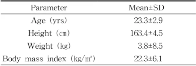
Parameter Mean±SD
Age (yrs) 23.3±2.9
Height (㎝) 163.4±4.5
Weight (㎏) 3.8±8.5
Body mass index (㎏/㎡) 22.3±6.1
Table 1. General characteristics of subjects (N=20) signal collected was processed to the root mean
square. The maximal voluntary isometric contraction (MVIC) described by Kendall et al (2005) was used to determine the reference contraction.
Force plate3)
A force plate2) was used to measure COP (.4×.6
㎡). The sampling rate was 120 ㎐, and the collected data were analyzed using a Kistler control unit (Kistler Bioware Software, Kistler Instruments, Winterthur Wülflingen, Switzerland). The mean COP velocity in the antero-posterior and medio-lateral di- rections was then calculated.
Procedure
Each subject was asked to stand on the force plate with his feet shoulder width apart. Following a verbal command from the principal investigator, the subject flexed the non-dominant hip to 60° and re- laxed the knee joint to hang vertically while bearing his weight on the dominant lower limb. The arms were folded across the chest, and subjects were di- rected to maintain a forward-looking position. While balance was maintained for 10 seconds in the single leg standing position, muscle activity and the mean velocity of COP in the antero-posterior and the me- dio-lateral directions were measured. If the subject moved the dominant foot to maintain balance or was unable to maintain the single leg standing position, the data were not included in subsequent analyses.
The initial and final 2 second were discarded, and the remaining 6 second of data were recorded. The mean of three trials was calculated, with a 5 minute resting period between trials to minimize fatigue.
Following data collection in the preferred single leg standing position, lumbar stabilization training was undertaken using a pressure biofeedback unit with subjects in the supine, sidelying, and prone positions. Lumbar stabilization training was per- formed for a 30 minute period each day for 2 days.
The pressure biofeedback unit was placed between
the lumbar region and the treatment table for the supine and sidelying positions and between the ab- dominal region and the treatment table in the prone position. The target pressure was set to 70 ㎜Hg in each position, and the subject was instructed to watch the pressure gauge while performing hip flex- ion in the supine position, hip abduction in the side lying position, and hip extension in the prone position. Subjects were asked to perform each hip movement while keeping the pressure gauge within the limit of 5 ㎜Hg. If a pressure change of >5 ㎜ Hg occurred, the lumbar stabilization was considered to have failed. After two days of training, all sub- jects were able to maintain lumbar stabilization comfortably. Following the training regime, the data for the lumbar stabilization condition of the single leg standing position were collected using a proce- dure identical to the initial trials.
Statistical Analysis
Statistical analysis was performed using the SPSS ver. 13.0 software. A paired t-test was used to com- pare the muscle activity and the mean velocity of COP between the two conditions (preferred single leg standing vs. lumbar stabilization single leg standing).
A p-value of ≤.05 was deemed significant.
Results
Characteristics of Subjects
A sample of 20 male subjects was recruited for this study. The general characteristics of subjects are presented in Table 1.
2) Kistler force plate, Kistler Instruments, Winterthur Wülflingen, Switzerland.
Preferred single leg standing Lumbar stabilization single leg standing
Tibialis anterior 14.62±.34a 12.35±.39
Medial gastrocnemius 26.56±2.20 24.68±.21
Rectus femoris 29.62±1.01 27.24±1.08
Biceps femoris 14.63±.82 16.51±1.51
Gluteus maximus 30.40±8.34 42.94±13.47*
Gluteus medius 27.84±39.49 36.85±19.37*
Rectus abdominis 6.64±2.65 13.52±3.46*
Internal oblique 9.69±3.01 26.58±2.84*
External oblique 17.67±5.34 25.54±4.98*
Erector spinae 32.62±3.94 24.82±2.12*
aMean±SD (%MVIC), *Significant difference compared to preferred single leg standing.
Table 2. Comparison of muscle activity between conditions (N=20)
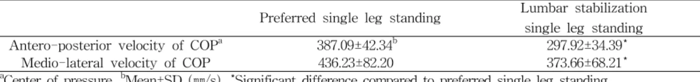
Preferred single leg standing Lumbar stabilization single leg standing Antero-posterior velocity of COPa 387.09±42.34b 297.92±34.39*
Medio-lateral velocity of COP 436.23±82.20 373.66±68.21*
aCenter of pressure, bMean±SD (㎜/s), *Significant difference compared to preferred single leg standing.
Table 3. Comparison of mean velocity of COP between conditions (N=20) Comparison of Muscle Activity Between
Conditions
The muscle activity of the erector spinae de- creased significantly under the lumbar stabilization single leg standing position compared with the pre- ferred single leg standing position (p<.05). The mus- cle activity of the rectus abdominis, external oblique, internal oblique, gluteus maximus, and gluteus med- ius increased significantly in the lumbar stabilization single leg standing position compared with the pre- ferred single leg standing position (p<.05). No sig- nificant differences were found in the muscle activity of the tibialis anterior, medial gastrocnemius, rectus femoris, and biceps femoris between the preferred single leg standing and lumbar stabilization single leg standing positions (p>.05) (Table 2).
Comparison of Mean Velocity of COP Between Conditions
The mean velocity of COP in the medio-lateral and antero-posterior directions decreased significantly
in the lumbar stabilization single leg standing posi- tion compared with the preferred single leg standing position (p<.05) (Table 3).
Discussion
The present study was performed to investigate the effects of lumbar stabilization on the muscle activity and mean velocity of COP during single leg standing in healthy subjects. The results pre- sented in this study showed that the EMG activity of the erector spinae (global muscle) decreased significantly, whereas the activity of the internal oblique (deep muscle) increased significantly during single leg standing following lumbar stabilization training. Additionally, the muscle activity of the rectus abdominis, external oblique, gluteus max- imus, and gluteus medius increased significantly during single leg standing with lumbar stabiliza- tion, whereas no differences were observed in the
tibialis anterior, medial gastrocnemius, rectus fem- oris, and medial hamstrings.
The increased muscle activity of the internal obli- que can be attributed to the successful implementation of the lumbar stabilization training program im- plemented in this study. It is well known that deep stabilizing muscles, such as the transversus and in- ternal oblique, can be selectively activated through training with a pressure biofeedback unit (Cynn et al, 2006; Oh et al, 2007). Thus, it can be stated that the deep muscles were selectively recruited for segmental stabilization, as lumbopelvic regional stability is re- quired during the unstable posture induced by single leg standing. Furthermore, activity in the global mus- cles such as the rectus abdominis and external oblique increased significantly, which did not support our re- search hypothesis. It was observed that the global muscles, including the rectus abdominis and external oblique, were activated concurrently with the local muscles during the various exercises (Arokoski et al, 2004; Jull et al, 1993). Furthermore, it was difficult to isolate local muscle activation from global muscle co-activation (Beith et al, 2001; Stevens et al, 2007).
Therefore, the increased muscle activity in the rectus abdominis, external oblique, and internal oblique in this study is in accordance with previous studies, and both deep and global muscles contributed to the sta- bility of the single leg standing position.
When maintaining the single leg standing position, both gluteus maximus and gluteus medius activity are required to sustain a stable posture. The lumbar stabilization applied in this study did not aim to strengthen the gluteus maximus and gluteus medius, although the muscle activities of both these muscles during single leg standing increased. Previous reports have demonstrated that an abdominal drawing-in maneuver performed using a pressure biofeedback unit successfully increased gluteus maximus muscle activity in the prone position and gluteus medius muscle activity in the side lying position in healthy control subjects (Cynn et al, 2006; Oh et al, 2007).
Our findings can be therefore be interpreted as in-
dicating that performance of lumbar stabilization us- ing a pressure biofeedback unit activates the gluteus maximus and gluteus medius muscles, and this in- creased activation contributes to the postural control capability during single leg standing.
The mean velocity of the antero-posterior and me- dio-lateral COP in the lumbar stabilization single leg standing position significantly decreased compared with that in the preferred single leg standing position, implying improved postural control capability with lumbar stabilization. COP parameters are regarded as representing the level of postural control (Geurts et al, 1993; Geurts et al, 1996; Winter, 1995). In previous studies, the displacement, velocity, or acceleration of COP has been measured to assess postural control capability (Garland et al, 2003; Pyöriä et al, 2004).
Raymakers and colleagues (2005) argued that the mean velocity of COP is a discriminating variable that determines the level of postural control among COP parameters. The location of COP represents neural control of the muscles (Winter, 1995). In the present study, although the COP parameters and the mean velocity of COP in the antero-posterior and the me- dio-lateral direction decreased significantly, the muscle activity of the lower leg did not decrease in the lum- bar stabilization single leg standing position.
Furthermore, the muscle activity of the proximal mus- cles (gluteus maximus and gluteus medius) increased significantly. This relationship between the mean ve- locity of COP and the muscle activity of the proximal and distal muscles therefore requires clarification in future studies of the single leg standing position.
Maintaining an unstable posture is required to per- form normal activities of daily living, in addition to the important purposes of physical therapy intervention.
Because a short period of lumbar stabilization training with a pressure biofeedback unit improved postural control and increased muscle activity in this study, it is suggested that lumbar stabilization be implemented for those who require lumbopelvic stability, particularly patients suffering from lower back pain.
The present study is not without limitations. First,
we could not completely control the learning and testing effect, as comparisons were performed with- out randomization of subjects. Second, the subjects in this study were healthy young male adults, limiting the study’s generalizability to the wider population.
Third, the kinematic data of the trunk and limbs were not collected. Thus, further studies with symptomatic samples are warranted to examine the long term ef- fects of lumbar stabilization on the general population.
Conclusion
The present study examines the effects of lumbar stabilization on both trunk and lower limb muscle activity and the mean velocity of COP during single leg standing using surface electromyography and a force plate. The EMG activity of the erector spinae decreased significantly, whereas the activity of the rectus abdominis, internal oblique, external oblique, gluteus maximus, and gluteus medius increased sig- nificantly during single leg standing following lumbar stabilization training. In contrast, no significant dif- ferences were observed in the tibialis anterior, medial gastrocnemius, rectus femoris, and medial hamstrings with lumbar stabilization during lumbar stabilization single leg standing compared with the preferred sin- gle leg standing position. In addition, the mean ve- locity of COP in the antero-posterior and medio-lat- eral directions during lumbar stabilization decreased significantly compared with that in the initial pre- ferred single leg standing position. This study high- lights lumbar stabilization as an alternative approach to both improve postural control capability and in- duce co-activation of the local and global muscles during single leg standing.
References
Akuthota V, Nadler SF. Core strengthening. Arch Phys Med Rehabil. 2004;85(3 Suppl 1):S86-92.
Arokoski JP, Valta T, Kankaanpaa M, et al.
Activation of lumbar paraspinal and abdominal muscles during therapeutic exercises in chronic low back pain patients. Arch Phys Med Rehabil.
2004;85(5):823-832.
Barnett F, Gilleard. W. The use of lumbar spinal stabilization techniques during the performance of abdominal strength exercise variations. J Sports Med Phys Fitness. 2005;45(1):38-43.
Beith ID, Synnott E, Newman A. Abdominal muscle activity during the abdominal hollowing ma- noeuvre in the four-point kneeling and prone positions. Man Ther. 2001;6(2):82-87.
Cairns M, Harrison K, Wright C. Pressure biofeed- back: A useful tool in the quantification of ab- dominal muscular dysfunction? Physiotherapy.
2000;86(3):127-138.
Cram JR, Kasman GS, and Holtz J. Introduction to Surface Electromyography. Maryland, Aspen, 1998.
Cynn H, Oh J, Kwon O, Yi C. Effects of lumbar stabilization using a pressure biofeedback unit on muscle activity and lateral pelvic tilt during hip abduction in sidelying. Arch Phys Med Rehabil. 2006;87(11):1454-1458.
Fritz JM, George S. The use of a classification ap- proach to identify subgroups of patients with acute low back pain. Interrater reliability and short-term treatment outcomes. Spine.
2000;25(1):106-114.
Garland SJ, Willems DA, Ivanova VTD, et al.
Recovery of standing balance and functional mobility after stroke. Arch Phys Med Rehabil.
2003;84(12):1753-1759.
Geurts AC, Nienhuis B, Mulder TW. Intrasubject variability of selected force-platform parameters in the quantification of postural control. Arch Phys Med Rehabil. 1993;74(11):1144-1150.
Geurts AC, Ribbers GM, Knoo JA, et al.
Identification of static and dynamic postural in- stability following traumatic brain injury. Arch Phys Med Rehabil. 1996;77(7):639-644.
Herrington L, Davies R. The influence of pilates
This article was received September 10, 2010, and was accepted October 18, 2010.
training on the ability to contract the transversus abdominis muscle in asymptomatic individuals. J Bodywork Mov Ther. 2005;9(1):52-57.
Hungerford B, Gilleard W, and Hodges P. Evidence of altered lumbopelvic muscle recruitment in the presence of sacroiliac joint pain. Spine.
2003;28(14):1593-1600.
Jonsson E, Seiger A, Hirschfeld H. One-leg stance in healthy young and elderly adults: A measure of postural steadiness? Clin Biomech (Bristol, Avon). 2004;19(7):688-694.
Jull G, Richardson C, Toppenberg R, et al. Towards a measurement of active muscle control for lum- bar stabilization. Aust J Phys. 1993;39:187-193.
Kendall FP, McCreary EK, Provance PG, Muscles:
Testing and function with posture and pain. 5th ed. Baltimore, Williams & Wilkins, 2005.
Kisner C, Colby LA, Therapeutic Exercise:
Foundations and techniques. 4th ed. Philadelphia, F.A. Davis, 2002.
Liebenson C. Sensory-motor training-an update. J Bodyw Mov Ther. 2005;9(2):142-147.
Mills JD, Taunton JA, Mills WA. The effect of a 10-week training regimen on lumbo-pelvic sta- bility and athletic performance in female ath- letes: A randomized-controlled trial. Phys Ther Sport. 2005;6(2):60-66.
Oh J, Cynn H, Won J, et al. Effects of performing an abdominal drawing-in maneuver during prone hip extension exercises on hip and back extensor muscle activity and amount of anterior pelvic tilt.
J Orthop Sports Phys Ther. 2007;37(6):320-324.
Panjabi MM. The stabilizing system of the spine.
Part I. Function, dysfunction, adaptation, and enhancement. J Spinal Disord. 1992a;5(4):383-389.
Panjabi MM. The stabilizing system of the spine.
Part II. Neutral zone and instability hypothesis.
J Spinal Disord. 1992b;5(4):390-397.
Pyöriä O, Era P, Talvitie U. Relationships between standing balance and symmetry measurements in patients following recent strokes or older strokes. Phys Ther. 2004;84(2):128-136.
Raymakers JA, Samson MM, Verhaar HJJ. The as- sessment of body sway and the choice of the stability parameter(s). Gait Posture.
2005;21(1):48-58.
Richardson C, Hodges P, Hides J. Therapeutic Exercise for Lumbopelvic Stabilization: A motor control approach for the treatment and pre- vention of low back pain. 2nd ed. Philadelphia, Churchill Livingstone, 2004.
Richardson CA, Jull GA. Muscle control-pain control.
What exercises would you prescribe? Man Ther.
1995;1(1):2-10.
Richardson C, Jull G. Hodges P, et al. Therapeutic exercise for spinal segmental stabilization in low back pain. London, Churchill Livingstone, 1999.
Richardson C, Jull G, Toppenberg R, et al.
Techniques for active lumbar stabilization for spinal protection: A pilot study. Aust J Phys.
1992;38:105-112.
Saal JA, Saal JS. Nonoperative treatment of herni- ated lumbar intervertebral disc with radiculopathy. Spine. 1989;14(4):431-437.
Stevens VK, Coorevits PL, Bouche KG, et al. The in- fluence of specific training on trunk muscle re- cruitment patterns in healthy subjects during sta- bilization exercises. Man Ther. 2007;12(3):271-279.
Szklut SE, Breath DM. Learning disabilities. In:
Umphred DA. ed. Neurological Rehabilitation. 4th ed. Mosby, St Louis, 2001.
Tidstrand J, Horneij E. Inter-rater reliability of three standardized functional tests in patients with low back pain. BMC Musculoskelet Disord.
2009;10:58.
Winter DA. Human balance and posture control dur- ing standing and walking. Gait Posture.
1995;3(4):193-214.
Wohlfart D, Jull G, Richardson C. The relationship between dynamic and static function of the ab- dominal muscles. Aust J Phys. 1993;39(1):9-13.