5)
∙ Received: February 28, 2019 Accepted: March 2, 2019
∙ Corresponding author: Sang-Gyu Kim
∙ Department of Nuclear Medicine, Severance Hospital, Yonsei University Health System,
50-1 yonsei-ro, Seodaemun-gu, Seoul, 120-752, Korea Tel: +82-2-2228-8359, Fax: +82-2-312-0578 E-mail: [email protected]
서 론
폐 환기 검사는 폐의 환기를 평가하기 위하여 직접 방사성 기체를 흡입한 후 이를 영상화한다.1) 따라서 흡입과 관련한 여 러 요소들에 의해 영향을 받게 되며, 그 세부 요소로는 호흡횟 수와 지속시간, 호흡량, 호흡방법 등이 있다.2) 그러나 실제 폐
Original Article
Technegas를 이용한 폐환기 검사와 폐기능 검사의상관관계에 관한 고찰
연세의료원, 세브란스병원, 핵의학과
김상규·김진구·백송이·강천구·김재삼
A Study on the Correlation between Lung Ventilation Scan using Technegas and Pulmonary Function Test in Patients with COPD
Sang-Gyu Kim, Jin-Gu Kim, Song-EE Baek, Chun-Koo Kang and Jae-Sam Kim Department of Nuclear Medicine, Severance Hospital, Yonsei University Health System, Seoul, Korea
Purpose Lung Ventilation Scan(LVS) images directly inhaled radiation gas to evaluate lung ventilation ability.
Therefore, it is influenced by various factors related to inhalation, including number of breaths, respiratory duration, respiration rate, and breathing method. In actual LVS examinations, it is difficult for objectify the patient's ability to inhale, and there is currently no known index related to inhalation. Therefore, this study confirms the correlation between counts per second(cps) in LVS and the results of pulmonary function test(PFT) and evaluate its usefulness as an objective indicator of inhalation.
Materials and Methods From October 2010 to September 2018, 36 Chronic Obstructive Pulmonary Disease(COPD) patients who had both LVS and PFT were classified by severity(Mild, Moderate, Severe). LVS was performed by creating Technegas with Vita Medical's Technegas Generator and inhaling it to the patient. LVS images were acquired with Philips's Forte equipment., and PFT used Carefusion's Vmax Encore 22. The correlation between the cps measured by setting the region of interest(ROI) of both lungs on the LVS and the forced vital capacity(FVC), forced expiratory volume in one second(FEV1), FEV1/FVC of the results of PFT was compared and analyzed.
Results We analyzed the correlation between cps of LVS using Technegas and the results of PFT by classifying COPD patients according to severity. Correlation coefficient between FEV1/FVC and cps was Severe -0.773, Moderate -0.750, and Mild -0.437. The Severe and Modulate result values were statistically significant(P<0.05) and Mild was not significant(P=0.155). On the other hand, the correlation coefficient between FVC and cps was statistically significant only in Mild and it was 0.882(P<0.05).
Conclusion According to the study, we were able to analyze correlation between cps of LVS using Technegas and the results of PFT in COPD Patients. Using this result, when performing a LVS, the results of PFT can be used as an index of inhaling capacity. In addition, it is thought that it will be more effective for the operation of the exam rooms.
Key Words COPD, LVS, PFT, FVC, FEV1, FEV1/FVC
환기 검사 시 활용할 수 있는 흡입과 관련한 지표는 널리 알려 진 바가 없으며, 또한 검사자가 환자의 흡입 능력을 객관화하 는 것에는 다소 어려움이 있다. 특히, 폐 환기 검사 시 폐기종과 만성기관지염을 동반하는 만성폐쇄성폐질환(Chronic Obstructive Pulmonary Disease, COPD) 환자에게서 참고할 만한 근거나 평가 기준이 존재하지 않기 때문에 방사성 기체의 흡입으로 목표한 계수율까지 도달하는데 걸리는 시간 예측이 불가하며, 이로 인해 비효율적인 검사실 운영의 문제점이 발생한다.3-4) 따라서 본 연구를 통해 COPD 환자에서 폐 환기 검사 시 계수 율과 폐 기능 검사 결과 값과의 상관관계를 확인하고, 흡입과 관련한 객관적인 지표로써 그 유용성을 알아보고자 한다.
실험재료 및 방법
Table 1. Severity classification of obstructive ventilatory disorders Degree of severity (% of predicted FEV1) GOLD
Mild ≥ 80%
Moderate 50~79%
Severe ≤ 49%
2013년 10월부터 2018년 9월까지의 환자 중 폐 환기 검사와 폐 기능 검사를 모두 시행한 COPD 환자 36명을 대상으로 하 였다. 여자 10명, 남자 26명이며 평균 나이는 59±13.8세이다.
일초율(Forced Expiratory Volume at 1 second, FEV1/Forced Vital Capacity, FVC) 70% 미만인 환자를 대상으로 GOLD (Global Initiative for Chronic Obstructive Lung Disease)기준 에 의거하여 폐쇄 환기장애의 중증도(Mild, Moderate, Severe)에 따라 12명씩 3개의 그룹으로 구분하였다(Table 1).5)
Fig. 1. The equipment used in this study is shown.
폐 기능 검사에 사용된 장비는 Vmax Encore 22 (Carefusion Corporation, USA)이고, Technegas생성에는 Technegas Generator (Vita Medical Ltd., Australia)가 사용되었다. 그리 고 폐 환기 검사에는 Dual-Head Forte (Philips Medical
Systems, Netherlands)감마카메라를 사용하였다(Fig. 1).
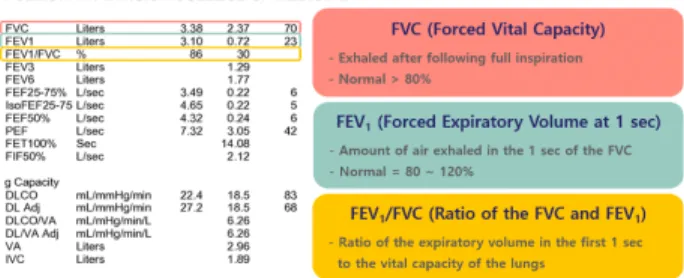
Fig. 2. The pulmonary function test results are as follows, and the indicators used in this study are explained.
폐 기능 검사 결과 값, FVC (Forced Vital Capacity)는 노력 폐활량으로 최대 흡기 후 노력 호기 중에 배출되는 유량을 측 정한 값이다. FEV1 (Forced Expiratory Volume at 1 second)은 1초 노력 호기량으로 최대 흡기 후 처음 1초 간 배출되는 유량 을 측정한 값이다. FVC분의 FEV1은 일초율로 노력 폐활량에 서 1초 노력 호기량이 차지하는 비율을 정상 참고치에 대한 백 분율로 표시한다(Fig. 2). 이를 폐 환기 검사의 계수율과 비교 분석 하였다. 정상인의 경우에 노력 폐활량의 약 70% 이상을 1초에 내쉴 수 있으며, 이 기준은 기도의 폐쇄 정도를 감별하 는데 유용한 지표로 사용되고 있다.5) 이러한 지표는 신장, 체 중, 인종, 성별, 연령 등의 여러 요인에 의하여 정상 참고치가 정해지기 때문에 보다 정확하고 객관적인 값을 제공한다.6) 본 원에서는 우리나라에서 주로 사용하는 ‘Morris 아시아인 퀀 저식’을 정상 참고치에 사용하였다.7)
Fig. 3. It is the process of converting Tc-99m into gas particle using Technegas Generator.
방사성동위원소로 Tc-99m, 555~740 MBq/0.2 ml를 Technegas 생성 장비 내에 준비한 탄소봉 안에 주입하고, 아르 곤 가스 충전 후 약 2500 ℃에서 가열하여 입자 크기 0.005~0.2 μm의 Technegas를 생성한다. 생성된 Technegas는 입자의 응 집을 방지하기 위해 10분 이내로 사용하며, 검사 기준 1500~2000 계수율(cps)에 도달 할 때까지 장비와 연결된 흡입 마스크를 통하여 환자에게 흡입을 유도하였다(Fig. 3).
Fig. 4. The scan conditions and protocol of lung ventilation scan are shown.
폐 환기 검사에 사용된 조준기는 LEHR (Low Energy High Resolution)이며, 화소(Matrix) 수는 256×256이고, 30만 계수 설정으로 앙와위 상태에서 전면상, 후면상, 양측 사위상, 측면 상을 획득하였다(Fig. 4). 구강과 식도, 위 등에 집적된 계수를 제외한 폐 실질의 계수율을 측정하기 위하여 5년차 이상 방사 선사 3명이 폐의 관심영역(Region Of Interest, ROI)을 설정하 여 폐 실질 계수율을 평균화하였다.
통계 프로그램은 SPSS Ver. 20.0으로 정규성 검정 후, 피어 슨 상관관계를 이용하여 분석하였다.
결 과
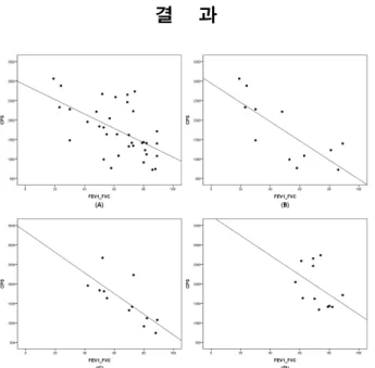
Fig. 5. The correlation between the cps of lung ventilation scan and the result of pulmonary function test. (A) is a correlation for all patients, classified by severity (B) is a correlation of the severe group, (C) is a correlation of the moderate group, (D) is a mild shows the correlation of groups.
폐 환기 검사에서 양측 폐의 실질 계수율과 폐 기능 검사에 서 일초율(FEV1/FVC) 사이의 음의 상관관계를 확인할 수 있 으며, COPD 질환의 중증도가 심할수록 부적상관이 강하게
관련됨을 확인 할 수 있다.
Table 2. Correlation and significance between the FEV1/FVC of PFT by COPD severity and LVS cps
FEV1/FVC All Severe Moderate Mild
Correlation -0.569* -0.773* -0.750* -0.437
P 0.000 0.003 0.005 0.155
N 36 12 12 12
* : Correlation is significant at the 0.01 level
전체 36명 환자를 대상으로 상관계수는 –0.569이며, P값 유 의성은 0.001 미만으로 나타났다. 12명씩 중증도 별로 확인한 결과 Severe의 상관계수는 –0.773이고, P값 유의성은 0.003 미 만으로 나타났다. 이와 더불어 Moderate의 상관계수는 – 0.750, P값 유의성은 0.005 미만으로 나타났다. Mild의 상관계 수는 –0.437로 통계적으로 유의하지 않았다(Table 2).
고 찰
폐는 산소를 섭취하고 체내에서 생긴 탄산가스를 배출하는 역할을 하며, 폐 기능 검사는 환자가 최대한 숨을 들이마신 후 내쉴 수 있는 공기량을 측정하는 검사법으로 폐의 기능적인 측면을 객관화한 지표로 사용된다.8) 이러한 지표 중에 FEV1/ VC와 폐 환기 검사 사이의 상관관계를 확인한 바와 같이 다른 주요 지표와는 어떠한 상관관계가 있는지 확인해 볼 필요성이 존재한다. 또한 특정 지표의 변화는 질환의 경중을 반영하고, 질환에 따른 고유한 특징적인 양상이 있어 질환을 조기에 진 단하거나 임상적 진행을 단계별로 평가하는데 도움을 준다.9) 이에 폐 기능 검사에서 FEV1지표를 폐쇄 환기장애의 중증도 인 GOLD기준으로 분류하여 중증도가 Severe에 가까울수록 음의 상관관계가 강하게 나타남을 확인 할 수 있었다.
폐 기능 검사에서 측정된 FEV1, FVC는 측정치와 함께 정상 참고치의 백분율로 표시되며, 판독하기 위해서는 환자에서 측정된 수치와 건강한 정상인 군에서 측정된 수치 간의 비교 가 필요하다.10) 또한 정상 참고치는 인종, 성별, 연령, 신장과 체중 등의 신체적인 요건과 측정 조건, 통계적 방법 및 사회 경 제적 또는 역학적 요건에 따라 달라질 수 있다.10) 단적으로 연 령이 증가할수록 FEV1/FVC가 감소하므로 위험에 노출된 적 이 없는 정상 고령에서 폐쇄 환기장애가 있다고 잘못 진단되 는 위양성율이 높아질 수 있다.11) 반면 같은 기준을 젊은 연령 에게 적용하였을 때에는 오히려 폐쇄 환기장애가 있으나 정상 으로 진단할 우려가 있다.12) 그러므로 향후에는 연령을 고려
하여 폐 환기 검사와의 상관관계를 확인 할 필요가 있다. 더 나 아가 폐섬유화 및 간질성 폐질환, 기관지 확장증 등에 대한 연 구도 필요하며, 폐 환기 검사 시 계수율과 질환별 호흡시간, 호 흡횟수와의 상관관계를 확인하는 연구 역시 진행되어야 할 것 으로 생각된다.
결 론
만성폐쇄성폐질환 환자를 대상으로 시행한 폐 환기 검사의 계수율과 폐 기능 검사 결과 값 사이의 상관관계를 확인할 수 있었다. 이를 통해 폐 환기 검사 시 호흡력에 대한 지표로써 폐 기능 검사 결과 값을 활용할 수 있을 것으로 기대되며, 비교적 정확한 검사 소요시간을 안내함으로써 환자의 적극적인 검사 참여를 유도할 수 있을 것이다. 또한 검사실 측면에서도 보다 효율적인 운영에 도움이 될 것으로 생각된다.
요 약
폐 환기 검사는 직접 방사성 기체를 흡입한 후 이를 영상화 한다. 그러나 실제 폐 환기 검사 시 활용할 수 있는 흡입과 관련 한 지표는 널리 알려진 바가 없다. 따라서 본 연구를 통해 흡입 과 관련한 객관적인 지표로써 폐 환기 검사 계수율과 폐 기능 검사 결과 값의 상관관계를 확인하고 그 유용성을 알아보고자 한다. 2010년 10월부터 2018년 9월까지 본원에서 폐 환기 검 사와 폐 기능 검사를 모두 시행한 만성폐쇄성 폐질환 환자 36 명을 대상으로 중증도(Mild, Moderate, Severe)별로 분류하였 다. 폐 환기 검사는 Technegas Generator (Vita Medical Ltd, Australia)로 Technegas를 생성하여, 환자에게 흡입하게 하였 다. 영상은 Forte (Philips Medical Systems, Netherlands) 장비 를 사용하였으며, 폐 기능 검사는 Vmax Encore 22 (Carefusion Corporation, USA)를 사용하였다. 폐 환기 검사에서는 양측 폐에 관심 영역을 설정하여 계수율을 측정하였고, 이와 함께 폐 기능 검사의 결과 값 중 노력 폐활량(FVC), 1초 노력 호기량 (FEV1), 일초율(FEV1/FVC) 사이의 상관관계를 비교 분석하 였다. 만성폐쇄성폐질환 환자의 폐 환기 검사 계수율과 폐 기 능 검사 결과 값의 상관관계를 분석한 결과, 중증도별 FEV1/FVC와 계수율 사이의 상관계수는 Severe에서 -0.773, Moderate에서 -0.750, Mild에서 -0.437이였다. Severe, Moderate 결과 값은 통계적으로 유의하였으며(P<0.05), Mild 는 유의하지 않았다(P=0.155). 반면에 FVC와 계수율 사이의 상관계수는 Mild에서만 0.882로 통계적으로 유의한 것을 확 인하였다(P<0.05). 만성폐쇄성폐질환 환자를 대상으로 시행
한 폐 환기 검사 시 계수율과 폐 기능 검사 결과 값의 상관관계 를 확인할 수 있었다. 이를 통해 폐 환기 검사 시 호흡력에 대한 지표로써 폐 기능 검사 결과 값을 활용할 수 있을 것으로 생각 된다.
REFERENCES
1. M. Bajc, J. B. Neilly, M. Miniati, C. Schuemichen, M.
Meignan, B. Jonson. EANM guidelines for ventilation/
perfusion scintigraphy. Eur J Nucl Med Mol Imaging.
2009;36:1356-1370.
2. Mills NL, Amin N, Robinson SD, Anand A, Davies J, Patel D, et al. Do Inhaled Carbon Nanoparticles Translocate Directly into Circulation in humans? Am J Respir Crit Care Med. 2006;173:426-431.
3. Sinzinger H, Rodrigues M, Kummer F. Ventilation/
perfusion lung scintigraphy. Multiple applications besides pulmonary embolism. Hell J Nucl Med. 2013;16(1):50-55.
4. Jogi J, Jonson B, Ekberg M, Bajc M. Ventilation–Perfusion SPECT with 99mTc-DTPA Versus Technegas: A Head-to-Head Study in Obstructive and Nonobstructive Disease. J Nucl Med. 2010;51:735-741.
5. Rabe KF, Hurd S, Anzueto A, Barnes PJ, Buist SA, Calverley P, et al. Global strategy for the diagnosis, management, and prevention of chronic obstructive pulmonary disease: GOLD executive summary. Am J Respir Crit Care Med 2007;176:532-555.
6. Choi JK, Paek D, Lee JO. Normal predictive values of spirometry in Korean population. Tuberc Respir Dis 2005;58:230-242.
7. Morris JF, Koski A, johnson LC. Spirometric standards for healthy nonsmoking adults. Am Rev Respir Dis 1971;
103:57-67.
8. The BTS COPD consortium. Spirometry in practice. A practical guide to using spirometry in primary care. Available at:
https://www.brit-thoracic.org.uk/document-library/ delivery- of-respiratory-care/spirometry/spirometry-in-practice; Accessed December 6, 2017.
9. Gold WM, Koth LL. In: Murray JF, editor. Murray and Nadel’s Textbook of Respiratory Medicine saunders, Elsevier; 2010.
10. American Thoracic Society. Lung function testing:
selection of reference values and interpretative strategies.
Am Rev Respir Dis 1991;144:1202-1218.
11. Swanney MP, Ruppel G, Enright PL, Pederson OF, Crapo RO, Miller MR, et al. Using the lower limit of normal for the FEV1/FVC ratio reduces the misclassification of airway obstruction. Thorax 2008;63:1046-1051.
12. Cerveri I, Corsico AG, Acoordini S, Niniano R, Ansaldo E, Antó JM, et al. Underestimation of airflow obstruction among young adults using FEV1/FVC <70% as a fixed cut-off: a longitudinal evaluation of clinical and functional outcomes. Thorax 2008;63:1040-1045.