저선량 흉부전산화단층촬영의 흡기 영상과 폐기능이 정상인 성인에서 호기 말 폐 감쇄도 증가 정도와 연령 및 흡연과의 연관성
분당서울대학교병원 1폐센터, 2내과, 3영상의학과, 4한림대학교 성심병원 내과 권성연1,2, 황용일4, 윤호일1,2, 이재호1,2, 이춘택1,2, 이경원1,3
Correlation between Expiratory Increase of Lung Attenuation and Age and Smoking in the Subjects with Normal Inspiratory Low Dose CT and Pulmonary Function Test
Sung-Youn Kwon, M.D.
1,2, Yong-Il Hwang, M.D.
4, Ho-Il Yoon, M.D.
1,2, Jae-Ho Lee, M.D.
1,2, Choon-Taek Lee, M.D.
1,2, Kyung Won Lee, M.D.
1,31
Respiratory Center, Departments of
2Internal Medicine,
3Radiology, Seoul National University Bundang Hospital, Seongnam,
4
Department of Internal Medicine, Hallym University Sacred Heart Hospital, Seoul, Korea
Background: The attenuation of the lung parenchyma increases on expiration as a consequence of decreased air in the lung. Expiratory CT scans have been used to show air trapping in patients with chronic airway disease and diffuse parenchymal disease and also in asymptomatic smokers. Although there have been several reports investigating the regional air trapping on a expiratory CT scan, there have been only a few reports evaluating the changes of whole lung attenuation with considering its clinical significance, and especially in healthy subjects.
The purpose of this study was to evaluate the correlation of an expiratory increase of lung attenuation with age and smoking in healthy subjects.
Methods: Asymptomatic subjects who underwent a low dose chest CT scan as part of a routine check-up and who showed normal spirometry and a normal inspiratory CT scan were recruited for this study. We excluded the subjects with significant regional air trapping seen on their expiratory CT scan. Lung attenuation was measured at 24 points of both the inspiratory and expiratory CT scans, respectively, for 100 subjects. The correlations between an expiratory increase of the lung attenuation and the amount of smoking, the patient's age and the results of spirometric test were assessed.
Results: There were 87 men and 13 women included in this study. Their median age was 49.0 years old (range:
25∼71). Sixty current smokers, 24 ex-smokers and 16 non-smokers were included. As age increased, the expiratory increase of lung attenuation was reduced at every measuring points (r=−0.297∼−0.487, Pearson correlation).
The statistical significance was maintained after controlling for the effect of smoking. Smoking was associated with a reduction of the expiratory increase of lung attenuation. But the significance was reduced after controlling for the patient's age. The FEV1, FVC, FEV1/FVC and FEF25∼75% were not associated with an expiratory increase of lung attenuation.
Conclusion: The expiratory increase of lung attenuation in subjects with a normal inspiratory CT scan was negatively correlated with age. It was also reduced in heavy smokers. It may reflect aging and the smoking related changes.
(Tuberc Respir Dis 2008;65:457-463)
Key Words: Lung attenuation, Expiratory CT, Smoking, Age
Address for correspondence: Kyung Won Lee, M.D.
Department of Radiology, Seoul National University Bundang Hospital, 300, Gumi-dong, Bundang-gu, Seong- nam 463-707, Korea
Phone: 82-31-787-7604, Fax: 82-31-787-4011 E-mail: [email protected]
Received: Nov. 16, 2008 Accepted: Nov. 25, 2008
서 론
일반적인 흉부 전산화단층촬영은 흡기 시에 영상을 얻 게 되지만, 최근 호기 시의 영상을 추가적으로 촬영함으로 써 호흡 주기에 따른 폐 밀도의 변화를 이용하는 것이 폐 의 기능적 영상을 얻는 검사법으로 많이 사용되고 있다1.
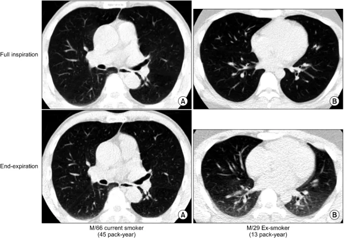
Figure 1. Examples of expiratory CT findings. (A) Paired inspiratory and expiratory CT scan of 66 years old current smoking male with 45 pack-year of smoking amount. Expiratory CT scan shows minimal change of lung density (bottom).
(B) Paired inspiratory and expiratory CT scan of 29 years old ex-smoking male with 13 pack-year of smoking amount.
Expiratory CT scan shows increased lung density compared with inspiratory CT scan (bottom).
정상적인 호기 시에는 폐 안의 공기 용적이 감소하게 되므 로 호기 말(end-expiration)에 전산화단층촬영을 시행하 게 되면 폐 감쇄도(lung attenuation)가 전반적으로 증가 되어2,3 흡기 시보다 고음영이 나타난다. 해부학적으로 의 존적인 부위(dependent portion)에서 이런 현상이 더 뚜 렷하게 나타나는 것으로 알려져 있다(Figure 1)4. 이에 비 해 공기 가둠(air trapping) 현상은 기도 폐쇄나 국소적인 폐 탄성도의 변화로 인해 호기 시에 과다한 공기가 폐의 일부에 축적되어 나타나는 현상으로, 호기 영상에서 폐 감쇄도의 증가가 없거나 아주 적어서 공기 가둠이 없는 주변 영역과 구별되는 저음영의 구역으로 관찰된다5. 호기 전산화단층활영에서 폐 감쇄도가 균일하지 않을 때 미만성 폐질환의 진단에 보조적인 도움을 줄 수 있다는 보고도 있지만6 호기 전산화단층촬영을 이용한 연구는 주 로 공기 가둠 현상에 초점이 맞추어져 왔고4,7. 만성 기도
질환8, 유육종증9,10 등에서 보고된 바 있다. 또한 이런 공 기 가둠 현상이 무증상의 흡연자에서도 관찰되므로, 이것 이 흡연과 연관된 초기 변화라는 보고가 있고2,3,11-13 폐기 능과 음의 상관 관계를 보이며8 연령과14 연관성이 있다는 보고도 있다.
그러나, 전산화단층촬영을 이용하여 국소적인 공기 가 둠이 아닌 전반적인 폐음영의 변화 정도를 고찰한 연구는 많지 않다. 정상적인 호기말 폐 감쇄도의 증가는 부위에 따라 80∼300 HU 정도로 제시되어 있는데15, 폐쇄성 환기 장애가 있는 경우에 이 증가 정도가 유의하게 감소 되어 있고 폐기종 환자에서는 폐 기능과 연관성이 있다는 보고 가 있으나16, 호흡기 질환이 없고 폐기능이 정상인 사람을 대상으로 하여 호기말의 폐 감쇄도 변화의 의미를 고찰한 연구는 드물다.
이에 본 연구에서는 호흡기 질환이 없고, 저선량 흉부
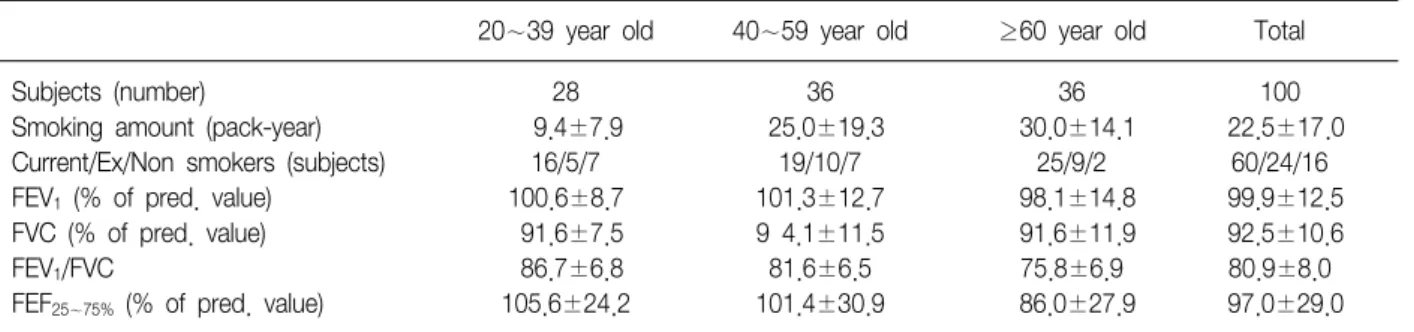
Figure 2. Example of lung attenuation measurement.
Table 1. Baseline characteristics of the study subjects
20∼39 year old 40∼59 year old ≥60 year old Total
Subjects (number) 28 36 36 100
Smoking amount (pack-year) 9.4±7.9 25.0±19.3 30.0±14.1 22.5±17.0
Current/Ex/Non smokers (subjects) 16/5/7 19/10/7 25/9/2 60/24/16
FEV1 (% of pred. value) 100.6±8.7 101.3±12.7 98.1±14.8 99.9±12.5
FVC (% of pred. value) 91.6±7.5 9 4.1±11.5 91.6±11.9 92.5±10.6
FEV1/FVC 86.7±6.8 81.6±6.5 75.8±6.9 80.9±8.0
FEF25∼75% (% of pred. value) 105.6±24.2 101.4±30.9 86.0±27.9 97.0±29.0
전산화단층촬영의 흡기 시 영상이 정상이며, 호기 시 영상 에서 국소적인 공기 가둠 현상이 없는 대상군에서 호기말 폐 감쇄도의 증가 정도와 연령 및 흡연과의 연관성을 알아 보고자 하였다.
대상 및 방법
1. 연구 대상
2004년 2월부터 4월까지 분당서울대학교병원 건강검진 센터를 방문하여 저선량 흉부전산화단층촬영과 폐기능 검사를 시행 받은 20세 이상 성인 중 호흡기계 증상과 폐 질환의 병력이 없고 폐기능 검사에서 일초율(FEV1/FVC) 이 70% 이상, 강제폐활량이 80% 이상인 정상 소견을 보이 며, 흉부 전산화단층촬영에서는 흡기 영상이 정상이면서
호기 영상에서 국소적 공기 가둠 소견이 없는 100명을 선 정하여 연구를 수행하였다.
2. 연구 방법
폐기능 검사와 전산화단층촬영은 같은 날 시행되었으 며 대상군의 의무기록을 통해 나이, 성별, 흡연력 등의 인 구학적 특성과 동반 질환의 유무를 확인하였다. 대상군은 40세와 60세를 기준으로 연령에 따라 세 군으로 나누었고 흡연량에 따라서는 비흡연자군, 20갑년 이하 흡연군, 그리 고 20갑년 초과 흡연군의 세 군으로 나누었다.
연구에 사용된 전산화단층촬영 기기는 MX 8000 IDT (Philips)로 16 channel이었다. 폐 실질 영상에 대해서 폐 창(lung window setting)에서 높이(window level) −650 HU, 너비(window width) 1,500 HU을 적용하여 관심 영 역에서의 폐 감쇄도를 HU값으로 측정하였다. 영상의학과 전문의가 각 대상마다 총 24개의 관심 영역을 선정하고 혈관 부위를 피하여 0.1 cm2의 관심 영역(region of inter- est, ROI)을 표시하였는데, 대동맥궁(aortic arch), 기관분 지부(carina), 하폐정맥(interior pulmonary artery) 수준의 영상을 상(upper), 중(middle), 하(lower) 수준으로 정하 고, 각 수준에서 좌폐와 우폐에 대하여 각각 복측 (ventral), 배측(dorsal), 내측(medial), 외측(lateral) 지점 을 늑막에서 1 cm 떨어진 곳에 표시하였다(Figure 2). 폐 감쇄도의 증가 정도는 선정된 관심 영역에서 호기말의 감 쇄도에서 흡기 시의 감쇄도를 뺀 값을 HU로 표시하였다.
분석 시에는 24개의 관심 영역을 해부학적 위치에 따라 좌우 구분 없이 복측(ventral), 배측(dorsal), 내측(medial), 외측(lateral), 상(upper), 중(middle), 하(lower)의 7개 해 부학적 부위로 재분류하여, 포함된 관심영역의 폐 감쇄도 증가값의 평균을 구하여 각 부위의 감쇄도 값으로 사용하 였다.
통계 분석은 윈도우형 SPSS version 12.0을 이용하였
Table 2. Lung attenuation change during inspiration and expiration
Inspiration Expiration Difference
(HU) (HU) (HU)
Ventral −903.9±18.1 −843.5±30.8 60.4±26.6 Medial −889.8±22.1 −770.0±70.7 79.0±33.1 Lateral −882.4±23.2 −808.7±36.0 73.7±31.1 Dorsal −869.9±28.6 −751.7±51.9 122.2±52.1 Upper −884.4±21.0 −803.7±40.1 80.7±36.8 Middle −888.7±22.3 −808.0±36.7 80.7±32.9 Lower −886.1±25.1 −795.9±37.5 90.2±36.0
Table 3. Expiratory increase of lung attenuation according to smoking groups
Nonsmoker ≤20 pack-year >20 pack-year p-value
(n=16) (HU) (n=33) (HU) (n=51) (HU) (one way ANOVA)
Ventral 69.5±24.2 71.4±30.5 50.3±20.6 0.000
Medial 77.4±28.5 91.5±32.7 71.4±32.8 0.023
Lateral 75.7±23.7 86.0±34.4 62.2±28.5 0.010
Dorsal 118.4±44.5 141.3±59.2 111.1±46.6 0.031
Upper 84.6±27.5 94.9±41.3 70.2±33.3 0.009
Middle 83.6±27.0 95.0±37.9 70.4±27.7 0.003
Lower 87.5±31.8 102.7±38.5 82.9±33.9 0.044
Table 4. Correlation between expiratory increase of lung attenuation and smoking amount
Correlation coefficient (r) Smoking Smoking amount
amount (Age adjusted)
Ventral −0.412* −0.257*
Medial −0.276* −0.160
Lateral −0.320* −0.223*
Dorsal −0.250* −0.108
Upper −0.325* −0.196
Middle −0.347* −0.207*
Lower −0.249* −0.126
*p<0.05.
다. 각 연령군, 흡연에 따른 분류군 사이에 호기말 감쇄도 증가 정도에 차이가 있는가를 일원배치 분산분석으로 확 인하였다. 호기말 폐 감쇄도의 증가와 연령, 흡연량, 폐기 능과의 상관관계를 알아보기 위하여 Pearson 상관분석으 로 이변량 상관계수를 구하였고 각각의 변수를 통제변수 로 이용하여 편상관계수를 구하였다. 0.05 미만의 p값을 통계적으로 유의한 것으로 판단하였다.
결 과
1. 대상군의 특성
연구 대상 100명 중 남자 87명, 여자 13명이었고, 연령의 중앙 값은 49.0세(범위 25∼71세)였다. 연령군별로는 20
∼39세가 28명, 40∼59세가 36명, 60세 이상이 36명이었 다. 현재 흡연자 60명, 과거 흡연자가 24명, 비흡연자가 16명이었으며 평균 흡연량은 22.5±17.0갑년이었다 (Table 1). 강제폐활량(forced vital capacity, FVC), 1초간 강제날숨량(forced expiratory volume in 1 second, FEV1) 의 정상 예측치에 대한 백분율은 각 연령군이나 흡연군 사이에 차이는 없었으며 FEV1/FVC는 높은 연령군일수록
낮았다. 흡연량에 따른 연령군 분포는 비흡연자, 20 갑년 이하 흡연자, 20갑년 초과 흡연자가 각각 20∼39세군에서 7명, 19명, 2명, 40∼59세군에서 7명, 8명, 21명, 그리고, 60세 이상군에서 2명, 6명, 28명이었다.
2. 흡기-호기 시의 폐 감쇄도의 변화
7개의 영역에서 구한 평균 폐 감쇄도의 증가값은 Table 2와 같았으며 기존에 알려진 바와 같이 폐의 의존성 부위 (dorsal and lower) 부분에서 비의존성 부위보다 폐 감쇄 도의 증가가 더 많았다.
3. 흡연과 폐 감쇄도 변화
각 흡연군에 따른 폐 감쇄도의 증가 정도를 비교하였을 때, 20갑년을 초과하는 흡연력을 가진 군에서 모든 영역 에서의 폐 감쇄도 증가 정도가 적었다(일원배치 분산분석, Table 3). 사후 분석(post-hoc analysis)을 시행하였을 때 는 20갑년을 초과하는 흡연력을 가진 군이 20갑년 이하 흡연군보다 모든 관심 영역에서 유의하게 호기말 폐 감쇄
Table 6. Correlation between expiratory increase of lung attenuation and age
Correlation coefficient (r) Age Age (smoking adjusted)
Ventral −0.487* −0.376*
Medial −0.325* −0.238*
Lateral −0.297* −0.186
Dorsal −0.371* −0.302*
Upper −0.376* −0.277*
Middle −0.411* −0.310*
Lower −0.329* −0.254*
*p<0.05.
Table 5. Expiratory increase of lung attenuation according to age group
20∼39 year old 40∼59 year old ≥60 year old p-value
(n=28) (HU) (n=36) (HU) (n=36) (HU) (one way ANOVA)
Ventral 78.1±28.3 60.9±25.8 46.0±15.9 0.000
Medial 95.6±34.6 78.1±31.7 67.0±28.2 0.002
Lateral 85.6±32.0 75.7±30.6 62.6±27.6 0.010
Dorsal 145.2±62.1 128.7±48.5 97.7±35.5 0.001
Upper 98.1±41.6 83.5±35.9 64.2±25.9 0.001
Middle 98.5±36.1 82.2±31.0 65.2±24.5 0.000
Lower 106.8±39.2 92.0±34.8 75.4±28.5 0.002
도 증가가 적었다.
갑년으로 표시한 흡연량과 호기말 폐 감쇄도의 증가 사 이의 상관 관계를 알아보았을 때에는 유의한 음의 상관 관계가 관찰 되었으며, 연령으로 보정하여도 폐의 복측 (ventral), 외측(lateral), 중간부(middle)에서 유의성이 유 지 되었다(Table 4).
4. 연령에 따른 폐 감쇄도 변화
대상군을 연령을 기준으로 20∼39세, 40∼59세, 60세 이상의 세 군으로 나누어 폐 감쇄도 증가 정도를 비교하였 을 때 유의한 차이가 있었고(일원배치 분산분석, Table 5), 사후 분석에서는 60세 이상군이 20∼39세군보다 모든 관심 영역에서 유의하게 호기말 폐 감쇄도 증가가 적었다.
Pearson 상관분석을 시행했을 때, 연령과 폐 감쇄도 증 가 사이에 유의한 음의 상관관계가 관찰되었고 흡연량으 로 보정하였을 때에도 폐의 외측부(lateral portion)를 제 외한 모든 부위에서 유의한 상관관계를 보였다(Table 6).
고 찰
본 연구에서 호기말 평균 폐 감쇄도의 증가 정도는 평 가 부위에 따라서 60.4±26.6 HU에서 122.5±52.1 HU까 지 측정되었다. 기존에 알려진 대로 의존성 부위인 폐의 하부와 배측부에서의 상부, 복측부에 비하여 더 큰 폭의 감쇄도 증가가 관찰되었다. 이는 호기말 평균 폐 감쇄도 의 증가 정도를 폐의 의존성 부위와 비의존성 부위에서 각각 219±28.2 HU와 171±16.9 HU로 보고한 Webb 등17 의 연구나 폐의 상부와 하부에서 각각 100.9±50.2 HU, 133.3±56.1 HU로 보고한 Tanaka 등18의 연구보다는 낮 게 측정된 것이다. 그러나, Webb 등의 연구는 대상군의 나이가 24∼31세로 젊어서 연령 증가에 의한 변화를 반영
하지 못하였고, Tanaka 등의 연구는 6개의 관심 영역에서 만 폐 감쇄도를 측정하였기 때문에 폐 전체의 용적을 반영 하기에 부족하였다. 또한 두 연구 모두 국소적인 공기 가 둠 현상이 각각 40%와 64%에서 관찰되었다고 보고하였 다. 따라서 60세 이상 고령자를 포함한 다양한 연령층을 대상으로 하고, 국소적 공기 가둠 현상이 없는 대상만을 분석하였던 본 연구와는 차이가 있을 것으로 생각된다.
흡연량에 따라 평균 호기말 폐 감쇄도의 증가 정도를 알아 보았을 때 각 흡연군 사이의 일원배치 분산분석에서 통계적인 유의성이 관찰되었다. 이는 정상 폐기능을 보이 는 무증상 성인을 대상으로 한 기존의 연구에서 호기말 평균 폐 감쇄도 증가가 흡연량에 따라 차이가 없다는 결과 와18 차이를 보이는 것이다. 본 연구의 대상자 중 20갑년 이하의 흡연군에는 젊은 연령자가 더 많이 포함되어 있고 20갑년 초과 흡연군에는 고령자가 더 많이 포함되어 있어 연령의 효과로 인한 차이를 고려할 수 있다. 그러나 상관 분석에서 흡연량과 호기말 평균 폐 감쇄도의 증가 정도 사이에 관찰된 음의 상관관계가 연령 보정 후에도 유의하
게 유지되었으므로 흡연 자체로 인한 영향을 배제할 수는 없다고 생각된다.
기존의 연구에서는 대상군의 연령이 비교적 젊고17 대 상군의 수가 적어서17,18 평균 호기말 폐 감쇄도의 증가 정 도와 연령에 대한 분석이 이루어지지 못하였다. 본 연구 에서는 60세를 기준으로 구분하였을 때 폐 감쇄도 증가의 차이가 뚜렷하게 나타났다. 또한 호기말 폐 감쇄도 증가 와 연령 사이에 유의한 음의 상관관계가 관찰되었고, 이러 한 상관성은 흡연량으로 보정하였을 때도 그 경향을 유지 하였으므로, 호기말 폐 감쇄도 증가 정도의 감소가 노화와 관련된 변화일 수 있음을 시사한다고 할 수 있다. 기존 연구 중 호기 전산화단층촬영이 노화나 흡연에 의한 변화 를 추적 관찰할 때 유용성이 적다는 보고도 있었으나19, 대상군에 국소적 공기 가둠 현상이 보이는 경우를 배제하 지 않아 대상군의 차이로 인한 다른 결과일 수 있겠다.
그 동안 호기말 흉부 전산화단층촬영과 폐기능에 대한 연구는 주로 공기 가둠 현상과 폐기능 사이의 관계에 대해 서 연구되어 왔다. 폐쇄성 환기장애가 있을 때는 폐 감쇄 도 증가 정도가 작고16, 폐기능이 정상일 때에는 호기말 폐 감쇄도의 증가 정도와 폐기능 사이에 유의한 상관관계 를 보이지 않는다는 보고가 있다20. 본 연구에서는 질환이 없고 폐기능과 흡기 시 전산화단층촬영이 정상 소견이며 국소적 공기 가둠 현상이 없는 대상만을 가지고 기존의 연구에서 자주 분석되지 않았던 연령과 폐 감쇄도 증가 정도 사이의 관계를 확인함으로써 노화와 관련된 변화를 고찰하였다는 의미가 있다.
본 연구는 몇 가지 제한점을 지닌다. 먼저, 각 흡연군별 로 연구 대상의 연령 분포가 동일하지 않아 흡연에 의한 영향이 축소 평가되었을 가능성이 있다. 또한 연령과의 상관관계 평가 시, 흡연에 의한 영향을 통계적으로 배제하 기는 하였지만, 전체 대상군 중에서 비흡연자의 숫자가 상대적으로 적고 또한 후향적 단면적 연구여서 흡연에 의 한 효과가 실질적으로 배제되는 데에는 제한이 있었다고 생각된다. 향후 흡연군과 연령군을 통제한 대상군에 대한 전향적인 연구가 필요할 것으로 판단된다.
향후 추가적 연구가 필요하겠으나, 호기말 폐 감쇄도 증가가 연령 증가에 따라 저하되는 것으로 판단되며, 향후 노화 과정에 대한 추적에 있어서 유용한 기능적 영상 검사 법이 될 수 있을 것으로 기대된다.
요 약
연구배경: 호기말에 흉부 촬영을 시행하면 폐 내의 공 기의 부피가 감소되어 폐 감쇄도가 증가한다. 이런 폐 감 쇄도의 변화가 국소적으로 저하되는 공기 가둠 현상에 대 해서는 일부 연구들이 있었으나 전체 폐의 전반적인 감쇄 도 변화에 대한 연구는 거의 없고, 특히 정상인을 대상으 로 한 연구는 거의 없다. 저자들은 호흡기 질환이 없으며 폐기능 및 흡기 전산화단층촬영이 정상 소견이고 호기 영 상에서 국소적인 공기 가둠 현상이 없는 100명을 대상으 로 연구를 수행하였다.
방 법: 흉부 전산화단층촬영 영상에서 24개의 지점을 정하고, 흡기 및 호기 시 각 지점에서 폐 음영도를 측정한 후 호기말 폐 감쇄도 증가와 흡연량, 연령 사이의 상관 관계를 알아보았다.
결 과: 연구 대상 100명의 연령은 중앙값 49.0세(범위 25∼71세)였고, 평균 흡연량은 22.5±17.0갑년이었다. 연 령과 폐 감쇄도의 증가 사이에는 유의한 음의 상관관계가 있었으며 이는 흡연의 효과를 보정하였을 때에도 유지되 었고, 연령을 기준으로 세 군으로 나누어 폐 감쇄도의 증 가 정도를 비교하였을 때에도 유의한 차이가 있었다. 흡 연량과 호기말 폐 감쇄도의 증가 사이에는 유의한 음의 상관관계가 관찰되었으나 연령을 보정하였을 때에는 유 의성이 감소하였다.
결 론: 흡기-호기 시의 폐의 전반적인 감쇄도 변화 정 도는 연령과 상관관계가 있으며, 노화를 반영할 가능성이 있다.
참 고 문 헌
1. Naidich DP, Webb WR, Grenier PA, Harkin TJ, Gefter WB. Functinal imaging of the airways. In: Imaging of the airways. Functional and Radiologic correlation. 1st ed. Philadelphia: Lippincott Williams & Wilkins; 2005.
p. 177-211.
2. Hansell DM. Small airway diseases: detection and in- sights with computed tomography. Eur Respir J 2001;
17:1294-313.
3. Kazerooni EA. High-resolution CT of the lungs. AJR Am J Roentgenol 2001;177:501-19.
4. Grenier PA, Beigelman-Aubry C, Fetita C, Preteux F, Brauner MW, Lenoir S. New frontiers in CT imaging of airway disease. Eur Radiol 2002;12:1022-44.
5. Austin JH, Muller NL, Friedman PJ, Hansell DM,
Naidich DP, Remy-Jardin M, et al. Glossary of terms for CT of the lungs: recommendations of the Nomen- clature Committee of the Fleischner Society. Radiology 1996;200:327-31.
6. Arakawa H, Webb WR, McCowin M, Katsou G, Lee KN, Seitz RF. Inhomogeneous lung attenuation at thin-sec- tion CT: diagnostic value of expiratory scans. Radiol- ogy 1998;206:89-94.
7. de Jong PA, Muller NL, Pare PD, Coxson HO. Comput- ed tomographic imaging of the airways: relationship to structure and function. Eur Respir J 2005;26:140-52.
8. Lucidarme O, Coche E, Cluzel P, Mourey-Gerosa I, Howarth N, Grenier P. Expiratory CT scans for chronic airway disease: correlation with pulmonary function test results. AJR Am J Roentgenol 1998;170:301-7.
9. Terasaki H, Fujimoto K, Muller NL, Sadohara J, Uchida M, Koga T, et al. Pulmonary sarcoidosis: comparison of findings of inspiratory and expiratory high-reso- lution CT and pulmonary function tests between smok- ers and nonsmokers. AJR Am J Roentgenol 2005;185:
333-8.
10. Arakawa H, Niimi H, Kurihara Y, Nakajima Y, Webb WR. Expiratory high-resolution CT: diagnostic value in diffuse lung diseases. AJR Am J Roentgenol 2000;175:
1537-43.
11. Spaggiari E, Zompatori M, Verduri A, Chetta A, Bna C, Ormitti F, et al. Early smoking-induced lung lesions in asymptomatic subjects: correlations between high reso- lution dynamic CT and pulmonary function testing.
Radiol Med 2005;109:27-39.
12. Verschakelen JA, Scheinbaum K, Bogaert J, Demedts M, Lacquet LL, Baert AL. Expiratory CT in cigarette smok- ers: correlation between areas of decreased lung at- tenuation, pulmonary function tests and smoking history. Eur Radiol 1998;8:1391-9.
13. Mastora I, Remy-Jardin M, Sobaszek A, Boulenguez C, Remy J, Edme JL. Thin-section CT finding in 250 volun- teers: assessment of the relationship of CT findings with smoking history and pulmonary function test results. Radiology 2001;218:695-702.
14. Lee KW, Chung SY, Yang I, Lee Y, Ko EY, Park MJ.
Correlation of aging and smoking with air trapping at thin-section CT of the lung in asymptomatic subjects.
Radiology 2000;214:831-6.
15. Webb WR, Muller NL, Naidich DP. Normal lung anatomy. In: High-Resolution CT of the Lung. 3rd ed.
Philadelphia: Lippincott Williams & Wilkins; 2001. p.
49-69.
16. Kauczor HU, Hast J, Heussel CP, Schlegel J, Mildenberger P, Thelen M. CT attenuation of paired HRCT scans obtained at full inspiratory/expiratory posi- tion: comparison with pulmonary function tests. Eur Radiol 2002;12:2757-63.
17. Webb WR, Stern EJ, Kanth N, Gamsu G. Dynamic pul- monary CT: findings in healthy adult men. Radiology 1993;186:117-24.
18. Tanaka N, Matsumoto T, Miura G, Emoto T, Matsunaga N, Ueda K, et al. Air trapping at CT: high prevalence in asymptomatic subjects with normal pulmonary function. Radiology 2003;227:776-85.
19. Soejima K, Yamaguchi K, Kohda E, Takeshita K, Ito Y, Mastubara H, et al. Longitudinal follow-up study of smoking-induced lung density changes by high-reso- lution computed tomography. Am J Respir Crit Care Med 2000;161:1264-73.
20. Tanaka N, Matsumoto T, Suda H, Miura G, Matsunaga N. Paired inspiratory-expiratory thin-section CT find- ings in patients with small airway disease. Eur Radiol 2001;11:393-401.