Oxya Chinensis Sinuosa Mishchenko Extract: Potent Glycosidase Inhibitor Alleviates Postprandial Hyperglycemia in Diabetic Mice
Jae Eun Park and Ji Sook Han*
Department of Food Science and Nutrition, Pusan National University, Busan 46241, Korea Received October 12, 2020 /Revised November 2, 2020 /Accepted November 3, 2020
This study was designed to investigate whether extracts from Oxya chinensis sinuosa Mistshenk (an edi- ble insect considered a grasshopper) could inhibit the activity of carbohydrate digestive enzymes and alleviate postprandial hyperglycemia in diabetic mice. Oxya chinensis sinuosa Mistshenk was extracted with 80% ethanol (OEE) or water (OWE) and then concentrated. The carbohydrate digestive enzyme- inhibiting activity of the resulting extracts was evaluated by examining α-glucosidase and α-amylase.
The IC
50values of OEE against α-glucosidase and α-amylase were 0.229 mg/ml and 0.106 mg/ml, respectively. This result indicated that OEE has stronger inhibitory effects than OWE and positive control. The blood glucose levels of the diabetic control mice increased after one meal. However, when OEE (300 mg/kg) was added to starch, this increase in postprandial blood glucose levels was sig- nificantly suppressed. The area under the curve also significantly decreased following the administration of OEE, which exhibited no cytotoxicity. These results indicate that OEE is more efficacious than OWE and may be used as a carbohydrate digestive enzyme inhibitor, delay carbohydrate digestion and glu- cose absorption, and thus alleviate postprandial hyperglycemia caused by dietary carbohydrates.
Key words : α-amylase, α-glucosidase, grasshopper, Oxya chinensis sinuosa Mishchenko, postprandial
hyperglycemia
*Corresponding author
*Tel : +82-51-510-2836, Fax : +82-51-583-3648
*E-mail : [email protected]
This is an Open-Access article distributed under the terms of the Creative Commons Attribution Non-Commercial License (http://creativecommons.org/licenses/by-nc/3.0) which permits unrestricted non-commercial use, distribution, and reproduction in any medium, provided the original work is properly cited.
Introduction
It has been estimated that the number of people with type 2 diabetes will increase globally from 405·6 million in 2018 to 510·8 million by 2030[9]. Diabetes mellitus is a chronic metabolic disease characterized by increased blood glucose levels, due to changes in insulin resistance and blood insulin levels. Chronic hyperglycemia causes abnormalities in lipid and protein metabolism, leading to various complications such as decreased kidney function, arteriosclerosis, decreased vision due to retinal hemorrhage, foot ulcers, and peripheral neuropathy [33].
Fasting and postprandial hyperglycemia are the main fea- tures of type 2 diabetes [12]. Clinical studies show that post- prandial hyperglycemia is an independent and direct risk factor for cardiovascular disease in diabetic patients [17].
Therefore, its early identification and effective control is im- portant for the treatment of diabetes and prevention of dia-
betes-related complications [8, 34]. One way to control post- prandial blood glucose level is to slow glucose absorption in the intestine, by inhibiting the action of certain carbohy- drate hydrolases (glycosidases), namely pancreatic α-amy- lase and intestinal α-glucosidase [2]. With this aim, synthetic inhibitors of these enzymes, such as acarbose and voglibose, have been developed to control hyperglycemia [25]. However, treatment of diabetes with drugs over the long term is asso- ciated with the manifestation of side effects. Efforts have been made, in recent years, to prevent excessive absorption of glucose and to treat diabetes by using functional foods that do not have any side effects [40].
Global interest in edible insects has also been increasing, and the market size of the global insect industry is expected to continue to grow. Oxya chinensis sinuosa Mishchenko (O.
Mishchenko) has a long history of use as a medicine in several
countries, and is traditionally used to treat diabetes, in- flammation and liver disease [37]. O. Mishchenko is an edible insect belonging to the grasshopper family (Acrididae) and is widely distributed in Korea, Japan and China, where it is consumed as a nutritious insect, abundant in protein and unsaturated fatty acids such as linoleic (C18:2) and oleic acids (C18:1) [31]. Recent studies have reported that O.
Mishchenko extract has various bioactivities, including an-
ti-oxidant, anti-inflammatory, anti-microbial and cellular pro-
Table 1. IC50 values of O. Mishchenko extracts for α-glucosidase and α-amylase
Sample IC50 (mg/ml)1)
α-glucosidase α-amylase
OEE OWE Acarbose
0.229±0.016c 0.283±0.015a 0.272±0.008b
0.106±0.014c 0.259±0.006a 0.226±0.009b
1)IC50 value is the concentration of a sample required for 50%
inhibition. Each value is expressed as the mean±SD of tripli- cate experiments. Values with different superscript letters within a column are significantly different (p<0.05) based on Duncan’s multiple range tests. OEE: O. Mishchenko 80% etha- nol extract; OWE: O. Mishchenko water extract; Acarbose: pos- itive control.
tective effects [27, 38, 44].
However, to our knowledge, none of the previous studies have investigated the use of O. Mishchenko extract in the treatment of postprandial hyperglycemia, in vitro or in vivo.
This study investigated its effect on α-glucosidase and α- amylase in vitro and verified the same in a diabetic animal model.
Materials and Methods
Preparation of O. Mishchenko extract
A sample of O. Mishchenko was collected from Gangwon, Korea and washed with water to remove foreign substances.
Wings and legs were carefully removed, lyophilized, and pulverized into a powder form using a grinder (Shinhan Science & Technology Co., Kyunggi, Korea). The sample was extracted three-times with 10x volume of either 80% ethanol or water, for 12 hr at room temperature. The filtered samples were vaporized by vacuum (BUCHI Co., Flawil, Switzer- land) to obtain O. Mishchenko ethanol (OEE) and water (OWE) extracts, and were stored in a deep freezer.
Measurement of cytotoxicity
Cell viability in response to the extracts was analyzed us- ing 3T3-L1 cells (Korean Cell Line Bank, Seoul, Korea) and 3-(4,5-dimethylthiazol-2-yl)-2,5-diphenyltetrazolium bromide (MTT, Sigma-Aldrich Chemical Co., St. Louis, Missouri, USA). 3T3-L1 cells were seeded in 96 well plates at a density of 1×10
4cells/well and preincubated at 37℃ under an at- mosphere containing 5% CO
2for 24 hr. After 24 hr, the cells were treated independently with various concentrations of OEE and OWE (0.00, 0.02, 0.10, 020, 0.30, 0.50 and 1.00 mg/ml) for 24 hr. Following treatment, a filtered MTT sol- ution (100 μl) was added to each well and incubated for an additional 4 hr at 37℃. Formazan was carefully aspirated and DMSO (100 μl) was added to each well. The absorbance of the DMSO solution was assayed at 540 nm on a micro- plate reader (Model 680, Bio-Rad Laboratories Inc., Hercules, CA, USA).
Inhibition of α-glucosidase by O. Mishchenko extract in vitro
The inhibition of α-glucosidase activity was analyzed by a chromogenic process using yeast enzymes [43]. Simply, yeast α-glucosidase (100 U; Sigma-Aldrich Chemical Co.) was dissolved in a 100 mM phosphate buffer (pH 7.0) con-
taining 0.2 g/l sodium azide. A 5 mM solution of p-nitro- phenyl-α-D-glucopyranoside (≥99%; Sigma-Aldrich Chemical Co.) was dissolved in the same PBS (pH 7.0) to obtain the substrate solution. Then, the α-glucosidase (50 μl) and OEE or OWE (10 μl), dissolved in dimethyl sulfoxide (DMSO; Bio Basic Inc., Markham, Ontario, Canada), were blended in a microtiter plate, and the absorbance was recorded at 405 nm using a microplate reader at 0 min. The mixture was then incubated for 5 min and the substrate solution (50 μl) was added and incubated at room temperature for another 5 min, before the increase in absorbance was measured. The in- hibitory activities of OEE and OWE at various concen- trations were indicated as absorbance changes, relative to those in the vehicle control (%). The IC
50values (i.e., the concentrations of OEE and OWE resulting in 50% inhibition of the maximum activity) were calculated.
Inhibition of α-amylase by O. Mishchenko extract in vitro
Inhibition of α-amylase was assayed using the same meth- od specified above, using α-amylase from porcine pancreas (100 U; Sigma-Aldrich Chemical Co..) and p-nitrophenyl-α- D-maltopentoglycoside (≥99%; Sigma-Aldrich Chemical Co.).
In vivo experiments
Four-week-old male ICR mice were purchased from Orient Inc. (Seoul, Korea) and acclimatized for 2 weeks be- fore being randomly assigned into the experimental groups.
The animals were housed in individual cages with free ac-
cess to water, in a room with a 12:12 hr light/dark cycle,
temperature of 24±1℃, and humidity of 55±5%. The mice
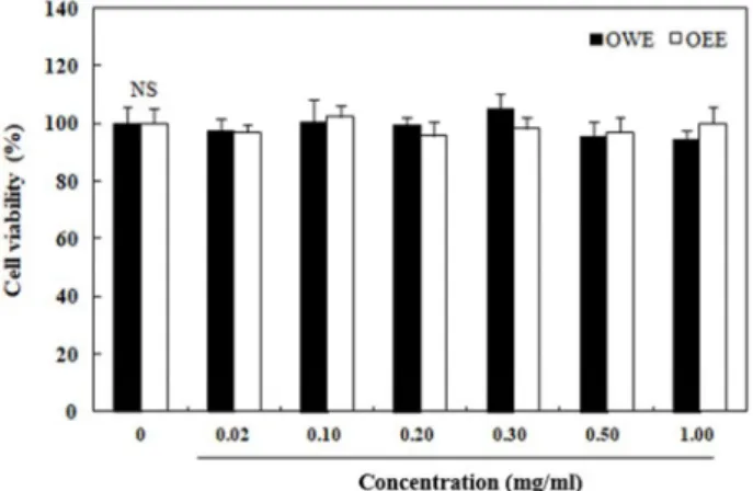
Fig. 1. Effect of O.Mishchenko extracts on cytotoxicity in 3T3-L1 cells. 3T3-L1 cells were treated with various concen- trations (0.02, 0.10, 0.20, 0.30, 0.50, and 1.00 mg/ml) of OEE and OWE for 20 hr, and cytotoxic effects were measured by the MTT assay. Each value is expressed as the mean ± SD of triplicate experiments. NS: not sig- nificant. OEE: O. Mishchenko 80% ethanol extract; OWE:
O. Mishchenko water extract; Acarbose: positive control.
were fed a pelleted feed (5L79; Orient, Inc., Seoul, Korea).
After an adaptation period, diabetes was induced as de- scribed below. All procedures involving in the handling and care of mice complied with the current international laws and policies (National Institutes of Health Guide for the Care and Use of Laboratory Animals), and were approved by the Animal Ethics Committee of the University (PNU-2019-2468).
Induction of experimental diabetes in a mouse model Diabetes was induced by an intraperitoneal injection of streptozotocin (STZ; 60 mg/kg, Sigma-Aldrich Chemical Co.) dissolved in citrate buffer (0.1 M, pH 4.5). The fasting blood glucose level was checked at 7 days post injection to confirm the induction of diabetes, using blood from the tail vein with a glucose meter (Roche Diagnostics GmbH, Mann- heim, Germany) and glucose strips. Mice with fasting blood glucose level above 250 mg/dl were regarded as being dia- betic.
Measurement of blood glucose levels
The mean blood glucose level in each group (normal mice and diabetic mice) was similar, and each group was divided into four sub-groups of seven mice. A total of 8 groups were used and the following were orally administered after over- night fasting: 1) control: soluble starch (2 g/kg of body weight [BW]), 2) OEE: soluble starch with OEE (300 mg/kg of BW), 3) OWE: soluble starch with OWE (300 mg/kg of BW), 4) acarbose: soluble starch with acarbose (100 mg/kg of BW). Blood samples were collected from the tail vein at 0, 15, 30, 60, and 120 min, and blood glucose was checked using the blood glucose meter. Areas under the concen- tration-time curves (AUCs) were identified using the tra- pezoidal rule.
Statistical analysis
Data are represented as the mean ± standard deviation (SD). Statistical analysis was conducted using SAS software ver. 9.1 (SAS Institute, Inc., Cary, NC, USA). The t-test was utilized to compare the control and sample groups. Dissimi- larity between groups was assessed by one-way analysis of variance, followed by Duncan’s post-hoc multiple range tests. A p-value < 0.05 was regarded as being significant.
Results Cytotoxic effect of O. Mishchenko
The viability of 3T3-L1 cells treated with various concen- trations of OEE and OWE, as described above, was eval- uated using the MTT assay. OEE and OWE did not affects cell viability till a concentration of 1.00 mg/ml, the highest used in this study (Fig. 1).
Inhibition of α-glucosidase by O. Mishchenko extracts in vitro
Alpha-glucosidase converts the carbohydrates degraded by α-amylase into glucose [8]. Inhibition of this enzyme is thought to prevent the rapid rise of blood glucose after meals by delaying glycolysis and absorption. Therefore, the inhibitory activity of OEE and OWE on α-glucosidase (EC 3.2.1.20) was measured. The inhibitory activity of OEE was found be dose-dependent in nature, i.e., 20.53, 33.47, 37.84, 46.87, and 57.42% at 0.02, 0.05, 0.10, 0.20, and 0.30 mg/ml, respectively (Fig. 2.), same was the case with OWE, and the levels of inhibition were 19.04, 28.63, 36.99, 42.11, and 51.52%
at concentrations of 0.02, 0.05, 0.10, 0.20, and 0.30 mg/ml, respectively. In particular, OEE showed significantly higher α-glucosidase inhibitory activity than OWE. IC
50values for OEE with respect to α-glucosidase activity were 0.229 mg/
ml, and the same was 0.283 mg/ml for OWE. Acarbose, a commercial hypoglycemic pharmaceutical product, inhibited enzyme activity by 52.45% at a concentration of 0.30 mg/ml.
Thus, at the same concentration (0.30 mg/ml), OEE showed
markedly higher inhibitory activities than acarbose.
Fig. 2. Effect of O. Mishchenko extracts on α-glucosidase inhibi- tory activities. Each value is expressed as the mean ± SD of triplicate experiments. Values with different su- perscript letters are significantly different (p<0.05) based on Duncan’s multiple range tests. The concentration of acarbose, used as a positive control, was 0.30 mg/ml.
OEE: O. Mishchenko 80% ethanol extract; OWE: O.
Mishchenko water extract; Acarbose: positive control.
Fig. 3. Effect of O. Mishchenko extracts on α-amylase inhibitory activities. Each value is expressed as the mean ± SD of triplicate experiments. Values with different superscript letters are significantly different (p<0.05) based on Duncan’s multiple range tests. The concentration of acar- bose, used as a positive control, was 0.30 mg/ml. OEE:
O. Mishchenko 80% ethanol extract; OWE: O. Mishchenko water extract; Acarbose: positive control.
Inhibition of α-amylase by O. Mishchenko extracts in vitro
The inhibitory activities of OEE and OWE against α-amy- lase are shown in Fig. 3. OEE inhibited α-amylase by 20.69, 35.37, 49.03, 63.42, and 65.98% at concentrations of 0.02, 0.05,
0.10, 0.20, and 0.30 mg/ml, respectively. The inhibitory effect of OWE against α-amylase was also concentration-depend- ent, and the levels of inhibition were 16.55, 22.99, 40.54, 45.37 and 53.11% at concentrations of 0.02, 0.05, 0.10, 0.20, and 0.30 mg/ml, respectively. OEE inhibited α-amylase more ef- fectively than OWE. The inhibitory effect of OEE against α- amylase was markedly higher than that of acarbose at the same concentration (0.30 mg/ml). The IC
50value of OEE against α-amylase was 0.106 mg/ml, and that of OWE and acarbose was 0.259 and 0.226 mg/ml, respectively, indicat- ing that OEE had significantly higher inhibitory activity than acarbose.
Effect of O. Mishchenko extract on blood glucose levels in vivo
OWE (300 mg/kg body weight, BW), OEE (300 mg/kg BW), and acarbose (100 mg/kg BW) were orally adminis- tered to mice, with soluble starch (2 g/kg BW), to confirm the inhibitory effect of the O. Mishchenko extracts on the lev- els of postprandial blood glucose. After administration of the extracts, blood was collected from the tail vein of normal mice and STZ-induced diabetic mice at 0, 15, 30, 60, and 120 min, and the change in postprandial blood glucose levels was measured. In normal mice, blood glucose levels in- creased to 242.33 mg/dl at 30 min administration of starch (Fig. 4A). Normal mice administered starch with OEE (155.00, 179.40, 188.80, and 127.40 mg/ml at 15, 30, 60, and 120 min, respectively) or OWE (172.00, 192.50, 190.83, and 139.67 mg/ml at 15, 30, 60, and 120 min, respectively) showed significantly decreased levels of blood glucose. In diabetic mice, the blood glucose level in the control group, administered with only soluble starch, increased to 406.50, 431.50, and 473.50 mg/ml after 15, 30, and 60 min, re- spectively dropping to 460.00 mg/ml after 120 min (Fig. 4B).
The results with OEE and soluble starch were 327.00, 361.00,
358.67, and 291.00 mg/ml after 15, 30, 60, and 120 min,
respectively. When OWE was administered with starch,
postprandial hyperglycemia also decreased (374.50, 381.00,
397.00, and 365.00 mg/ml after 15, 30, 60, and 120 min, re-
spectively), but this was not as pronounced as that observed
with OEE. Peak postprandial blood glucose was also sig-
nificantly lower in the normal (non-diabetic) group, follow-
ing starch and OEE administration, than in those adminis-
tered starch alone. The AUC of the diabetic group treated
with OEE (663.58±78.62 mg/ml) was significantly lower than
that of the diabetic control group (880.68±53.77 mg/ml)
A. normal mice B. diabetic mice
Fig. 4. Blood glucose levels after administration of O.Mishchenko extracts to streptozotocin-induced diabetic mice and normal mice.
Control (distilled water), OEE (300 mg/kg), OWE (300 mg/kg), or acarbose (100 mg/kg) were orally co-administered with starch (2 g/kg). Each value is expressed as the mean ± SD of seven mice per group. Values with different superscript letters are significantly different (p<0.05) based on Duncan’s multiple range tests. OEE: O. Mishchenko 80% ethanol extract; OWE:
O. Mishchenko water extract; Acarbose: positive control.
Table 2. Areas under the concentration–time curves (AUCs) of postprandial glucose responses in normal and streptozotocin-induced diabetic mice
Group1) AUC (mg ・ hr/dl)
Normal mice Diabetic mice Control
OEE OWE Acarbose
400.04±39.25a 323.42±43.87c 340.56±51.76b 287.81±56.39d
880.68±53.77a 663.58±78.62c 749.43±51.52b 602.25±53.11c
1)OEE (300 mg/kg), OWE (300 mg/kg), acarbose (100 mg/kg), and distilled water (control) were orally co-administered with starch (2 g/kg). Each value is expressed as the mean±SD of seven mice. Values with different superscript letters within a column are significantly different (p<0.05) based on Duncan’s multiple range tests. OEE: O. Mishchenko 80% ethanol extract;
OWE: O. Mishchenko water extract; Acarbose: positive control.
(Table 2). The acarbose (oral hypoglycemic agent used as a positive control) and OEE groups showed similar patterns for blood glucose levels and AUCs.
Discussion
Diabetes is a metabolic disease characterized by hypergly- cemia. Chronic hyperglycemia causes complications such as macrovascular and microvascular issues, diabetic neuro- pathy, and kidney disease [6, 20, 28]. The treatment of dia- betes is generally a combination of medication, diet and exercise. The goal of treatment is to maintain ideal blood
glucose, to prevent or delay diabetic complications [18, 35].
It is critical to maintain the postprandial and fasting blood glucose levels as close as possible to the normal levels [6].
It has been reported that postprandial hyperglycemia re- duces insulin sensitivity, impairs pancreatic function, and re- duces insulin secretion, thereby exacerbating the diabetic condition and causing macrovascular and microvascular complications [20]. In general, starch ingested via the diet is digested into small sugars by α-amylase, and then into glucose by α-glucosidase on the brush border of the mucosa in the small intestine, which leads to an increase in blood glucose after absorption. In this respect, inhibition of carbo- hydrate hydrolases can play an important role in controlling blood glucose and provide effective anti-diabetic control by targeting postprandial hyperglycemia [45].
Alpha-glucosidase is an enzyme that breaks down carbo- hydrates in the diet and converts them into glucose, and α-glucosidase inhibitors slow down the increase in post- prandial blood glucose by delaying carbohydrate digestion and absorption [15, 16]. Long-term use of α-glucosidase in- hibitors may cause side effects such as bloating, vomiting and diarrhea in some patients, which may limit their use.
Therefore, research is under way to search for hypoglycemic agents from among natural products that have few side ef- fects [23].
As natural products, edible insects have been used to treat
various diseases. Oxya chinensis sinuosa Mishchenko) has
long been used, orally, as a medicine, particularly in Korea.
In recent years, it has been recognized as a non-polluting, nutritional foodstuff, and its supply has increased annually.
O. Mishchenko contains abundant amounts of energy, pro-
tein, unsaturated fatty acids, trace nutrients—such as zinc, phosphorus, and calcium—and pigments, such as chlor- ophyll, carotenoids, and polyphenols [30]. It has been used in traditional anti-diabetic medicines [13]. Nevertheless, there is no experimental data demonstrating a relationship between the suppression of glucose absorption in the intes- tine, postprandial blood glucose levels and O. Mishchenko.
Thus, this study aimed to investigate whether supplementa- tion by O. Mishchenko inhibits α-glucosidase. We investigated the effect of OEE and OWE on the activity of α-glucosidase.
The results showed that both OEE and OWE inhibited α- glucosidase, in particular OEE showed higher inhibitory ac- tivity than acarbose, a commercial inhibitor of α-glucosidase.
O. Mishchenko has a higher content of unsaturated fatty
acids, chlorophyll, carotenoids, and polyphenols than other edible insects. In the case of freeze-dried samples, poly- phenols, such as flavonoids, terpenoids, and phenolic acids are present at 14.06-17.61 mg/100 g [30, 31]. Polyphenols are known to exhibit physiological activities, such as anti- oxidant, anti-obesity, anti-inflammatory, and anti-diabetic ef- fects, and play an important role in the inhibition of glyco- sidase enzymes [1, 22]. Phenolic acids are generally classified into two major groups, i.e., benzoic acids containing 7 carbon atoms and cinnamic acids containing 9 carbon atoms.
Cinnamic acid and its derivatives are a major group of mole- cules that are ubiquitously distributed in fruits, vegetables, and edible insects [3, 5, 11]; they have been investigated for their potential as inhibitors of carbohydrate hydrolases.
Studies have reported that the hydroxyl groups of cinnamic acid can play an important role in the inhibition of enzymes such as pancreatic α-amylase. Inhibition of α-amylase is largely due to the presence of hydroxyl groups at the para- and meta-positions of the cinnamic acid molecule [3, 4, 36].
Buszewska-Forajra [11] detected cinnamic acid derivatives as components of grasshoppers, using gas chromatog- raphy-tandem mass spectrometry (GC-MS/MS). Cinnamic acid and its derivatives are one of the most abundant groups of compounds found in grasshoppers, and may contribute, at least in part, to the inhibition of α-glucosidase and α-amy- lase in vitro. In addition, when extracting ethanol, it is ex- pected that the carbohydrate digestive enzyme inhibitory ac- tivity of the ethanol extract rich in polyphenol content would be higher than that of the water extract because polyphenols
are more polar than water [5, 26]. Some studies have shown that the anti-inflammatory, apoptosis-protective effects, and antioxidant activities of the ethanol extract of O. Mistshenk were more potent than those of the water extract, which was consistent with our results [38, 44].
In patients with type 2 diabetes, blood glucose increases rapidly after eating, and if higher levels of blood glucose persist, various diabetic complications occur [42]. There are two major factors that control diabetes, one is blood glucose regulation through insulin secretion, and the other is de- layed digestion and absorption of carbohydrates resulting in suppression of the rapid increase in blood glucose after eating [32]. In this study, the focus was on the latter, and it is an important factor in controlling diabetes, which regu- lates fasting blood glucose and postprandial blood glucose levels. In particular, it is more important to control blood glucose levels after eating than to control fasting blood glu- cose [7]. In this study, to verify the results of the in vitro test, we used an animal model in which hyperglycemia was induced with STZ to confirm the ability of the extracts to control postprandial blood glucose levels.
The hypoglycemic effect of OEE was greater than that of OWE, after starch loading in the animal model. OEE sig- nificantly reduced postprandial hyperglycemia when ad- ministered to diabetic mice. These results suggest that the OEE supplementation slows postprandial hyperglycemia by delaying the absorption of starch. Acarbose is an oral hyper- glycemic agent that lowers blood glucose levels after meals, and significantly reduces the AUC value [23]. OEE also sig- nificantly reduced the AUC value in our study, in addition to reducing the maximum blood glucose level. Our results indicate that OEE alleviates postprandial hyperglycemia by delaying the absorption of dietary carbohydrates, due to the inhibitory activity of OEE on carbohydrate digestive en- zymes.
Postprandial hyperglycemia is an independent contrib-
utor to diabetes complications as well as being a feature of
diabetes [19]. Various epidemiological studies have sug-
gested that postprandial hyperglycemia may be more closely
correlated with cardiovascular morbidity and mortality than
fasting hyperglycemia [10]. Pharmaceutical agents, espe-
cially acarbose, can reduce postprandial hyperglycemia by
alleviating blood glucose levels after eating. However, these
are usually associated with side effects, such as weight gain,
abdominal discomfort, and diarrhea [14, 21]. Our results in-
dicate that O. Mishchenko can improve postprandial hyper-
glycemia and help prevent the occurrence of diabetic complications.
Recently, there have been an increased number of studies investigating the anti-diabetic effects of edible insects. For example, an extract of Gryllus bimaculatus effectively in- hibited the occurrence of diabetes by protecting pancreatic islet function, and significantly reduced blood glucose in a diabetic animal model [41]. In another example, the increase in plasma membrane GLUT4 expression upon administra- tion of the mealworm extract has been known to promote the uptake of blood glucose into cells and relieved hyper- glycemia in diabetic C57BL/Ksj-db/db mice [29]. In another study, Bombycis corpus normalized the blood glucose and se- rum insulin levels in diabetic rats [24]. Silkworms also ex- hibit therapeutic potential for reducing the plasma glucose levels in db/db mice, and show inhibitory activity against α-glycosidase [39].
In summary, in our study we were able to demonstrate that O. Mishchenko extract strongly inhibits the activities of α-glucosidase and α-amylase. Comparison of the two ex- tracts shows that OEE has a stronger inhibitory effect than OWE in vitro. Administration of OEE or OWE with starch delayed the digestion of carbohydrates and absorption of glucose, resulting in alleviation of postprandial hyper- glycemia in vivo. OEE co-administration alleviated post- prandial blood glucose levels more than OWE alone in dia- betic mice. Thus, OEE seems to be a better candidate than OWE as a potential functional food to decrease postprandial hyperglycemia.
Acknowledgement
This research was supported by the Global Ph.D. Fellow- ship Program through the National Research Foundation of Korea (NRF-2019H1A2A1074826).
The Conflict of Interest Statement
The authors declare that they have no conflicts of interest with the contents of this article.
References
1. Abbas, M., Saeed, F., Anjum, F. M., Afzaal, M., Tufail, M.
T., Bashir, M. S., Ishtiaq, A., Hussain, S. and Suleria, H. A.
2016. Natural polyphenols: an overview. Int. J. Food Prop.
20, 1689-1699.
2. Abid, S., Lekchiri, A., Mekhfi, H., Ziyyat, A., Legssyer, A., Aziz, M. and Bnouham, M. 2014. Inhibition of α-glucosidase and glucose intestinal absorption by Thymelaea hirsuta frac- tions. J. Diabetes 6, 351-359.
3. Adisakwattana, S., Sookkongwaree, K., Roengsumran, S., Petsom, A., Ngamrojnavanich, N., Chavasiri, W., Deesamer, S. and Yibchok-anun, S. 2004. Structure-activity relation- ships of trans-cinnamic acid derivatives on alpha-glucosi- dase inhibition. Bioorg. Med. Chem. Lett. 14, 2893-2896.
4. Adisakwattana, S., Chantarasinlapin, P., Thammarat, H. and Yibchok-Anun, S. 2009. A series of cinnamic acid derivatives and their inhibitory activity on intestinal alpha-glucosidase.
J. Enzyme Inhib. Med. Chem. 24, 1194-1200.
5. Adisakwattana, S. 2017. Cinnamic acid and its derivatives:
mechanisms for prevention and management of diabetes and its complications. Nutrients 9, 163.
6. Asano, N., Tomioka, E., Kizu, H. and Matsui, K. 1994. Sugars with nitrogen in the ring isolated from the leaves of Morus bombycis. Carbohydr. Res. 253, 235-245.
7. Avignon, A., Radauceanu, A. and Monnier, L. 1997. Non- fasting plasma glucose is a better marker of diabetic control than fasting plasma glucose in type 2 diabetes. Diabetes Care 20, 1822-1826.
8. Baron, A. D. Postprandial hyperglycaemia and α-glucosi- dase inhibitors. 1998. Diabetes Res. Clin. Pract. 40, 51-55.
9. Basu, S., Yudkin, J. S., Kehlenbrink, S., Davies, J. I., Wild, S. H., Lipska, K. J., Sussman, J. B. and Beran, D. 2018.
Estimation of global insulin use for type 2 diabetes, 2018-30:
a microsimulation analysis. Lancet Diabetes Endocrinol. 7, 25- 33.
10. Bonora, E. and Muggeo, M. 2001. Postprandial blood glu- cose as a risk factor for cardiovascular disease in type Ⅱ diabetes; the epidemiological evidence. Diabetologia 44, 2107- 2114.
11. Buszewska-Forajta, M., Struck-Lewicka, W., Bujak, R., Siluk, D. and Kaliszan, R. 2014. Determination of water-soluble components of abdominal secretion of grasshopper (Chor- thippus spp.) by GC/MS/MS in search for potential wound healing agents. Chromatographia 77, 1091-1102.
12. Chinenye, S. and Young, E. E. 2012. Isolated postprandial hyperglycemia in type 2 diabetic patients in a Nigerian Tertiary Health Center. Indian J. Endocrinol. Metab. 16, 604- 608.
13. Chung, M. Y., Kwon, E. Y., Hwang, J. S., Goo, T. W. and Yun, E. Y. 2013. Pretreatment conditions on the powder of Tenbrio molitor for using as a novel food ingredient. J. Seric.
Entomol. Sci. 51, 9-14.
14. Clissold, S. P. and Edwards, C. 1988. Acarbose: a prelimi- nary review of its pharmacodynamic and pharmacokinetic properties, and therapeutic potential. Drugs 3, 214-243.
15. Forman, L. J., Estilow, S., Lewis, M. and Vasilenko, P. 1986, Streptozotocin diabetes alters immunoreactive beta-endor- phin levels and pain perception after 8 wk in female rats.
Diabetes 35, 1309-1313.
16. Fujita, H., Yamagami, T. and Ohshima, K. 2001. Efficacy and safety of Touchi extract, α-glucosidase inhibitor derived
from fermented soybeans, in non-insulin-dependent dia- betic mellitus. J. Nutr. Biochem. 12, 351-356.
17. Gerich, J. E. 2003. Clinical significance, pathogenesis, and management of postprandial hyperglycemia. Arch. Intern.
Med. 163, 1306-1316.
18. Goldmann, A., Milat, M. L. and Ducrot, P. H. 1990. Tropane derivatives from Calystegia sepium. Phytochemistry 29, 2125- 2128.
19. Grundy, S. M., Benjamin, I. J., Burke, G. L., Chait, A., Eckel, R. H. and Howard, B. V. 1999. Diabetes and cardiovascular disease: a statement for healthcare professionals from the American Heart Association. Circulation 100, 1134-1146.
20. Haller, H. 1998. The clinical importance of postprandial glucose. Diabetes Res. Clin. Pract. 40, 43-49.
21. Hanefeld, M. 1998. The role of acarbose in the treatment of non-insulin-dependent diabetes mellitus. J. Diabetes Complications 12, 228-237.
22. Hanhineva, K., Törrönen, R. and Bondia-Pons, I. 2010. Impact of dietary polyphenols on carbohydrate metabolism. Int. J.
Mol. Sci. 11, 1365-1402.
23. Inoue, I., Takahashi, K., Noji, S., Awata, T., Negishi, K. and Kataya-ma, S. 1997. Acarbose controls postprandial hyper- proinsulinemia in non-insulin-dependent diabetes mellitus.
Diabetes Res. Clin. Pract. 36, 143-151.
24. Jeong, B. M., Hyun, M. K., Sin, W. Y., Kim, M. R., Shin, H. C., Yoon, C. H. and Jeong, J. C. 2004. Effects of Bombycis corpus on streptozotocin-induced diabetic rats. J. Internal.
Kor. Med. 25, 288-297.
25. Kalita, D., Holm, D. G., LaBarbera, D. V., Petrash, J. M. and Jayanty, S. S. 2018. Inhibition of alpha-glucosidase, alpha- amylase, and aldose reductase by potato polyphenolic compounds. PLoS One 13, 0191025.
26. Kim, D. S., Choi, M. H. and Shin, H. J. 2018. Polyphenol contents and antioxidant activities of domestic bamboo leaves with different extraction solvents. J. Adv. Engin.
Technol. 11, 7-13.
27. Kim, H. J., Kang, S. J., Kim, S. G., Kim, J. E., Koo, H. Y., Park, J. H. and Choi, H. C. 2015. Antioxidant activity and antimicrobial activity of the grasshopper, Oxya chinensis sinuosa. J. Seric. Entomol. Sci. 53, 130-134.
28. Kim, J. S., Kwon, C. S. and Son, K. H. 2000. Alpha-glucosi- dase inhibitory activities of some wild vegetable extracts.
J. Food Sci. Nutr. 5, 174-176.
29. Kim, S. Y., Park, J. E. and Han, J. S. 2019. Tenebrio molitor (Mealworm) extract improves insulin sensitivity and alle- viates hyperglycemia in C57BL/Ksj-db/db mice. J. Life Sci.
5, 570-579.
30. Kim, T. S., Lee, J. H., Choi, B. D. and Ryu, H. S. 1987. Nutri- tional value of dried paddy grasshopper, Oxya chinensis formosana. J. Kor. Soc. Food Nutr. 16, 98-104.
31. Kim, Y. S. and Kwon, T. D. 2018. Effects of grasshopper diet and treadmill exercise performance of blood lipid pro- file and antioxidant enzyme in rats. Kor. J. Sports Sci. 27, 1605-1614.
32. Krentz, A. J. and Bailey, C. J. 2005. Oral antidiabetic agents:
current role in type 2 diabetes mellitus. Drugs 65, 385-411.
33. Laakso, M. 1999. Hyperglycemia and cardiovascular disease in type 2 diabetes. Diabetes 48, 937-942.
34. Li, Y., Wen, S., Kota, B. P., Peng, G., Li, G. Q., Yamahara, J. and Roufogalis, B. D. 2005. Punica granatum flower extract, a potent alpha-glucosidase inhibitor, improves postprandial hyperglycemia in Zucker diabetic fatty rats. J. Ethnopharma- col. 99, 239-244.
35. Madar, Z. 1984. Effect of brown rice and soybean dietary fiber on the control of glucose and lipid metabolism in rats.
Am. J. Clin. Nutr. 43, 388-396.
36. Narita, Y. and Inouye, K. 2009. Kinetic analysis and mecha- nism on the inhibition of chlorogenic acid and its compo- nents against porcine pancreas α-amylase isozymes I and II. J. Agric. Food Chem. 57, 9218-9225.
37. Paek, M. K., Hwang, J. M., Jung, K. S., Kim, T. W., Kim, M. C., Lee, Y. J., Cho, Y. B., Park, S. W., Lee, H. S., Ku, D. S., Jeong, J. C., Kim, K. G., Choi, D. S., Shin, E. H., Jwang, J. H., Lee, J. S., Kim, S. S. and Bea, Y. S. 2010. Checklist of Korean Insects. Nature and Ecology. Pub. Seoul, Korea.
36.
38. Park, J. Y, Heo, J. C., Woo, S. U., Yun, C. Y., Kang, S. W., Hwang, J. S. and Lee, S. H. 2006. Anti-inflammatory and cel- lular protective effects on hydrogen peroxide induced cytotox- icity of grasshopper extracts. Kor. J. Food Preserv. 13, 796-802.
39. Ryu, K. S., Lee, H. S., Kim, K. Y., Kim, M. J., Kang, P. D.
and Chun, S. N. 2012. Anti-diabetic effects of the silkworm (Bombyx mori) extracts in the db/db mice. Planta Med. 78, 458.
40. Salimifar, M., Fatehi-Hassanabad, Z. and Fatehi, M. 2013.
A review of natural products for controlling type 2 diabetes with an emphasis on their mechanisms of action. Curr.
Diabetes Rev. 9, 402-411.
41. Taek, C. H., Sang, S. K., Yeona, K., Han, C. M., Taewan, K., Hwan, L. S., Lee, D. H. and Ho, K. J. 2019. Anti-diabetic activity of edible insect Gryllus bimaculatus extracts in in- sulin-deficient diabetic mice. J. Kor. Soc. Food Sci. Nutr. 10, 1165-1171.
42. Tai, E., Lim, S. C., Tan, B. Y., Chew, S. K., Heng, D. and Tan, C. E. 2000. Screening for diabetes mellitus-a two-step approach in individuals with impaired fasting glucose im- proves detection of those at risk of complications. Diabet.
Med. 17, 771-775.
43. Watanabe, J., Kawabata, J., Kurihara, H. and Niki, R. 1997.
Isolation and identification of alpha-glucosidase inhibitors from tochucha (Eucommia ulmoides). Biosci. Biotechnol. Bio- chem. 61, 177-178.
44. Yoon, Y. I., Chung, M. Y., Hwang, J. S., Goo, T. W., Ahn, M. Y., Lee, Y. B., Han, M. S. and Yun, E. Y. 2014. Anti-in- flammatory effect of Oxya chinensis sinuosa ethanol extract in LPS-induced RAW 264.7 cells. J. Life Sci. 24, 370-376.
45. Young, I. R. and Stout, R. W. 1987. Effects of insulin and glucose on the cells of the arterial wall: Interaction of insulin with dibutyryl cyclic AMP and low density lipoprotein in arterial cells. Diabete Metab. 13, 301-306.
초록:당뇨 모델을 이용한 벼메뚜기( O. Mistshenk ) 추출물의 식후 고혈당 완화 효과
박재은․한지숙*
(부산대학교 식품영양학과)