Phellinus linteus Extract Regulates Macrophage Polarization in Human THP-1 Cells
Sang-Yull Lee
2†, Sul-Gi Park
1†, Sun-Nyoung Yu
1, Ji-Won Kim
1, You-Lim Hwang
1, Dong-Seob Kim
3and Soon-Cheol Ahn
1*
1Department of Microbiology & Immunology, Pusan National University School of Medicine, Yangsan 50612, Korea
2Department of Biochemistry, Pusan National University School of Medicine, Yangsan 50612, Korea
3Department of Food Science & Technology, College of National Resources & Life Science, Pusan National University, Milyang 50463, Korea
Received January 18, 2020 /Revised February 21, 2020 /Accepted February 21, 2020
Macrophages are initiators for regulating a host’s defenses to eliminate pathogens and trigger tissue repair. Macrophages are classified into two types: classically (M1) activated macrophages and alter- natively (M2) activated macrophages. M1-phenotype macrophages directly or indirectly kill infectious organisms and tumor cells via pro-inflammatory responses, whereas M2-phenotype macrophages re- model wounded tissue through anti-inflammatory responses. In this paper, we investigated how Phellinus linteus hot water extract passed from Diaion HP-20 resin (PLEP) regulates polarization of M1-like or M2-like macrophages in human THP-1 cells. PLEP did not have cytotoxicity at a high con- centration of 300 μg/ml. We observed morphological alteration of the THP-1 cells, which are stimu- lated by PLEP, LPS/INF-γ (M1 stimulators) or IL-4/IL13 (M2 stimulators). PLEP exposure induced morphology contiguous with LPS/INF-γ. qPCR was also performed to determine whether PLEP influ- ences M1 or M2 polarization-related genes. M1-phenotype macrophage–specific genes, such as TNF-α, IL-1β, IL-6, IL-8, CXCL10 and CCR7, were enhanced by PLEP in a dose-dependent manner similar to LPS/INF-γ. Conversely, M2-phenotype–specific genes, such as MRC-1, DC-SIGN, CCL17 and CCL22, were suppressed by PLEP. PLEP also significantly up-regulated secretory inflammation cytokines re- lated to M1 polarization of macrophages, including TNFα, IL-1β and IL-6, which was similar to the gene expression. Further, MAPK and NF-κB signaling were increased by treatment with PLEP, result- ing in enhancement of cytokine secretion. PLEP might therefore be used as a promising booster of pro-inflammatory responses through M1 polarization of human THP-1 cells.
Key words : Human THP-1 cells, MAPK signaling, NF-κB signaling, Phellinus linteus extract, polarization
of macrophage
†Authors contributed equally.
*Corresponding author
*Tel : +82-51-510-8092, Fax : +82-55-382-8090
*E-mail : [email protected]
This is an Open-Access article distributed under the terms of the Creative Commons Attribution Non-Commercial License (http://creativecommons.org/licenses/by-nc/3.0) which permits unrestricted non-commercial use, distribution, and reproduction in any medium, provided the original work is properly cited.
Journal of Life Science 2020 Vol. 30. No. 2. 113~121 DOI : https://doi.org/10.5352/JLS.2020.30.2.113
Introduction
Macrophages are leukocyte-derived cells which are well known for eliminating pathogens via phagocytosis. In the past, macrophages were categorized by means of organs, however, current classification has been changed from or- gan-specific macrophages to M1- and M2-polarized macro- phages [6]. M1-polarized (classically activated) macrophages are usually activated by lipopolysaccharides (LPS) and in- terferon-γ (INF-γ) and directly kill contagious organisms
and tumor cells against pathological status through secre- tion of cytokines and chemokines or indirectly attract other immune cells to remove them. M2-polarized (alternatively activated) macrophages are stimulated by interleukin-4 (IL-4) and interleukin-13 (IL-13) and repair injured-tissue and build extracellular matrix [13]. M1-polarized macro- phages are inclined to produce not only pro-inflammatory cytokines (TNF-α, IL-1β and IL-6), but also representing chemokine receptors (CCL21, CCL24, CCR7 and CXCL10) [26]. On the other hand, M2-polarized macrophages tend to restrain a secretion of pro-inflammatory cytokines and produce anti-inflammatory cytokines, such as IL-10, CCL17, CCL18 and CCL22 [24]. Also, they express various receptors, such as mannose receptor (MRC-1; CD206), dendritic cell- specific intercellular adhesion molecule-3-grabbing non-in- tegrin (DC-SIGN; CD209) and scavenger receptor (CD163) on their surface [15].
Mitogen-activated protein kinases (MAPKs) are mainly
related with the transduction of externally originated signals.
The kinases regulate cellular homeostasis which is involved in cell growth, differentiation and death [18]. Interestingly, the extracellular-signal regulated protein kinase (ERK), the c-Jun N-terminal protein kinase (JNK) and the p38 MAPK signaling pathways are activated in macrophages by lip- opolysaccharide (LPS), which is constituent of Gram-neg- ative bacterial outer membrane [23]. And also other modu- lator, INF-γ, activates MAPKs signaling pathway in a nu- merous cellular models including macrophages [22]. It has been demonstrated that the production of TNF-α and IL-1β is inhibited by suppression of phosphorylation of ERK in macrophages [21]. And expression of other chemokines and inducible NO synthase is substantially up-regulated by this kind of phosphorylation [22].
NF-κB is involved in several cellular responses in an im- mune cell such as regulation of survival, activation and differentiation. It is crucial mediator of pro-inflammatory re- sponses and regulates induction of cytokine and chemokine [12]. This family consists of five members, containing homo- and heterodimers of p50 (also known as NF-κB1), p52 (also known as NF-κB2), p65 (also known as RelA), RelB and c-Rel, and controls transcriptional activities of target genes [19]. Inhibitors-of-kappaB (IκB), which has four different iso- forms like IκBα, IκBβ, IκBε and IκBδ, disturbs the nucleus translocation and transcriptional activity of Rel/ NF- κB [3, 25]. Also, other researchers have been demonstrated that degradation of IκB is important factor to activate NF-κB [9].
Many therapeutic drugs have been developed from natu- ral sources mostly like plants. Medicinal plants, which are widely used in Asian countries, have various compounds as potential pharmaceutical agents and have been used as a resources to discover the novel drugs [2]. Also, Javed Iqbal et al. summarized the anti-cancer materials which are de- rived from plants [10]. Phellinus linteus is one of the popular medicinal products and consists of several bioactive comple- ments like polysaccharides, triterpenoids, polyphenols and furans [4]. Many researchers have reported that extract of P. linteus have pharmaceutical activities such as anti-oxida- tion, anti-microbe, anti-cancer, immunomodulatory effect, anti-diabetes and neuroprotection [4]. Most published pa- pers have consistently asserted that extracts from the fruiting body of P. linteus have anti-inflammatory activity [11, 14, 17]. Our previous research has shown that extract of P. lin- teus induced pro-inflammatory responses in murine RAW 264.7 cells [20]. Therefore, we further explored whether ex-
tract of P. linteus promotes inflammatory or anti-inflamma- tory responses and which signals are related to immune re- sponse in human THP-1 cells through their macrophage polarization.
Materials and Methods
Chemicals and Materials
Phellinus linteus hot water extract passed from Diaion HP-20 resin (PLEP) was used to research the polarization of human THP-1 cells. Procedure of extraction and purifica- tion was described in our previous paper [20]. Briefly, hot water extract of P. linteus was passed through a Diaion HP-20 resin, which binds the lipophilic elements, to remove hydrophobic elements and active anti-inflammatory compo- nents. Lipopolysaccharide (LPS) was purchased from Sig- ma-Aldrich Co. (St. Louis, MO, USA). To differentiate mono- cytic THP-1 cells to macrophagic THP-1 cells, phorbol 12- myristate 13-acetate (PMA, Cayman Chemical, Ann Arbor, MI, USA) was used. And other immune stimulators, INF-γ, IL-4 and IL-13, were purchased from Peprotech (Seoul, Korea). 3-(4,5-Dimethylthiazol-2-yl)-2,5-diphenyl tetrazolium bromide (MTT, Duchefa Biochemie, Haarlem, Netherland) was used to examine cell viability. Antibodies against p- ERK, p-JNK, p-p38, p-NF-κB and p-IκBα were purchased from Cell Signaling Technology (Beverly, MA, USA) and β- Actin from Santa Cruz Biotechnology (Dallas, TX, USA).
And goat-anti-rabbit or -mouse IgG secondary antibodies were purchased from Enzo Life Science (Farmingdale, NY, USA). Western blot was performed by using Amersham
TMECL
TMPrime western blotting detection reagents (GE Healthcare, Chicago, IL, USA). Total RNA was purified by RiboEx solution purchased from GeneAll
®(Seoul, Korea).
Cell line and culture
Human monocytic cell line THP-1 cells were cultured in
Roswell Park Memorial Institute (RPMI) 1640 media supple-
mented with 10% fetal bovine serum and 100 unit/ml of
penicillin and streptomycin. All of culturing stuff were pur-
chased from Welgene (Gyeongsan, Korea). Fig. 1 shows how
to stimulate the differentiation and polarization of THP-1
cells, modified referring to Marie Genin et al [7]. Briefly,
THP-1 cells were treated with PMA for 24 hr to differentiate
monocytes to macrophages. To polarize into M1-like pheno-
type, 10 ng/ml of LPS and 20 ng/ml INF-γ (LPS/INF-γ)
were treated for 24 hr. For polarization of M2, 20 ng/ml
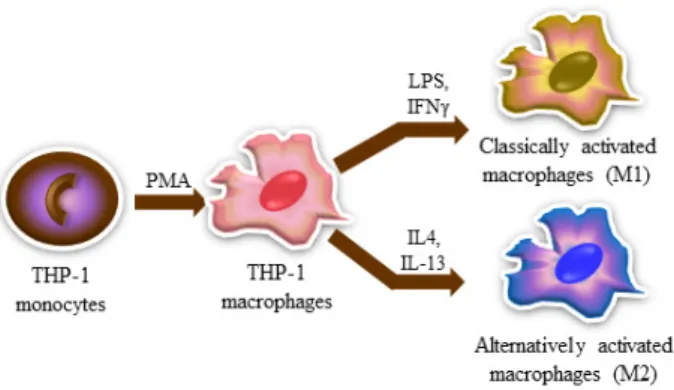
Fig. 1. Scheme of the process of differentiation and polarization on human THP-1 cells. To differentiate from monocytic THP-1 cells to M0-like macrophages, 50 nM of PMA was treated for 24 hr. To polarize from M0-like macrophages to M1-like macrophages, 10 ng/ml of LPS and 20 ng/ml of INF-γ were treated for 24 hr. And to polarize from M0-like macrophages to M2-like macrophages, 20 ng/ml of IL-4 and 20 ng/ml of IL-13 were treated for 24 hr.
of IL-4 and IL-13 (IL-4/IL-13) were added for 24 hr. To exam- ine whether PLEP influences polarization of THP-1 cells, various concentrations of PLEP were used (10, 30 and 100 μg/ml).
Analysis of cell viability
The cell viability was measured using MTT assay. Human monocytic THP-1 cells were seeded and treated with PMA for 24 hr. Then, PMA-derived (M0) THP-1 cells, were ex- posed by several concentrations of PLEP for 24 and 48 hr.
0.5 mg/ml MTT solution was added and incubated at 37℃
for 1.5 hr. The supernatant was discarded by suction and the precipitated formazan was dissolved in dimethyl sulf- oxide (DMSO, Junsei Chemical, Chuo-ku, Japan). The optical density was carried out at 570 nm using by SpectraMAX M2e (Molecular Devices, San Jose, CA, USA).
Analysis of quantitative real-time polymerase chain reaction (qPCR)
Total RNA was extracted with RiboEx solution, followed by modified TRIZOL method [5]. cDNA synthesis and qPCR were performed by using TOPscript™ RT DryMIX (Enzyno- mics, Daejeon, Korea) and TOPreal ™ qPCR 2X PreMIX (Enzynomics), respectively, according to manufacturer's in- structions. Primer sequences were as follows: TNFα-ss, 5’- CAGGCAGTCAGATCATCTTCTC-3’ and TNFα-as, 5’-ACT CGGCAAAGTCGAGATAGTC-3’; IL-1β-ss, 5’-CTCTCTCAC CTCTCCTACTCAC-3’ and IL-1β-as, 5’-ACACTGCTACTTC TTGCCCC-3’; IL-6-ss, 5’-AGTGAGGAACAAGCCAGA-GC-
3’ and IL-6-as, 5’-GTTGGGTCAGGGGTGGTTAT-3’; IL-8-ss, 5’-GCAGAGGGTTGTGGAGA-AGT-3’ and IL-8-as, 5’-CCCT ACAACAGACCCACACA-3’; CCR7-ss, 5’-TGGTGGTGGCT CTCCTTGTC-3’ and CCR7-as, 5’-TGTGGTGTTGTCTCCGA TGTAATC-3’; CXCL10-ss, 5’-GAAAGCAGTTAGCAAGGA AAGGTC-3’ and CXCL10-as, 5’-ATGTAGGGAAGTGATGG GAGAGG-3’; MRC-1-ss, 5’-CAGCGCTTGTGATCTTCATT- 3’ and MRC-1-as, 5’-TACCCCTGCTCCTGGTTTT-3’; DC- SIGN-ss, 5’-TCAAGCAGTATTGGAACAGAGGA-3’ and DC- SIGN-as, 5’-CAGGAGGCTGCGGACTTTTT-3’; CCL17-ss, 5’- CGGGACTACCTGGGACCTC-3’ and CCL17-as, 5’-CCTCA CTGTGGCTCTTCTTCG-3’; CCL22-ss, 5’-ATGGCTCGCCTA CAGACTGCACTC-3’ and CCL22-as, 5’-CACGGCAGCAGA CGCTGCTTCCA-3’; GAPDH-ss, 5’-GACAGGATGCAGAA GGAGAT-3’ and GAPDH-as, 5’-TTGCTGATCCACATCTGC TG-3’. First qPCR step was pre-incubation at 95℃ for 600 s and the amplification step was performed at 95℃ for 20 s, at 58℃ for 20 s and 72℃ for 20 s, followed by 40 cycles.
And last step of melt curve was processed at 95℃ for 10 s, at 65℃ for 30 s, at 97℃ for 1 s and finally a total of 45 cycles. All reactions were carried out in triplicate and nor- malization of mRNA expression was used to GAPDH genes.
Western blotting
THP-1 cells were seeded into 6-well plate at a density of 1×10
6cells/well. After 24 hr, monocytic THP-1 cells were incubated with PMA to differentiate into macrophages.
Before immune stimuli exposes, cells were washed twice with 1xPBS to remove PMA effects. Various stimuli were treated to M0-like macrophages for 24 hr; LPS/INF-γ, IL-4/
IL-13 and PLEP exposure. Cells were lysed in PRO-PREP solution (iNtRON Biotechnology, Seongnam, Korea) with phosphatase inhibitor cocktail (Sigma-Aldrich Co) following manufacturer's specifications. Bradford method was per- formed to quantify proteins from cell lysates. Proteins were separated using 10-15% sodium dodecyl sulfate-polyacryl- amide gel electrophoresis (SDS-PAGE) and transferred to polyvinylidene fluoride (PVDF) membrane (GE Healthcare).
The membranes were blocked with 5% skim milk in TBS-T buffer [150 mM NaCl, 20 mM Tris (pH 7.4), 0.1% Tween 20] at room temperature for 1 hr and probed with specific primary or secondary antibodies. Finally, ECL solution was used to detect specific proteins on the membranes.
Intensities of each bands were measured by using a fluo-
rescence scanner (Amersham Imager 680, GE Healthcare)
and analyzed with its software (GE Healthcare).
Fig. 2. Effect of PLEP on cell viability of THP-1 cells. Cells were treated with various concentrations of PLEP for 24 and 48 hr. After then, 0.5 mg/ml of MTT solution was added.
Insoluble crystalized-formazan was dissolved in DMSO.
The absorbance at 570 nm was determined.
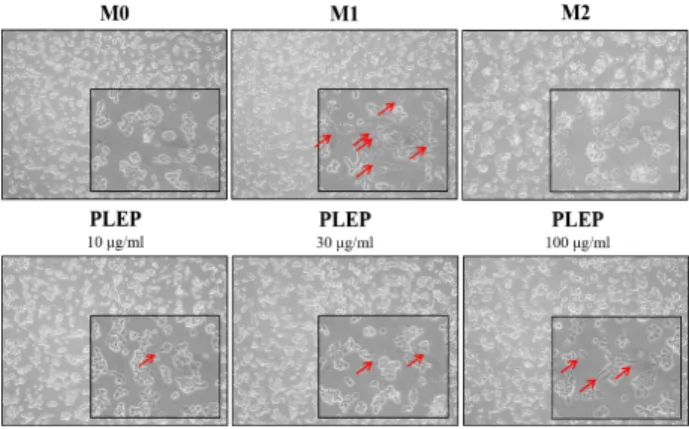
Fig. 3. Morphological changes of THP-1 cells after exposure of several immune stimuli. Morphological changes were observed via microscopy (100x and 400x). Cells were treated with 50 nM of PMA for 24 hr and then another 24 hr incubated with immunomodulators such as 10 μg/
ml of LPS and 20 ng/ml of INF-γ, IL-4 and IL-13 or PLEP. Various stimuli-induced morphological changes were observed in PMA-derived human THP-1 cells. Some morphological alterations were accentuated by red arrows.
M0, PMA-derived macrophages; M1, LPS/INFγ- stimu- lated macrophages; M2, IL-4/IL-13-stimulated macro- phages; PLEP, PLEP-stimulated macrophages.
Enzyme-linked immunosorbent assay (ELISA) Immuno-modulators were treated to THP-1 cells for vari- ous time points (3, 6, 12 and 24 hr). Supernatant from stimu- lated-THP-1 cells was collected to examine secretory cyto- kines related with pro-inflammation. ELISA was performed using human TNF-α, IL-1β and IL-6 ELISA kit (Invitrogen, Carlsbad, CA, USA) according to manufacturer's guidelines.
Statistical analysis
All experiments were repeated at least three times and the results were expressed as means ± SEM. Unless other- wise stated, the data are expressed as means ± SEM. Each experimental results were verified by one-way ANOVA, fol- lowed by Tukey-Kramer multiple comparison tests. Results were statistically significant at *p<0.05, **p<0.01 and ***p<
0.001.
Results
PLEP did not affect cell viability but induced mor- phological changes of THP-1 cells
At first, we carried out to confirm whether PLEP itself have a cytotoxic effect on macrophage-like (M0) THP-1 cells which are stimulated with PMA. Monocytic THP-1 cells were incubated with PMA for 24 hr to differentiate to macro- phage nature. Various concentration of PLEP from 10 to 300 μg/ml was treated to cells for 24 or 48 hr. As shown in Fig.
2, PLEP did not affect cell viability of THP-1 cells up to 300 μg/ml.
Morphological change indicates a sign of cellular alter-
ation procedure. Monocytic THP-1 cells basically presented a round shape and a suspended culture property, whereas M0 THP-1 cells had an attached culture property. When M0 THP-1 cells were stimulated with LPS/INF-γ (M1 stim- ulators) or IL-4/IL13 (M2 stimulators), they displayed char- acteristic flat, ramose and amoeboid shapes. PLEP-stimu- lated THP-1 cells showed some morphological changes, compared to M0 THP-1 cells. The flatted and branched shapes like typical M1 morphologies, were increased in PLEP-stimulated THP-1 cells as pointed by a red arrow (Fig.
3). Taken together, those results represented that PLEP had no effect on cytotoxicity, but induced morphological alter- ation from M0-like phenotype to M1-like phenotype in THP-1 cells.
PLEP influenced macrophage polarization of THP-1 cells
Previously we confirmed that PLEP promotes gene ex-
pressions, which are involved in pro-inflammation, of RAW
264.7 cells [20]. We further explored whether PLEP induces
pro-inflammatory or anti-inflammatory responses in macro-
phage-like THP-1 cells through their polarization. Macrop-
hages secrete inflammatory cytokines and chemokines to
regulate their host defense against external factors. Real-time
PCR was performed to quantify gene expressions related
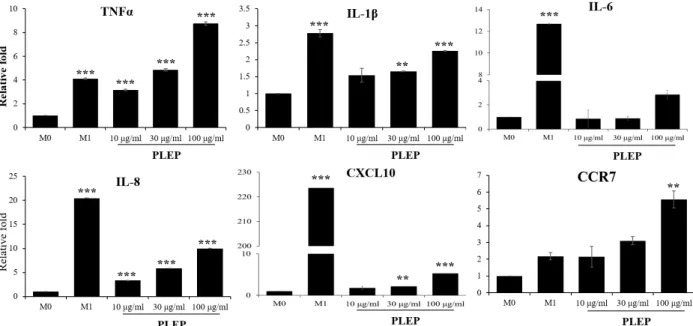
Fig. 4. Expression of M1 macrophages-related genes. After being stimulated with immune modulators (LPS/INF-γ or PLEP), cells were harvested and subjected to Trizol method to analyze M1 type inflammatory-related genes. The levels of mRNA ex- pression were performed using quantitative real-time PCR and normalized to GAPDH. Data are presented as mean ± SEM (n=3 in each group). **p<0.01, ***p<0.001 vs. M0 group. M0, PMA-derived macrophages; M1, LPS/INFγ-stimulated macrophages; PLEP, PLEP-stimulated macrophages.
with phenotypes of macrophages such as TNF-α, IL-1β, IL-6, IL-8, CXCL10 and CCR7 as biomarkers of M1-polarization and MRC-1 (CD206), DC-SIGN (CD209), CCL17 and CCL22 as those of M2-polarization. As shown in Fig. 4, PLEP specif- ically augmented M1-polarization indicating markers in a dose-dependent manner like as M1 type stimulators LPS/
INF-γ. Interestingly, PLEP at 100 μg/ml up-regulated those gene expressions, including TNF-α (8.5-fold, compared to control) and CCR7 (5.5-fold), which were higher than LPS/
INF-γ treatment (4.11- and 2.17-fold, respectively). LPS/INF- γ stimulated higher gene expression of IL-1β (2.77-fold), IL-6 (12.68-fold), IL-8 (20.46-fold) and CXCL10 (223.63-fold) than PLEP treatment (2.2-fold, 2.8-fold, 9.9-fold, 5.2-fold, respe- ctively). In case of M2-polarization, M2 type stimulators IL-4/IL-13 activated expression of their markers. However, PLEP significantly diminished their expression in a dose-de- pendent manner (Fig. 5). Our results suggested that PLEP enhances expression of M1 type specific genes, however re- duces expression of M2 specific genes in human THP-1 cells.
PLEP stimulated pro-inflammation status in THP-1 cells
TNF-α, IL-1β and IL-6 are well known for pro-in- flammation secretory cytokines by activating macrophages into M1 type. They are predominantly related with the en-
hancement of inflammatory responses. Therefore, we ana- lyzed protein level of these cytokines by ELISA assay. As shown in Fig. 6, PLEP significantly promoted secretory lev- els of TNF-α, IL-1β and IL-6 in a dose- and time-dependent manners. Those results were similar with the expression of those genes (Fig. 4). Also, the levels of cytokines were the highest at 12 hr treatment with PLEP but decreased at 24 hr.
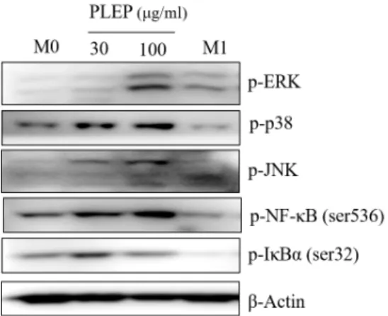
The MAPK signaling, which is modulated by p38, JNK and ERK, is the part of pathway to regulate inflammation process. This pathway stimulates a lot of pro-inflammatory cytokines and chemokines. Hence, we examined whether PLEP increases pro-inflammatory cytokines via activation of MAPKs signaling. PLEP substantially increased phosphor- ylation of JNK, ERK and p38 in a dose-dependent manner (Fig. 7), which means activation of MAPKs signaling to in- duce the expression of M1 polarized inflammatory response proteins. Also, the transcription factor NF-κB is a key media- tor of inflammatory responses, thereby inducing various in- flammation-related cytokines and chemokines. As shown in Fig. 7, phosphorylation of NF-κB was enhanced at 100 μg/ml of PLEP, whereas phosphorylation of IκBα was decreased.
All these results indicated that PLEP promotes expression
of M1-polarized immune responsive cytokines via activation
of MAPK and NF- κB signaling on THP-1 cells.
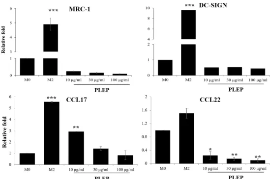
Fig. 5. Expression of M2 macrophages-related genes. After being stimulated with immune modulators (IL-4/IL-13 or PLEP), cells were harvested and implemented using Trizol method to analyze M2-like nature-related genes. The levels of mRNA expression were determined by quantitative real-time PCR and normalized to GAPDH. Data are presented as mean ± SEM (n=3 in each group). *p<0.05, **p<0.01, ***p<0.001 vs. M0 group. M0, PMA-derived macrophages; M2, IL-4/IL-13-stimulated macro- phages; PLEP, PLEP-stimulated macrophages.
Fig. 6. The level of inflammatory cytokines in M1-type macrophage THP-1 cells. After cells were exposed to immune modu- lators (LPS/INF-γ or PLEP), the supernatants werassay was performed to analyze pro-inflammatory cytokines. The lev- els of secretory protein expression were examined at various time points (3, 6, 12 and 24 hr). Data are presented as mean
± SEM (n=3 in each group). *p<0.05, **p<0.01, ***p<0.001 vs. M0 group. M0, PMA-derived macrophages; M1, LPS/
INFγ-stimulated macrophages; PLEP, PLEP-stimulated mac- rophages.
Discussion
Our previous research has demonstrated that Phellinus lin- teus has a paradoxical effect of immune responses in murine
RAW 264.7 cell line. Flow-through fraction of a Diaion HP-20
resin (PLEP) showed pro-inflammatory activity through
up-regulation of nitric oxide (NO) and inflammatory cyto-
kine, whereas fraction eluted from Diaion HP-20 resin
Fig. 7. Expression of MAPKs and NF-κB signaling proteins.
After being treated with immune modulators (LPS/
INF-γ or PLEP), cells were harvested and western blot- ting was performed to analyze pro-inflammatory-re- lated signaling proteins. The levels of protein expression were normalized to β-Actin. M0, PMA-derived macro- phages; M1, LPS/INFγ-stimulated macrophages; PLEP, PLEP-stimulated macrophages.
(PLEE) displayed anti-inflammatory activity, including sup- pression of NO production [20]. Thus, it was assumed that pro-inflammatory and anti-inflammatory agents must be separated from Diaion HP-20 resin. To further identify the role of PLEP in human THP-1 system, we carried out and characterized THP-1 cells after stimulating with several immunomodulators. Our finding proposed that PLEP has accelerated property of pro-inflammation unlike other pub- lished papers that extract of P. linteus has anti-inflammatory activity. As shown in Fig. 4, PLEP increased pro-inflamma- tory genes, such as TNF-α, CCR7, IL-β, IL-6, IL-8 and CXCL10, like as positive control LPS/INF-γ (M1 stim- ulators), similar with expressions in RAW 264.7 cells of our previous paper [20].
Furthermore, we carried out ELISA and western blot as- say to confirm the secretory protein level, which signaling pathway contributes under the PLEP treatment. We con- firmed up-regulation of secreted pro-inflammatory cyto- kines, such as TNF-α, IL-β and IL-6, from THP-1 cells ex- posed with M1 modulators or PLEP (Fig. 6). And inflamma- tion related-MAPK signaling was extremely increased by PLEP in a dose-dependent manner (Fig. 7). Once MAPKs are activated, they stimulate phosphorylation of various fac- tors, including ETS like-1 protein, c-Jun, activating tran- scription factor 2 and cAMP-response element binding pro- tein, and consequently control macrophage activity, such as cell growth, differentiation, polarization and inflammatory
responses through regulation of gene expression [16]. NF-κB is a major manipulator of the inflammatory responses to eliminate external pathogens and tumors. It is controlled by the IκB kinase, which facilitates the phosphorylation of IκB, causing degradation mediated by proteasome [1]. When IκB is degraded, the NF-κB is unconstrained to move into the nucleus, after then transcribing the pro-inflammatory genes [8]. Biologically active NF-κB was promoted by PLEP, whereas phosphorylation of IκBα was diminished (Fig. 6).
To identify whether PLEP affect an anti-inflammatory re- sponse, expression of M2 markers were observed using qPCR. Surface markers of M2 polarized macrophage (MRC-1 and DC-SIGN) and anti-inflammatory cytokines (CCL17 and CCL22) were suppressed by PLEP in a dose-dependent man- ner (Fig. 5).
In conclusion, PLEP has potential activity of enhancement of pro-inflammation via strengthening M1 polarization and suppressing M2 polarization on THP-1 cells.
Acknowledgment
This work was supported by a 2-year Research Grant of Pusan National University.
The Conflict of Interest Statement
The authors declare that they have no conflicts of interest with the contents of this article.
References
1. Arenzana-Seisdedos, F., Turpin, P., Rodriguez, M., Thomas, D., Hay, R. T., Virelizier, J. L. and Dargemont, C. 1997.
Nuclear localization of I kappa B alpha promotes active transport of NF-kappa B from the nucleus to the cytoplasm.
J. Cell. Sci. 110, 369-378.
2. Atanasov, A. G., Waltenberger, B., Pferschy-Wenzig, E. M., Linder, T., Wawrosch, C., Uhrin, P., Temml, V., Wang, L., Schwaiger, S., Heiss, E. H., Rollinger, J. M., Schuster, D., Breuss, J. M., Bochkov, V., Mihovilovic, M. D., Kopp, B., Bauer, R., Dirsch, V. M. and Stuppner, H. 2015. Discovery and resupply of pharmacologically active plant-derived nat- ural products: A review. Biotechnol. Adv. 33, 1582-1614.
3. Basak, S., Kim, H., Kearns, J. D., Tergaonkar, V., O'Dea, E., Werner, S. L., Benedict, C. A., Ware, C. F., Ghosh, G., Verma, I. M. and Hoffmann, A. 2007. A fourth IkappaB protein within the NF-kappaB signaling module. Cell 128, 369-381.
4. Chen, W., Tan, H., Liu, Q., Zheng, X., Zhang, H., Liu, Y.
and Xu, L. 2019. A Review: The bioactivities and pharmaco-
logical applications of Phellinus linteus. Molecules 24, pii:
E1888.
5. Chomczynski, P. and Mackey, K. 1995. Short technical reports. Modification of the TRI reagent procedure for iso- lation of RNA from polysaccharide- and proteoglycan-rich sources. Biotechniques 19, 942-945.
6. Das, A., Sinha, M., Datta, S., Abas, M., Chaffee, S., Sen, C.
K. and Roy, S. 2015. Monocyte and macrophage plasticity in tissue repair and regeneration. Am. J. Pathol. 185, 2596- 2606.
7. Genin, M., Clement, F., Fattaccioli, A., Raes, M. and Michiels, C. 2015. M1 and M2 macrophages derived from THP-1 cells differentially modulate the response of cancer cells to etoposide. BMC Cancer 15, 577.
8. Gilmore, T. D. 2006. Introduction to NF-kappaB: players, pathways, perspectives. Oncogene 25, 6680-6684.
9. Henkel, T., Machleidt, T., Alkalay, I., Kronke, M., Ben- Neriah, Y. and Baeuerle, P. A. 1993. Rapid proteolysis of I kappa B-alpha is necessary for activation of transcription factor NF-kappa B. Nature 365, 182-185.
10. Ijaz, S., Akhtar, N., Khan, M. S., Hameed, A., Irfan, M., Arshad, M. A., Ali, S. and Asrar, M. 2018. Plant derived anticancer agents: A green approach towards skin cancers.
Biomed. Pharmacother. 103, 1643-1651.
11. Kim, B. C., Jeon, W. K., Hong, H. Y., Jeon, K. B., Hahn, J. H., Kim, Y. M., Numazawa, S., Yosida, T., Park, E. H.
and Lim, C. J. 2007. The anti-inflammatory activity of Phellinus linteus (Berk. & M.A. Curt.) is mediated through the PKCdelta/Nrf2/ARE signaling to up-regulation of heme oxygenase-1. J. Ethnopharmacol. 113, 240-247.
12. Lawrence, T. 2009. The nuclear factor NF-kappaB pathway in inflammation. Cold Spring Harb. Perspect. Biol. 1, a001651.
13. Ley, K. 2017. M1 means kill; M2 means heal. J. Immunol.
199, 2191-2193.
14. Lin, C. J., Lien, H. M., Chang, H. Y., Huang, C. L., Liu, J. J., Chang, Y. C., Chen, C. C. and Lai, C. H. 2014. Biological evaluation of Phellinus linteus-fermented broths as anti-in- flammatory agents. J. Biosci. Bioeng. 118, 88-93.
15. Martinez, F. O., Helming, L. and Gordon, S. 2009. Alternative activation of macrophages: an immunologic functional per- spective. Annu. Rev. Immunol. 27, 451-483.
16. Neamatallah, T. 2019. Mitogen-activated protein kinase
pathway: A critical regulator in tumor-associated macro- phage polarization. J. Microsc. Ultrastruct. 7, 53-56.
17. Park, H. J. 2017. Anti-allergic and anti-inflammatory activity of Phellinus linteus grown on Panax ginseng. Food Sci.
Biotechnol. 26, 467-472.
18. Seger, R. and Krebs, E. G. 1995. The MAPK signaling cascade. FASEB J. 9, 726-735.
19. Sun, S. C., Chang, J. H. and Jin, J. 2013. Regulation of nuclear factor-kappaB in autoimmunity. Trends Immunol. 34, 282- 289.
20. Kang, T. W., An, H. H., Park, S. G., Yu, S. N., Hwang, Y. L., Kim, J. W. and Ahn, S. C. 2019. Conditioned media of RAW 264.7 cells stimulated with Phellinus linteus extract regulates the epithelial-mesenchymal transition in prostate cancer cells. J. Life Sci. 29, 904-915.
21. Valledor, A. F., Comalada, M., Xaus, J. and Celada, A. 2000.
The differential time-course of extracellular-regulated kin- ase activity correlates with the macrophage response to- ward proliferation or activation. J. Biol. Chem. 275, 7403- 7409.
22. Valledor, A. F., Sánchez-Tilló, E., Arpa, L., Park, J. M., Caelles, C., Lloberas, J. and Celada, A. 2008. Selective roles of MAPKs during the macrophage response to IFN-γ. J.
Immunol. 180, 4523-4529.
23. Valledor, A. F., Xaus, J., Comalada, M., Soler, C. and Celada, A. 2000. Protein kinase Cε is required for the Induction of mitogen-activated protein kinase phosphatase-1 in lip- opolysaccharide-stimulated macrophages. J. Immunol. 164, 29-37.
24. Van Ginderachter, J. A., Movahedi, K., Hassanzadeh Ghas- sabeh, G., Meerschaut, S., Beschin, A., Raes, G. and De Baetselier, P. 2006. Classical and alternative activation of mononuclear phagocytes: picking the best of both worlds for tumor promotion. Immunobiology 211, 487-501.
25. Verma, I. M., Stevenson, J. K., Schwarz, E. M., Van Antwerp, D. and Miyamoto, S. 1995. Rel/NF-kappa B/I kappa B fam- ily: intimate tales of association and dissociation. Genes Dev.
9, 2723-2735.
26. Xuan, W., Qu, Q., Zheng, B., Xiong, S. and Fan, G. H. 2015.
The chemotaxis of M1 and M2 macrophages is regulated by different chemokines. J. Leukoc. Biol. Suppl. 97, 61-69.
초록:상황버섯 추출물의 인간 유래 THP-1 단핵구 세포주의 분극화 조절
이상률
2†․박슬기
1†․유선녕
1․김지원
1․황유림
1․김동섭
3․안순철
1*
(1부산대학교 의학전문대학원 미생물학 및 면역학 교실, 2부산대학교 의학전문대학원 생화학 교실, 3부산대학교
생명자원과학대학 식품공학과)
대식세포는 특성에 따라 크게 classically activated macrophages (M1-phenotype macrophages)와 alternatively acti- vated macrophages (M2-phenotype macrophages) 두 가지의 형태로 나눌 수 있다. M1 대식세포의 경우 직∙간접적으 로 병원체, 감염된 조직 및 암세포 등을 제거하는 능력을 가진 반면, M2 대식세포의 경우 항염증 반응을 동반한 손상된 세포 조직의 복구 및 세포외기질의 생성에 관여하고 있다. 본 연구에서는 상황버섯 열수추출물을 합성 흡착제인 Diaion HP-20에 통과시켜 소수성 물질을 제거한 시료(PLEP)을 이용하여 인간 유래 THP-1 단핵구 세포주의 염증성 혹은 항염 증성 분극화 특성을 알아보았다. 먼저 PLEP 자체의 단핵구 세포에 대한 세포독성을 확인한 결과, 고농도의 300 μg/ml 에서 세포독성이 확인되지 않았다. 한편 세포의 형태학적 변화를 확인한 결과, PLEP의 농도가 증가함에 따라 M1- phe- notype 대식세포와 유사한 flatted and branched 형태가 증가하였다. 대식세포로 분화시킨 THP-1 세포주에 PLEP를 처 리한 후, M1 대식세포 분극화 관련 유전자인 TNF
![director [deg.]](data:image/gif;base64,R0lGODlhAQABAIAAAP///wAAACH5BAEAAAAALAAAAAABAAEAAAICRAEAOw==)