https://doi.org/10.1007/s43188-019-00025-1 ORIGINAL ARTICLE
Arylquin 1, a potent Par‑4 secretagogue, induces lysosomal membrane permeabilization‑mediated non‑apoptotic cell death in cancer cells
Kyoung‑jin Min1 · Sk Abrar Shahriyar1 · Taeg Kyu Kwon1
Received: 19 July 2019 / Revised: 16 August 2019 / Accepted: 28 August 2019 / Published online: 21 November 2019
© Korean Society of Toxicology 2019
Abstract
Arylquin 1, a small-molecule prostate-apoptosis-response-4 (Par-4) secretagogue, targets vimentin to induce Par-4 secre- tion. Secreted Par-4 binds to its receptor, 78-kDa glucose-regulated protein (GRP78), on the cancer cell surface and induces apoptosis. In the present study, we investigated the molecular mechanisms of arylquin 1 in cancer cell death. Arylquin 1 induces morphological changes (cell body shrinkage and cell detachment) and decreases cell viability in various cancer cells.
Arylquin 1-induced cell death is not inhibited by apoptosis inhibitors (z-VAD-fmk, a pan-caspase inhibitor), necroptosis inhibitors (necrostatin-1), and paraptosis inhibitors. Furthermore, arylquin 1 significantly induces reactive oxygen species levels, but antioxidants [N-acetyl-l-cysteine and glutathione ethyl ester] do not inhibit arylquin 1-induced cell death. Further- more, Par-4 knock-down by small interfering RNA confers no effect on cytotoxicity in arylquin 1-treated cells. Interestingly, arylquin 1 induces lysosomal membrane permeabilization (LMP), and cathepsin inhibitors and overexpression of 70-kDa heat shock protein (HSP70) markedly prevent arylquin 1-induced cell death. Therefore, our results suggest that arylquin 1 induces non-apoptotic cell death in cancer cells through the induction of LMP.
Keywords Arylquin 1 · Non-apoptotic cell death · Lysosomal membrane permeabilization · Prostate‐apoptosis‐response‐4 · Cell death
Introduction
Arylquin 1 is identified as a prostate‐apoptosis‐response‐4 (Par-4) secretagogue in normal cells. Arylquin 1 binds vimentin and releases vimentin-bound Par-4 for secretion [1, 2]. Par-4 was first identified by differential screening for genes up-regulated after the induction of programmed cell death in prostate cancer cells [3]. Most studies of Par-4 focus on the apoptotic effect mediated by intracellular Par-4. The apoptotic effects of Par-4 are involved in the activation of the Fas death receptor signaling pathway and inhibition of cellular pro-survival mechanisms [4]. Recently, Rangnekar et al. reported that Par-4 protein is secreted by normal cells, and extracellular Par-4 induces cancer cell-specific apop- tosis via interaction with the cell-surface receptor 78-kDa glucose-regulated protein (GRP78) [5].
Lysosomal membrane permeabilization (LMP) is defined as damage to the lysosomal membrane that causes the release of lysosomal contents into the cytosol and increase in cytosolic acidity [6]. Massive LMP induces necrotic cell death, while partial and selective LMP lead to apoptotic cell death [7]. Several papers have reported that tumor cell lysosomes are more fragile than normal lysosomes and are more susceptible to LMP [8]. Therefore, lysosomotropic agents induce LMP and result in lysosomal-dependent cell death, which may exert useful antitumor effects in apoptosis- resistant cells.
In this study, we investigated whether arylquin 1 induces cell death and identified the molecular mechanism of arylquin 1-induced cell death in human renal carcinoma Caki cells.
pISSN 1976-8257
* Taeg Kyu Kwon [email protected]
1 Department of Immunology, School of Medicine, Keimyung University, 1095 Dalgubeoldaero, Dalseo-Gu, Daegu 42601, South Korea
Materials and methods
Cell cultures and materialsAmerican Type Culture Collection (Manassas, VA, USA) supplied all human cancer cells (renal carcinoma: Caki and ACHN, hepatocellular carcinoma: SK-Hep1, glioma:
U87MG, breast carcinoma: MDA-MB-231) and normal mouse kidney cells (TCMK-1). Normal human kidney mesangial cells (MCs) were purchased from Lonza (Basel, Switzerland). Cells were grown in Dulbecco’s Modified Eagle Medium supplemented with 10% fetal bovine serum and 100 μg/mL gentamycin. R&D system (Minneapolis, MN, USA) supplied z-VAD-fmk, and Calbiochem (San Diego, CA, USA) supplied necrostatin-1 and N-acetyl- cysteine (NAC). Enzo Life Sciences (Plymouth Meeting, PA, USA) supplied pepstatin A, PD98059, and SP600125, and Cayman Chemical (Ann Arbor, MI, USA) supplied E64D. Santa Cruz Biotechnology supplied anti-apoptosis- inducing factor (AIF) (1:700, sc-5586), anti-cathepsin D (1:1000, sc-6486), anti-HSP70 (1:1000, sc-24), anti-Par-4 (1:1000, sc-1807), and anti-Lamp1 (1:700, sc-5570) anti- bodies (Dallas, TX, USA). Sigma Chemical Co. supplied anti-actin (1:10,000, A5441) antibody, glutathione ethyl ester (GEE), and arylquin 1 (St. Louis, MO, USA).
Cell viability assay
We performed an XTT assay to measure cell viability (WelCount™ Cell Viability Assay Kit, WelGENE, Daegu, Korea). After arylquin 1 treatment, 20 μL of XTT solution (1 mg/mL), containing phenazine methosulfate, was added to each well for 3 h and measured using a microtiter plate reader (Tecan Sunrise, Research Triangle Park, NC) at 450 nm.
Small interfering RNA
Santa Cruz Biotechnology supplied small interfering RNA (siRNA) (Par-4 and AIF) (Dallas, TX, USA), and Bioneer supplied GFP (control) siRNA (Daejeon, Korea). We used Lipofectamine® RNAiMAX Reagent (Invitrogen, Carlsbad, CA, USA) to transfect siRNA oligonucleotides.
Western blot analysis
Whole cell lysates were obtained as described previously using modified radioimmunoprecipitation assay lysis buffer [9]. Whole cell lysates were centrifuged at 13,000×g for 15 min at 4 °C, and the supernatant was collected into new tubes. Proteins were separated using sodium dodecyl sulfate
polyacrylamide gel electrophoresis and transferred to an Immobilon-P membrane. After blocking using 5% skimmed milk in tris-buffered saline with Tween 20, specific proteins were detected using enhanced chemiluminescence.
Measurement of reactive oxygen species
We used 2′,7′-dichlorodihydrofluorescein diacetate (H2DCFDA) to detect intracellular reactive oxygen spe- cies (ROS) [10]. After treatment, cells were stained with H2DCFDA for 10 min, and then washed with phosphate- buffered saline (PBS) twice. Fluorescence of cells in PBS was measured using a flow cytometer (BD Biosciences, San Jose, CA, USA).
Measurement of LMP
To monitor lysosomal destabilization, we used LysoTraker Red. Caki cells were treated with arylquin 1 for the indicated time periods; the cells were then incubated with 2.5 μM of LysoTracker Red (Molecular Probes Inc., Eugene, OR, USA) for 5 min at 37 °C. The cells were then trypsinized and resuspended in PBS, and fluorescence was measured at spe- cific time intervals using a flow cytometer (BD Biosciences, San Diego, CA, USA).
Fractionation of cytosol and membrane extracts Cells were washed with ice-cold PBS, resuspended in cyto- sol extraction buffer (250 mM sucrose, 10 mM KCl, 1.5 mM MgCl2, 1 mM EDTA, 1 mM EGTA, 20 mM HEPES) con- taining 250 μg/mL digitonin, and left on ice for 10 min;
lysate was then centrifuged at 13,000×g for 90 s. The super- natant (cytosol) was transferred to a new tube, and pellets (membrane fraction) were suspended with lysis buffer.
Lysates were centrifuged at 13,000×g at 4 °C for 15 min to obtain the supernatant fractions that were collected as the membrane extract.
Stable transfection in Caki cells
pEGFP-HSP70 was a gift from Lois Greene (Addgene plas- mid # 15215) [11]. The Caki cells were transfected in a sta- ble manner with the pEGFP-HSP70 using Lipofectamine™
2000 as prescribed by the manufacturer (Invitrogen, Carls- bad, CA, USA). After 48 h of incubation, transfected cells were selected in primary cell culture medium containing 700 μg/mL G418 (Invitrogen, Carlsbad, CA, USA). After 2 or 3 weeks, single independent clones were randomly iso- lated, and each individual clone was plated separately. After clonal expansion, cells from each independent clone were tested for HSP70 expression by immunoblotting.
Statistical analysis
The data were analyzed using one-way analysis of variance and post hoc comparisons (Student–Newman–Keuls) using the Statistical Package for Social Sciences 22.0 software (SPSS Inc., Chicago, IL, USA). The p values < 0.05 were considered significant.
Results
Effect of arylquin 1 on cell death in various cancer cells
Arylquin 1 was identified as a potent Par-4 secretagogue.
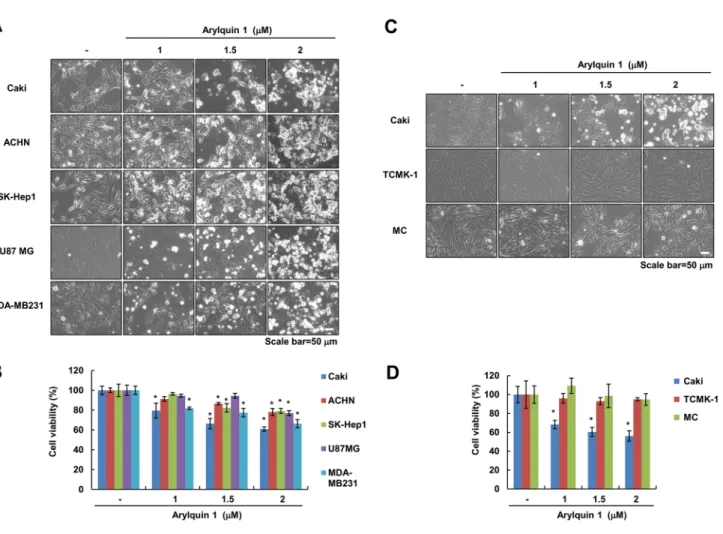
We examined whether arylquin 1 induces cell death in mul- tiple types of cancer cells. Arylquin 1 induced cell body shrinkage and cell detachment (Fig. 1a) and decreased cell viability in a dose-dependent manner (Fig. 1b). However, arylquin 1 had no effect on cell viability in normal cells
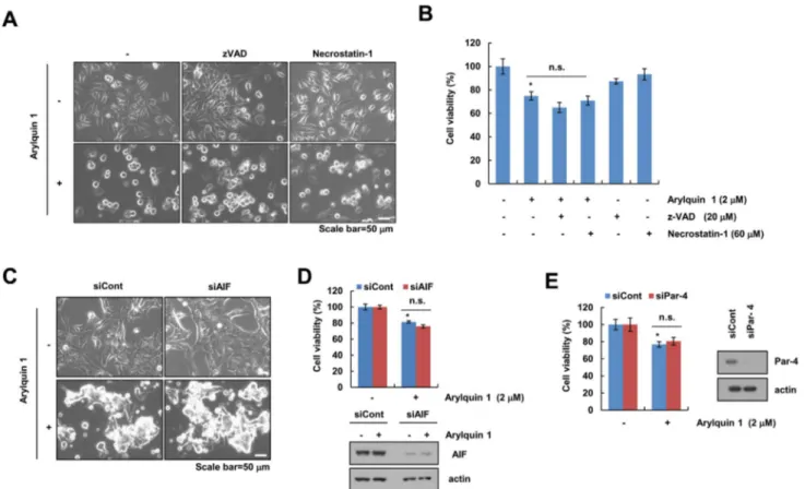
[normal mouse kidney cells (TCMK-1) and normal human kidney MCs] (Fig. 1c, d). We chose the 2 μM arylquin 1, which causes cell death of 25–35% to identify the cell death mechanisms. Next, to investigate whether arylquin 1-induced cell death is involved in apoptosis or necrop- tosis, we used z-VAD-fmk (pan-caspase inhibitor) and necrostation-1 (a selective inhibitor of necroptosis). Both inhibitors did not affect arylquin 1-induced morphologi- cal changes (cell body shrinkage and cell detachment) and reduction in cell viability (Fig. 2a, b). Pan-caspase inhibitor, z-VAD-fmk, did not block caspase-independent apoptosis. AIF is a critical regulator of caspase-independ- ent apoptosis [12, 13]. Knock-down of AIF expression by siRNA did not confer morphological changes and cyto- toxicity in arylquin 1-treated cells (Fig. 2c, d). Arylquin 1 binds vimentin, displaces Par-4 from vimentin for secre- tion, and triggers apoptosis of diverse cancer cells, but not normal cells [1]. Interestingly, we found that knock-down of Par-4 expression using siRNA had no effect on cyto- toxicity in arylquin 1-treated cells (Fig. 2e). Therefore,
Fig. 1 Arylquin 1 induces cell death in various cancer cells. a–d Cells were treated with the indicated concentrations of arylquin 1 for 24 h. Cell morphology was examined using interference light micros- copy (a, c). Cell viability was determined using the XTT assay (b, d).
The values in b, d represent the mean ± SEM from three independent samples. *p < 0.05 compared to the control. SEM standard error of the mean
these results indicate that arylquin 1 induces caspase- and Par-4-independent non-apoptotic cell death.
Influence of ROS signaling and paraptosis on arylquin 1‑induced cell death
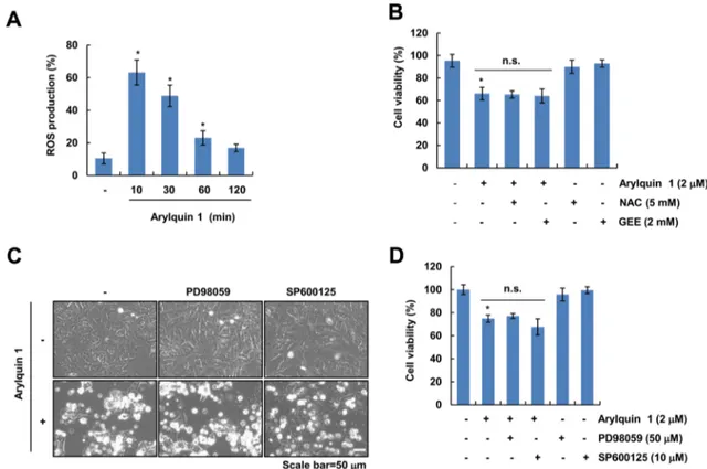
We investigated whether ROS signaling is involved in arylquin 1-induced cell death. Arylquin 1 transiently induced ROS generation and then gradually decreased until 2 h in Caki cells (Fig. 3a). Pretreatment with ROS scavengers (NAC and GEE) did not block arylquin 1-induced morpho- logical changes and cytotoxicity (Fig. 3b). Therefore, these results indicate that ROS signaling is not associated with arylquin 1-induced cell death. Paraptosis is a non-apoptotic cell death mode that is characterized by dilation of the endoplasmic reticulum and/or mitochondria [14]; protein synthesis and MAP kinases, including ERK and JNK, are associated with paraptosis [14, 15]. Pretreatment with pro- tein MAP kinase inhibitors [MEK inhibitor (PD98059) and JNK inhibitor (SP600125)] did not affect arylquin 1-induced
morphological changes and cytotoxicity (Fig. 3c, d). Taken together, these results indicate that paraptosis may not play a role in arylquin 1-induced cell death.
Effect of arylquin 1 on LMP
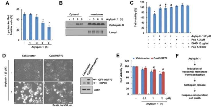
LMP can trigger lysosomal cell death such as non-pro- grammed necrosis, lysosomal apoptosis, or cell death with apoptosis-like features [16]. First, we examined whether arylquin 1 induces LMP. Arylquin 1 markedly induced loss of lysosomal membrane integrity and released cathepsin D into the cytosol (Fig. 4a, b). In addition, inhibitors of cath- epsins (pepstatin A and E64D) markedly inhibited arylquin 1-induced cytotoxicity (Fig. 4c), suggesting that the cell death effects of arylquin 1 depend on the cytosolic release of lysosomal cathepsins. Nylandsted et al. reported that HSP70 could inhibit LMP [17]. Overexpression of HSP70 inhibited the induction of morphological changes (cell body shrinkage and cell detachment) and reduction in cell viability (Fig. 4d,
Fig. 2 Arylquin 1-induced cell death is independent of caspase, AIF, and Par-4. a, b Caki cells were pretreated with 20 μM z-VAD and 60 μM necrostatin-2 for 30 min, and then treated with 2 μM arylquin 1 for 24 h. c, d Caki cells were transiently transfected with control siRNA (siCont) and AIF siRNA (siAIF). After 24 h, Caki cells were treated with 2 μM arylquin 1 for 24 h. e Caki cells were transiently transfected with control siRNA (siCont) and Par-4 siRNA (siPar-4).
After 24 h, Caki cells were treated with 2 μM arylquin 1 for 24 h.
The cell morphology was examined using interference light micros- copy (a, c). Cell viability was determined using the XTT assay (b, d, e). The protein expression levels of AIF, Par-4, and actin were determined by western blotting. The values in b, d, e represent the mean ± SEM from three independent samples. *p < 0.05 compared to the control
e). Taken together, these results indicate that LMP may play a role in arylquin 1-induced cell death.
Discussion
In this study, we identified that arylquin 1, a Par-4 secre- tagogue, induces LMP-mediated non-apoptotic cell death in various cancer cells. A previous study identified the potent cytotoxic activity of arylquin 1 through Par-4 secre- tion, which induces caspase-dependent apoptosis in a panel of cancer cell lines. In the present study, we identified that arylquin 1 specifically induced LMP and that it was associated with arylquin 1-mediated cell death. Arylquin 1-induced cell death was not inhibited by apoptosis inhibi- tors (z-VAD), necroptosis inhibitors (necrostatin-1), and paraptosis inhibitors. Interestingly, HSP70 overexpression and cathepsin inhibitors attenuated arylquin 1-induced cell death. Our results suggest that arylquin 1 induces LMP- dependent non-apoptotic cell death (Fig. 4f).
Low concentrations of arylquin 1 (500 nM) did not induce cell death in normal cells and cancer cells, except for PC3 cells [1]. However, arylquin 1 (500 nM) induced cell death in cancer cells when it was co-cultured with normal cells and cancer cells. Arylquin 1 induced paracrine apoptosis in cancer cells through Par-4 secreted by normal cells [1].
The localization of Par-4 showed different biological effects on cells. Intracellular Par-4 plays a role in the inhibition of pro-survival pathways [18] and activation of Fas-mediated apoptosis [4]. Interestingly, extracellular (secreted) Par-4 acts via the paracrine system, which binds to cell surface GRP78, leading to activation of the extrinsic apoptotic pathway [5]. Importantly, arylquin 1-induced cell death did not correlate with Par-4 expression/secretion in the pre- sent study because knock-down of Par-4 by siRNA did not affect cytotoxicity in arylquin 1-treated cells (Fig. 2e). We explored whether arylquin 1 directly induced cell death in various cancer cells. Pan-caspase inhibitor z-VAD, necrop- tosis inhibitors, paraptosis inhibitors, and knock-down of AIF by siRNA did not inhibit arylquin 1-induced cell death in human renal Caki cells (Figs. 2, 3). To clarify the role of
Fig. 3 Reactive oxygen species and paraptosis are not involved in arylquin 1-induced cell death. a Caki cells were treated with 2 μM arylquin 1 for the indicated time periods. After treatment, cells were stained with H2DCFDA dye. Fluorescence was detected using flow cytometry. b Caki cells were pretreated with 5 mM NAC and 2 mM GEE for 30 min, and then cells were treated with 2 μM arylquin 1 for 24 h. Cell viability was determined using the XTT assay. c, d Caki
cells were pretreated with 50 μM PD98059 and 10 μM SP600125 for 30 min, and then added with the 2 μM arylquin 1 for 24 h. Cell morphology was examined using interference light microscopy (c).
Cell viability was determined using the XTT assay (d). The values in a, b, d represent the mean ± SEM from three independent samples.
*p < 0.05 compared to the control
LMP in arylquin 1-induced cell death, lysosomal function was further assessed by measuring LMP. For the first time, we showed that arylquin 1 induced LMP, resulting in the release of the lysosomal enzyme cathepsin D in Caki cells (Fig. 4a, b). LMP can also be induced by various stimuli.
ROS are one of the major triggers of LMP. Pretreatment with ROS scavengers did not block arylquin 1-induced cyto- toxicity; thus, arylquin 1-induced ROS was not associated with arylquin 1-mediated cell death (Fig. 3b). It is difficult to determine the major source of LMP in arylquin 1-treated cells. Therefore, further investigation is required to under- stand the exact mechanism of arylquin 1-induced cell death.
In addition, it would be interesting to study how arylquin 1 induces LMP independently via ROS function.
In conclusion, our results support that arylquin 1 induces non-apoptotic cell death through the induction of LMP in human renal carcinoma cells.
Acknowledgements This work was supported by an NRF Grant funded by the Korea Government (MSIP) (2014R1A5A2010008 and NRF- 2019R1A2C2005921) and a 2018 Scholar Research Grant from Keim- yung University.
Compliance with ethical standards
Conflict of interest The authors declare no conflicts of interest.
References
1. Burikhanov R, Sviripa VM, Hebbar N, Zhang W, Layton WJ, Hamza A, Zhan CG, Watt DS, Liu C, Rangnekar VM (2014) Arylquins target vimentin to trigger Par-4 secretion for tumor cell apoptosis. Nat Chem Biol 10:924–926
2. Sviripa VM, Burikhanov R, Obiero JM, Yuan Y, Nickell JR, Dwo- skin LP, Zhan CG, Liu C, Tsodikov OV, Rangnekar VM, Watt DS (2016) Par-4 secretion: stoichiometry of 3-arylquinoline binding to vimentin. Org Biomol Chem 14:74–84
3. Sells SF, Wood DP Jr, Joshi-Barve SS, Muthukumar S, Jacob RJ, Crist SA, Humphreys S, Rangnekar VM (1994) Commonality of the gene programs induced by effectors of apoptosis in androgen- dependent and -independent prostate cells. Cell Growth Differ 5:457–466
4. Chakraborty M, Qiu SG, Vasudevan KM, Rangnekar VM (2001) Par-4 drives trafficking and activation of Fas and Fasl to induce prostate cancer cell apoptosis and tumor regression. Can Res 61:7255–7263
Fig. 4 Arylquin 1 induces lysosomal membrane permeabilization- mediated cell death. a, b Caki cells were treated with 2 μM arylquin 1 for the indicated time periods, and then cells were incubated with the LysoTracker Red fluorescent dye. The fluorescence intensity was detected using flow cytometry (a). Cytosol and membrane fractions (lysosome-rich fraction) were prepared, and the protein expression levels of cathepsin D and Lamp1 were determined by western blot- ting (b). c Caki cells were pretreated with 2 μM pepstatin A (Pep A) and/or 10 μg/mL E64D for 30 min, and then added with 2 μM arylquin 1 for 24 h. Cell viability was determined using the XTT
assay. d, e Caki/vector and Caki/HSP70 cells were treated with the indicated concentrations of arylquin 1 for 24 h. Cell morphology was examined using interference light microscopy (d). Cell viability was determined using the XTT assay (e). f Schematic diagram of arylquin 1-induced cell death in cancer cells. The protein expression levels of HSP70 and actin were determined by western blotting. The values in a, c, e represent the mean ± SEM from three independent samples.
*p < 0.05 compared to the control. #p < 0.05 compared to the arylquin 1
5. Burikhanov R, Zhao Y, Goswami A, Qiu S, Schwarze SR, Rang- nekar VM (2009) The tumor suppressor Par-4 activates an extrin- sic pathway for apoptosis. Cell 138:377–388
6. Boya P, Kroemer G (2008) Lysosomal membrane permeabiliza- tion in cell death. Oncogene 27:6434–6451
7. Guicciardi ME, Leist M, Gores GJ (2004) Lysosomes in cell death. Oncogene 23:2881–2890
8. Dielschneider RF, Henson ES, Gibson SB (2017) Lysosomes as oxidative targets for cancer therapy. Oxid Med Cell Longev 2017:3749157
9. Seo SU, Kim TH, Kim DE, Min KJ, Kwon TK (2017) NOX4- mediated ROS production induces apoptotic cell death via down- regulation of c-FLIP and Mcl-1 expression in combined treatment with thioridazine and curcumin. Redox Biol 13:608–622 10. Lee GH, Jin SW, Kim SJ, Pham TH, Choi JH, Jeong HG (2019)
Tetrabromobisphenol A induces MMP-9 expression via NADPH oxidase and the activation of ROS, MAPK, and Akt pathways in human breast cancer MCF-7 cells. Toxicol Res 35:93–101 11. Zeng XC, Bhasin S, Wu X, Lee JG, Maffi S, Nichols CJ, Lee
KJ, Taylor JP, Greene LE, Eisenberg E (2004) Hsp70 dynamics in vivo: effect of heat shock and protein aggregation. J Cell Sci 117:4991–5000
12. Susin SA, Lorenzo HK, Zamzami N, Marzo I, Snow BE, Brothers GM, Mangion J, Jacotot E, Costantini P, Loeffler M, Larochette N, Goodlett DR, Aebersold R, Siderovski DP, Penninger JM,
Kroemer G (1999) Molecular characterization of mitochondrial apoptosis-inducing factor. Nature 397:441–446
13. Joza N, Susin SA, Daugas E, Stanford WL, Cho SK, Li CY, Sasaki T, Elia AJ, Cheng HY, Ravagnan L, Ferri KF, Zamzami N, Wakeham A, Hakem R, Yoshida H, Kong YY, Mak TW, Zuniga- Pflucker JC, Kroemer G, Penninger JM (2001) Essential role of the mitochondrial apoptosis-inducing factor in programmed cell death. Nature 410:549–554
14. Sperandio S, de Belle I, Bredesen DE (2000) An alternative, nona- poptotic form of programmed cell death. Proc Natl Acad Sci USA 97:14376–14381
15. Sperandio S, Poksay K, de Belle I, Lafuente MJ, Liu B, Nasir J, Bredesen DE (2004) Paraptosis: mediation by MAP kinases and inhibition by AIP-1/Alix. Cell Death Differ 11:1066–1075 16. Aits S, Jaattela M (2013) Lysosomal cell death at a glance. J Cell
Sci 126:1905–1912
17. Nylandsted J, Gyrd-Hansen M, Danielewicz A, Fehrenbacher N, Lademann U, Hoyer-Hansen M, Weber E, Multhoff G, Rohde M, Jaattela M (2004) Heat shock protein 70 promotes cell survival by inhibiting lysosomal membrane permeabilization. J Exp Med 200:425–435
18. Nalca A, Qiu SG, El-Guendy N, Krishnan S, Rangnekar VM (1999) Oncogenic Ras sensitizes cells to apoptosis by Par-4. J Biol Chem 274:29976–29983