DOI http://dx.doi.org/10.7235/hort.2013.13067
흑색종세포의 멜라닌 생성억제로 인한 삼나물 추출물( Aruncus dioicus )의 미백효과
김동희1†ㆍ문용선2†ㆍ박태순1ㆍ황주영1ㆍ손준호1*
1한국한방산업진흥원 한방화장품팀, 2영남대학교 원예생명과학과
Potent Whitening Activity of Aruncus dioicus Extract in B16F10 Melanoma Cell by Suppression of Melanin Biosynthesis
Dong-Hee Kim1†, Yong-Sun Moon2†, Tae-Soon Park1, Ju-Young Hwang1, and Jun-Ho Son1*
1Team of Product Development, Daegu Gyeongbuk Institute for Oriental Medicine Industry, Gyeongsan 712-260, Korea
2Department of Horticulture, Yeungnam University, Gyeongsan 712-749, Korea
Abstract. Monoterpenoids were recently found as main biologically active compounds which is responsible for various physiological effect in goat’s beard (Aruncus dioicus). Ethyl acetate extract of A. dioicus (ADE) was treated to B16F10 melanoma cells for the examination of whitening activity. MTT assay was performed to evaluate cell toxicity and the result showed that slight cell toxicity (> 10%) by over 500 μg・mL-1. Thus, 0, 5, 10, or 50 μg・mL-1 ADE was used for further experiments. We found that tyrosinase activity was decreased according to ADE concentration, and the total melanin content was also dramatically reduced. Especially with 50 μg・mL-1 ADE treatment tyrosinase activity was reduced to 35.6%, and 58.8% of melanin content was lowered. In addition, whitening related proteins including tyrosinase, tyrosinase related protein 1 (TRP1), TRP2, microphthalmia associated transcription factor (MITF) and cAMP and protein kinase A (PKA) were reduced by ADE treatment. It caused decreased phosphorylation of cAMP response binding protein (CREB) but increased phosphorylation of extracellular signal related kinase (ERK). Therefore, in this paper we would like to suggest the potent usage of A. dioicus natively grown in Ulleungdo, Korea as materials of functional cosmetics by confirming whitening activity related with melanin content.
Additional key words: antioxidant effect, MAP kinase, melanocyte, ultraviolet rays
*Corresponding author: [email protected]
†These authors contributed equally to this work.
※ Received 1 May 2013; Revised 24 June 2013; Accepted 3 July 2013.
ⓒ 2013 Korean Society for Horticultural Science
서 언
자외선은 피부에 부정적인 영향을 주는 환경적 인자이 다. 과도한 자외선 노출은 멜라닌 생성을 증가시켜 색소침 착이 일어나고, 피부에 주름과 광노화를 촉진하며 피부염 증 유발과 심한 경우 피부암을 일으키는 요인이 되기도 한 다(Kligman, 1996). 일반적으로 멜라닌은 자외선과 같은 외 부 자극 요인으로부터 피부를 보호하기 위해 만들어 진다.
하지만 과도한 멜라닌 합성과 축적은 기미, 주근깨와 같은 시각적으로 좋지 않은 질병을 낳게 된다. 멜라닌 생성은 주 로 tyrosinase, tyrosinase related protein 1(TRP1), 그리고
TRP2의 세 효소에 의해 조절된다(Kameyama et al., 1995).
Tyrosine이 tyrosinase에 의해 L-3,4-dihydroxyphenylalanine (L-DOPA)과 DOPA quinone으로 변형되고, DOPA chrome 을 거쳐 중간체인 5,6-dihydroxyindole-2-carboxylic acid로 산화되어 eumelanin을 생성하게 된다(Kobayashi et al., 1994).
자외선에 의해 피부의 keratinocyte가 자극 받게 되면 - melanocyte stimulating hormone(-MSH), adrenocorricotropic hormone(ACTH) 등의 신호전달물질을 멜라닌 세포(melanocyte) 로 분비하게 된다(Hunt et al., 1994). 멜라닌 세포의 melanocortin type 1 receptor(MC1R)에서는 이러한 자극을 받아들여 세 포 내 cyclic adenosine monophosphate(cAMP)가 증가하고,
ISSN 1226-8763
Kor. J. Hort. Sci. Technol. 31(6):813-820, 2013
protein kinase A(PKA)를 활성화시킨다(Ha, 2009). Busca and Ballotti(2000) 그리고 Yao et al.(2013)의 연구결과에 의 하면 외부자극에 의한 전사인자인 microphthalmia associated transcription factor(MITF)의 발현은 cAMP의 활성화에 의 해 PKA와 cAMP response element binding protein(CREB) 의 발현이 증가되어야 하며, extracellular signal related kinase (ERK)의 인산화 발현이 감소되어야 한다고 보고하였다. MITF 의 발현은 결국 tyrosinase, TRP1, 그리고 TRP2 생합성을 촉진하여 멜라닌이 증가된다(Goding, 2000).
삼나물(Aruncus dioicus var. kamtschaticus)은 눈개승마 라고도 하며 울릉도에서 자생하는 장미과(Rosaceae)에 속하 는 다년생 식물이며, Jeong et al.(2011)은 삼나물의 주요 활 성 기능성 물질인 monoterpenoids를 최근 분리 동정하였으 며, 이런 기능성 물질들이 유해 산소를 막아 항산화, 항암, 면역증진, 간기능 강화 등에 효과가 있다고 보고되었다(Lee, 2003). 삼나물에 관한 연구는 삼나물 ethanol 추출물의 당뇨 에 관한 연구(Shin et al., 2008)와 항노화 효과가 검증 보고 된 바가 있다(Kim et al., 2012). 따라서 본 연구에서는 삼나 물 ethyl acetate 추출물을 이용하여 melanoma 세포 B16F10 에서 cAMP 경로를 통한 멜라닌 생합성 억제로 인한 미백효 과를 검증하고 미백소재로서의 가능성을 검증하였다.
재료 및 방법
식물재료, 시료추출 및 용매분획
본 실험에 사용한 삼나물(Aruncus dioicus)은 경상북도 울릉군 소재 ‘울릉웰빙식품’에서 식물분류가 검증된 재료를 구입하여 사용하였다. 실험재료인 건조 삼나물은 울릉도 서 면에서 60cm × 45cm 간격으로 가을에 노지에 직파하여 차 광재배하고, 2009년 4-5월경 수확 후 건조하였다. 삼나물 5kg을 70%(v/v) 아세톤에 넣고 상온에서 1주일간 3회 추출 한 다음 Whatman(No. 2) 여과지 Whatman international limited (Kent, England)로 여과하였다. 여액을 감압・농축한 뒤 동결・건조하여 최종 획득한 아세톤 추출물은 1,174g이였 다. Kim et al.(2012)의 방법으로 아세톤 추출물 1,174g을 증류수에 현탁 시킨 후 여러 용매를 첨가하여 단계적으로 분획하였다. 삼나물 아세톤 추출물과 n-hexane을 1:1 비율 로 분획 깔때기에 넣어 분획한 후 hexane 층을 다시 감압・농축 한 뒤 동결・건조하여 hexane 분획물 15.8g을 얻었다. 동일 한 과정을 통해 ethyl acetate 분획물 129.7g, butanol 분획물 182.9g, 및 물 분획물 854.6g을 순차적으로 얻어 실험 재료 로 사용하였다.
시약 및 기기
세포배양을 위해 dulbecco’s modified eagle medium(DMEM), fetal bovine serum(FBS), penicillin/streptomycin, 0.25% trypsin, 0.4% trypan blue stain은 gibco BRL(Franklin Lakes, NJ, USA)에서 구입하여 사용하였다. cAMP immunoassay kit는 Cayman(Ann Arbor, MI, USA)에서 구매하였으며, 1차 항 체 mouse anti-PKA, MITF, TRP1, TRP2, 그리고 goat anti- tyrosinase와 mouse-anti-goat IgG, rabbit-anti-mouse IgG 2 차 항체는 Santa Cruz Biotechnology Inc.(Dallas, TX, USA) 에서 구입하였다. 또한 면역형광 염색 분석을 위해 2차 항체 는 invitrogen사의 alexa flour 488(Carlsbad, CA)을 구입하 였다. 세포독성 측정에 사용된 세포주는 마우스 흑색종세포 멜라노마 세포 B16F10를 ATCC(Manassas, VA, USA)에서 구입하여 사용하였다. 3-[4,5-dimethylthiazol-2yl]-2,5-diphenyl- tetrazolium-bromide(MTT)와 ripa buffer는 Sigma-Aldrich Co.(St. Louis, MO, USA)에서 구입하였다. 총 폴리페놀 함 량(740nm), 세포독성 분석(540nm), tyrosinase 활성(490nm), 멜라닌 함량(405nm), cAMP 함량(415nm)은 microplate reader 기기(Sunrise, Tecan, Austria)를 이용하여 각각의 검출 파장 에서 분석하였다.
총 폴리페놀 함량 측정
총 폴리페놀은 100배 희석한 시료용액 3mL에 folin-ciocalteu phenol 시약 1mL를 가하고, 1N HCL 0.2mL을 넣은 후, 포 화용액 Na2CO3 1mL를 혼합한 후 1시간 실온에서 방치하 고, 740nm에서 흡광도를 측정하였다. 총 폴리페놀 함량은 표준물질인 탄닌산으로 미리 작성한 표준곡선의 흡광도 값 과 비교하여 산출하였다.
MTT Assay에 의한 세포독성 측정
세포독성 측정은 B16F10을 96 well plate에 5 × 10⁴cells/well 이 되게 180μL씩 분주하고 5% CO2 배양기에서 37°C로 24시 간 동안 안정화시킨 다음 시료를 농도별로 조제하여 20μL 씩 첨가하여 48시간 배양하였다. 배양 후 5mg・mL-1 농도로 제조한 MTT 용액 20μL를 첨가하여 4시간 반응시킨 뒤 배 양액을 제거하였으며, 각 well당 DMSO 150μL와 MTT 시 약이 염색된 B16F10 세포를 실온에서 10분간 반응시키고 540nm에서 흡광도를 측정하였다. 세포독성 측정은 시료용 액의 첨가군와 무첨가군의 흡광도 감소율로 나타내었다.
세포 내 Tyrosinase 활성 저해 분석 및 멜라닌 함량 측정 흑색종세포 B16F10에 시료를 처리하여 48시간 배양한 다음 배지를 제거하고 PBS buffer로 세척한 뒤 1 × 10⁶개
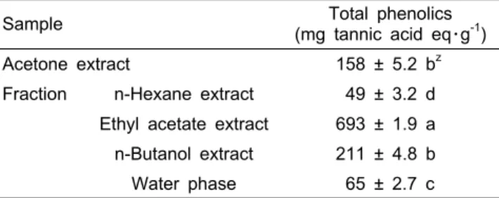
Table 1. Contents of total phenolics of each solvent fractions of dry leaves of Aruncus dioicus.
Sample Total phenolics
(mg tannic acid eq・g-1)
Acetone extract 158 ± 5.2 bz
Fraction n-Hexane extract 49 ± 3.2 d Ethyl acetate extract 693 ± 1.9 a n-Butanol extract 211 ± 4.8 b
Water phase 65 ± 2.7 c
Value of ± indicates standard deviation of triplicate.
zThe different letters express statistically significant difference by Duncan’s test (p < 0.05).
세포는 lysis buffer(67mM sodium phosphate buffer, 1%
triton X-100, 0.1mM phenylmethyl sulfonyl fluoride) 100μL 를 첨가하여 초음파 분쇄 후 얼음에 10분간 방치하였다. 실 험에 사용할 흑색종세포의 tyrosinase는 13,200rpm에서 20 분간 원심분리하여 얻어진 상층액을 이용하였으며, 세포의 pellet은 멜라닌 함량 측정에 사용하였다. 생성된 DOPA chrome의 양은 67mM phosphate buffer(pH6.8)에 녹인 8.0mM 의 L-DOPA 180μL를 기질로, 세포 상등액 20μL을 첨가하 여 37°C에서 60분간 반응시킨 후 490nm에서 측정하였다.
총 멜라닌 함량 측정은 상등액이 제거된 pellet을 건조시킨 뒤 1N NaOH 1mL를 넣어 80°C에서 1시간 반응시킨 뒤 405nm에서 흡광도를 측정하였다. 멜라닌 생합성 저해는 시료용액의 첨가구와 무첨가구의 흡광도 감소율로 나타내 었다.
세포 내 cAMP 농도 측정
세포 내 cAMP 농도 측정은 3 × 10⁵개의 B16F10 세포를 0.1M HCl에 용해하여 phosphodiesterase 활성을 억제시킨 뒤 상층액을 모아서 중화시키고 희석한 후 고정된 cAMP conjugate를 넣어 96well에서 cell lysate와 면역반응을 시켰 다. 효소의 활성 측정은 초과한 conjugate와 결합하지 않은 cAMP를 세척하고, substrate solution을 넣어 반응시킨 뒤 색의 변화가 멈추면 415nm에서 흡광도를 측정하였으며, 색 의 강도는 cell lysate 내에서 cAMP 농도와 역비례하였다.
Western Blot을 통한 단백질 발현량 측정
단백질 추출은 B16F10 세포를 100mm 조직배양접시에 cell seeding 후 24시간 동안 배양하여 안정화 시킨 세포(1 × 10⁶)에 ADE를 농도별로 처리한 배지에서 48시간 배양하여 상 등액을 제거한 후 RIPA buffer를 이용하여 수행하였다. 30μg 의 단백질은 변성 후 10%의 SDS-PAGE gel을 이용하여 전기 영동하여 분리하였다. 분리된 단백질은 semi-dry transfer 기기 (TE77 XP, Hoefer, USA)를 이용하여 high-quality polyvinylidene difluoride(PVDF) membrane에 옮긴 다음 실온에서 1시간 동안 blocking buffer(5% skim milk in TBST)를 처리하였 다. 1차 항체 mouse anti-PKA, MITF, TRP1, TRP2, 및 goat anti-tyrosinase는 1:1,000 비율로 희석하여 4°C에서 16시간 반응시킨 다음 10분 간격으로 TBST로 3회 세척하고 mouse anti-goat IgG-HRP와 rabbit anti-mouse IgG-HRP 두 가지 2차 항체는 1:1,000 비율로 희석하여 실온에서 2시간 동안 배양하였다. 단백질 발현량은 3회 세척한 뒤 X-ray 필름에 감광 하여 imagequant 기기(ImageQuant LAS4,000, GE healthcare life science, USA)를 이용하여 밴드를 확인하고 정량하였다.
Immuno-fluorescence Assay
면역형광분석을 위해 각 cell line은 Lab-tek chamber 8 well에 세포를 1 × 10³개로 분주하여 24시간 동안 안정시킨 뒤, ADE 시료를 농도별로 처리하여 준비하였다. 48시간 동 안 반응시킨 후 분석시료는 차가운 PBS buffer로 3회 세척 하고 0.5% triton X-100을 10분간 처리한 후 다시 3회 세척 한 다음 1% BSA를 넣어 1시간 동안 방치하였다. 면역형광 반응은 mouse anti-MITF 1차 항체를 4°C에서 16시간 반응 시킨 다음 3회 세척한 뒤 바로 invitrogen사의 alexa flour 488(Carlsbad, CA) goat anti-mouse IgG 2차 항체를 1:1,000 비율로 첨가하고 차광하여 1시간 동안 반응시켰다. 면역반 응을 하지 않은 여분의 2차 항체는 3회 세척하여 제거하고 형광 발현반응을 위해 기질인 4‘6-diamidino-2-phenylindole (DAPI)를 200μL씩 첨가하여 40분 동안 반응시켰다. 면역형 광 발현반응 다음 chamber를 제거하고 slide에 mounting solution을 첨가한 후 cover slip으로 고정하여 형광현미경 (Eclipse Ti-U, Nikon, Japan)으로 관찰하였다.
결과 및 고찰
총 폴리페놀 함량 분석
탄닌산을 기준물질로 하여 표준곡선을 그린 후 삼나물 아세톤 추출물과 여러 가지 유기용매 분획물의 폴리페놀 함 량을 측정한 결과, 삼나물 아세톤 추출물에서는 158mg・g-1 의 총 폴리페놀이 함유되어 있었다. 또한 삼나물 hexane 분 획물은 49mg・g-1, ethyl acetate 분획물은 693mg・g-1, butanol 분획물은 211mg・g-1, 그리고 물 분획물에는 65mg・g-1의 폴 리페놀 함량을 확인하였다. 그 중 ethyl acetate 분획물에 가장 많은 폴리페놀이 함유되어 있음을 확인할 수 있었다 (Table 1). 삼나물의 각 분획물의 미백효능을 확인하기 위해 in vitro 상에서 상용화된 버섯유래 tyrosinase의 활성 저해 결과는 ethyl acetate 분획물에서 가장 높은 미백효과가 있는 것을 확인하였고(자료 미제시), 이후 모든 실험은 삼나물 ethyl
Fig. 1. Cell viability of B16F10 melanoma cells by Aruncus dioicis ethyl acetate extract (ADE). 5 × 10⁴ cells were grown for 24 h at 37°C in 5% CO2 incubator. ADE was treated with various concentrations for cell toxicity analysis. Bars indicate mean ± standard deviation of triplicate. *Means statistically significant difference at p < 0.05.
A B
Fig. 2. Inhibitory effect on tyrosinase activity (A) and decrease of melanin contents (B) by Aruncus dioicis ethyl acetate extract (ADE) treatment in B16F10 melanoma cells. After 1 × 10⁶ cells were seeded in serum-free medium for 1 h, the cells were treated with 0, 5, 10 and 50 μg・mL-1 of ADE for 48 h. A, tyrosinase activity was analyzed using L-DOPA substrate; B, melanin content in the pellet was measured. Bars indicate mean ± standard deviation of triplicate. *,**Mean statistically significant difference at p < 0.05, p < 0.005, respectively.
Fig. 3. Decreased cAMP levels in B16F10 melanoma cells by Aruncus dioicis ethyl acetate extract (ADE) treatment. After 3 × 10⁵ cells were seeded in serum free medium for 1h, the cells were treated with 5, 10 and 50 μg・mL-1 of ADE for 48 h. cAMP content was measured by cAMP immuno-assay kit.
Bars indicate mean ± standard deviation of triplicate. *,**Mean statistically significant difference at p < 0.05, p < 0.005, respectively.
acetate 분획물(ADE)을 이용하여 실험하였다.
흑색종세포 B16F10의 세포독성 평가
기초실험 결과를 근거로 모든 실험에 이용될 ADE의 세포 독성을 평가하기 위해 MTT assay한 결과는 Fig. 1과 같았다.
ADE을 5, 10, 50, 100, 500, 그리고 1,000μg・mL-1 처리 후 세포 생존율을 측정한 결과 500μg・mL-1까지는 10% 이하의 낮은 세포독성을 나타내었다. 가장 고농도인 1,000μg・mL-1 은 약 80%의 세포 생존율을 나타내어 약 20%의 세포독성 을 확인하였다. 흑색종세포 B16F10에서의 tyrosinase 활성 도와 멜라닌 함량, 미백관련 신호전달 유전자들의 단백질 함량 측정을 위해 생존율이 100%에 가까운 농도이며 미백 소재로서의 가능성을 검증하기 위한 실험임으로 가능한 낮 은 농도를 최종 선택하여 5, 10, 그리고 50μg・mL-1의 농도 의 ADE를 처리하여 확인하였다.
Tyrosinase 활성 저해 및 멜라닌 함량 측정
버섯 유래 tyrosinase의 활성 저해를 측정한 결과는 ADE 농도 의존적 저해 효과를 나타내었으며, ADE 1,000μg・mL-1 에서 59.2%의 저해능을 나타내어 우수한 미백효능을 나타 내었다(자료 미제시). 본 실험에서는 50μg・mL-1의 농도에서 35.6%의 높은 tyrosinase 활성 저해효과를 확인하였다(Fig.
2A). 또한 ADE의 멜라닌 생합성 저해효과를 측정한 결과 같은 농도에서 58.8%의 멜라닌 함량 감소효과를 나타내어 삼나물의 미백효과가 매우 우수함을 확인하였다(Fig. 2B).
세포 내 Cyclic AMP(cAMP) 활성 측정
멜라닌 색소형성의 중심 기작은 cAMP의 과도한 발현에 의해 tyrosinase 유전자발현이 유도되고 멜라닌 생성이 증가
A B
ADE Cont. 5 10 50 ADE Cont. 5 10 50
MITF 0.97 0.92 0.90 0.38* PKA 2.70 1.82 1.65* 1.12**
Tyrosinase 1.42 1.50 1.32 0.72** pERK 0.21 0.30 0.42 0.67
TRP-1 2.43 2.12 1.99 1.54* ERK 2.69 2.65 2.80 2.79
TRP-2 1.30 1.16 0.78 0.61* pCREB 0.42 0.62 0.50 0.25**
CREB 1.36 1.42 1.41 1.44
Fig. 4. Inhibitory effects of Aruncus dioicis ethyl acetate extract (ADE) on whitening related proteins of melanogenesis in B16F10 melanoma cells. After 1 × 10⁶ were seeded in serum free medium for 1 h, the cells were treated with 0, 5, 10 or 50 μg・mL-1 of ADE for 48 h. Proteins were detected by Western blotting analysis with their own antibodies. Table showed normalization of proteins. All signals of proteins except pERK, pCREB were divided by β-actin’s signal. pERK and pCREB were normalized by ERK and CREB, respectively. A, protein expression involved to melanin biosynthesis; B, cAMP downstream proteins’ expression was evaluated by Western blot. *,**Mean statistically significant difference at p < 0.05, p < 0.005, respectively.
한다(Jung et al., 2009). cAMP 활성을 측정한 결과 ADE의 농도에 따른cAMP의 양의 변화를 나타내었다(Fig. 3). ADE를 처리 한 결과 5, 10μg・mL-1에서 각각 16%와 17%, 그리고 50μg・mL-1에서 37% cAMP 감소 효과를 확인하였다.
B16F10에서의 미백관련 인자들의 단백질 발현량 측정 ADE가 멜라닌 생합성에 관여하는 효소인 tyrosinase 활 성에 미치는 영향을 알아보기 위하여 mouse 유래 melanoma 세포 B16F10에 ADE 분획물 5, 10, 그리고 50μg・mL-1를 처 리한 후 48시간 뒤에 멜라닌 생합성 관련 단백질 발현량을 western blot으로 확인하였다. ADE를 농도별로 처리한 B16F10 군에서는 MITF, TRP1, TRP2, 및 tyrosinase 단백질의 발현 이 무처리군보다 감소하였다(Fig. 4A). 또한 MITF, TRP1, TRP2, 및 tyrosinase의 상위 단계 유전자인 PKA, ERK, 인 산화된 ERK, CREB, 및 인산화된 CREB 단백질 발현량을 측정한 결과, ADE 처리에 의해 CREB, ERK, 및 인산화된 ERK의 단백질량의 변화는 미비하지만, 멜라닌 생합성에 직 접 관여하는 상위 유전자인 PKA와 인산화된 CREB의 발현
량은 급격히 줄어드는 것을 확인하였다(Fig. 4B).
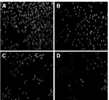
면역형광 염색 분석
B16F10 세포 내에서의 MITF의 발현 정도를 알아보기 위 하여 면역형광 염색법을 실시한 결과는 Fig. 5에 제시하였 다. ADE를 처리 하지 않은 군에서 MITF의 형광 발현이 많 이 일어났지만 ADE 농도가 증가할수록 MITF의 형광 발현 이 급감하는 것을 확인 할 수 있었다(Fig. 5).
본 연구에서는 삼나물 아세톤 추출물을 분획한 다음 얻어 진 ADE를 이용하여 멜라닌 생성, tyrosinase 활성 저해, 및 멜라닌 생합성에 관여하는 여러 유전자 발현에 미치는 영향 을 평가하였다. 멜라닌은 외부 스트레스로부터 피부를 방어 하는 기전의 일환으로 멜라닌 세포에서 합성되지만, 피부미 백에 부정적 영향을 주기 때문에 멜라닌 생합성을 차단하기 위한 다양한 시도가 있었다(Akiu et al., 1991; Mishima et al., 1988). 본 연구에서는 ADE를 이용하여 멜라닌 생성억제 효 과를 알아보기 위한 실험을 수행하였다. B16F10 세포에 ADE 를 5, 10, 50μg・mL-1로 처리한 결과 농도 의존적으로 멜라닌
A B
C D
Fig. 5. Reduced cellular expression of MITF transcription factor using immunofluorescence assay. 1 × 10³ B16F10 cells were seeded in serum free medium for 1 h at room temperature.
The cells were treated with Aruncus dioicis ethyl acetate extract (ADE) for 48 h. A, control; B, 5 μg・mL-1; C, 10 μg・mL-1; D, 50 μg・mL-1 of ADE treatment.
Fig. 6. Schematic diagram of cAMP dependent melanin biosynthesis pathway. Aruncus dioicis ethyl acetate extract (ADE) may block to the very upstream of melanin production in B16F10 melanoma cells. Left and right cells represent keratinocyte and melanocyte respectively. -MSH, -melanocyte sitmulating hormone; ACTH, adrenocorricotropic hormone; MC1R, melanocortin type 1 receptor; cAMP, cyclic adenosine monophosphae; PKA, protein kinase A; CREB, cAMP response element binding protein;
ERK, extracellular signal related kinase; MITF, microphthalmia associated transcription factor; TRP1, tyrosinase related protein 1; TRP2, tyrosinase related protein 2.
함량이 급감하는 것을 확인하였다(Fig. 2B). 멜라닌 생합성은 tyrosinase의 작용을 통해 이루어지며(Yamamura et al., 2002), 이 tyrosinase는 tyrosine(L-tyrosine)을 산화시켜 L-DOPA를 만드는 tyrosine hydroxylase로 작용하며, DOPA를 산화시켜 DOPA quinone을 만드는 DOPA oxidase로 작용하여 멜라
닌을 합성하는 중요한 효소로 알려져 있다(Lin et al., 2002).
그러므로 멜라닌 생성을 억제하는 피부 미백제 개발에는 tyrosinase의 활성을 억제하는 것이 필수적이다(Chin and Kim, 2005). Song(2008)은 적하수오 methanol 추출물을 B16F10 에 75μg・mL-1 처리한 결과 tyrosinase 활성이 22% 억제하는 것을 확인하였다. 이는50μg・mL-1 ADE에 의해 tyrosinase 활성이 35.6% 저해되고 멜라닌 함량이 58.8% 감소한 결과 를 근거로 삼나물이 적하수오보다 미백 효능이 더 우수할 것임을 추측할 수 있었다. 멜라닌 합성과 관련된 주요 효소 인 tyrosinase와 함께 TRP2는 DOPA chrome tautomerase로 알려져 있는 효소로서 DOPA chrome을 이용하여 멜라닌 전 구물질인 5,6-dihydroxy indole-2-carboxylic acid(DHICA) 를 생성한다. 또한 TRP1은 TRP2에 의해 생성된 DHICA를 산 화시켜 indole-2-carboxylic acid(IQCA)를 생성하는데, IQCA 는 흑갈색을 나타낸다(Oh, 2009). 또한 본 연구에서는 B16F10 세포에 ADE를 처리한 결과 tyrosinase, TRP1, 및 TRP2의 단백질 발현량이 감소하는 것을 확인하였고, tyrosinase 발 현을 조절하는 전사인자 MITF 발현 또한 ADE 처리에 의해 단백질 발현량이 줄어드는 것을 확인하였다. 모식화된 삼나 물 미백 효능에 관한 멜라닌 생합성 경로는 Fig. 6에 제시하 였다. 그러나 절패모와 목단피의 ethanol 추출물은 tyrosinase 활성 및 멜라닌 합성에는 40% 이하의 저해효능을 나타내었지 만, 멜라닌 합성관련 단백질인 tyrosinase, TRP1, 그리고 TRP2 의 발현을 억제하지 않음을 확인하였다(Ha, 2009; Na, 2009).
이는 절패모와 목단피에서는 알려지지 않은 또 다른 멜라닌 합성 억제 경로가 존재할 수 있을 것이라 예측할 수 있다.
Jung et al.(2009)은 cAMP는 cAMP 의존적 단백질 인산화 효소의 활성을 통하여 tyrosinase 유전자 발현을 유도하는 MITF의 발현을 증가시킨다고 보고하였다. MITF 발현에 영 향을 미치는 cAMP 발현여부를 측정한 결과 ADE에 의하여 cAMP의 양이 50μg・mL-1 농도에서 발현량이 37% 감소하는 것을 확인하였으며, 본 실험을 통하여 삼나물 ADE 분획물이 cAMP를 효과적으로 억제함으로써 이를 통해 tyrosinase 활 성뿐만 아니라 TRP1, TRP2 그리고 MITF의 발현을 효과적 으로 저해한다는 사실을 확인할 수 있었다. 또한 본 실험에서 삼나물 ADE 분획물 처리에 의해 cAMP의 down regulation 되는 PKA의 단백질 발현이 감소함을 확인할 수 있었다. 그 러므로 ADE에 의한 미백 효과는 멜라닌 생성에 있어서 분 자적 기작의 상위에 자리잡고 있는 신호전달을 억제함으로 써 기인하는 것으로 생각된다. 따라서 삼나물 ethyl acetate 분획물 50μg・mL-1 농도는 세포에는 매우 안전하지만 cAMP 가 급감함으로 인해 멜라닌 생합성에 관여하는 그 하위 유전 자들 발현을 효과적으로 저해하여 멜라닌 생성을 억제하는 것으로 추측된다. 이에 삼나물은 절패모와 목단피와 같은 약용식물 보다 효과적인 기능성화장품의 미백소재로 사용 가능할 것으로 예측한다.
초 록
울릉도 자생 삼나물(Aruncus dioicus)은 최근 monoterpenoids 생리활성 물질이 밝혀지면서 여러 가지 항산화와 관련된 생 리활성이 확인되고 있다. 삼나물 ethyl acetate 분획물에 의 한 미백효과를 검증하기 위하여 흑색세포종인 B16F10을 이 용하여 실험을 진행하였다. 삼나물 ethyl acetate 분획물(ADE) 의 세포독성을 측정하기 위하여 MTT assay를 실시한 결과, ADE 500μg・mL-1 이상의 농도에서 미비한 세포독성(10% 이 상)을 확인하였으며, 이후 실험에서는 5, 10, 50μg・mL-1 농 도가 세포 내에서 tyrosinase 활성과 멜라닌 양의 변화를 측 정하였다. 그 결과 ADE 농도에 따라 tyrosinase 활성이 감소 하고 총 멜라닌 함량이 감소하는 것을 확인하였다. 특히 50μ g・mL-1에서 35.6% tyrosinase 활성억제, 58.8% 멜라닌 함량 이 감소하는 것을 확인하였다. 또한 ADE에 의해 미백과 관 련된 tyrosinase, tyrosinase related protein 1(TRP1), TRP2, microphthalmia associated transcription factor(MITF) 및 그 상위 단계인 cAMP와 protein kinase A(PKA)의 단백질 양 이 감소하여 cAMP response binding protein(CREB)의 인 산화는 감소하고 extracellular signal related kinase(ERK)의
인산화는 증가하는 것을 확인하였다. 본 연구에서는 울릉도 자생 삼나물의 미백효과에 관한 효능을 확인하고 기능성화 장품의 소재로서의 활용 가능성이 있음을 확인하였다.
추가 주요어 : 항산화효과, MAP인산화효소, 멜라닌 세포, 자외선
인용문헌
Akiu, S., Y. Suzuki, T. Asahara, Y. Fujinuma, and M. Fukuda.
1991. Inhibitory effect of arbutin on melanogenesis-biochemical study using cultured B16 melanoma cells. Nihon Hifuka Gakkai Zasshi 101:609-613.
Busca, R. and R. Ballotti. 2000. Cyclic AMP a key messenger in the regulation of skin pigmentation. Pigm. Cell Res. 13:60-69.
Chin, J.E. and K.C. Kim. 2005. Effect of chestnut bark extracts on tyrosinase gene expression. Kor. J. Sanitation 20:10-16.
Goding, C.R. 2000. Mitf from neural crest to melanoma: Signal transduction and transcription in the melanocyte lineage. Genes Dev. 14:1712-1728.
Ha, T.K. 2009. Inhibitory effect of Fritillaria verticillata Willd.
var. thunbergii Baker ethanol extract on melanin biosynthesis.
MD Diss., Sangji Univ., Wonju, Korea.
Hunt, G., C. Todd, J.E. Cresswell, and A.J. Thody. 1994. Alpha- melanocyte stimulating hormone and its analogue Nle4DPhe7 alpha-MSH affect morphology, tyrosinase activity and melanogenesis in cultured human melanocytes. J. Cell Sci. 107:205-211.
Jeong, S.Y., D.Y. Jun, Y.H. Kim, B.S. Min, and M.H. Woo. 2011.
Monoterpenoids from the aerial parts of Aruncus dioicus var.
kamtschaticus and their antioxidant and cytotoxic activities.
Bioorg. Med. Chem. Lett. 21:3252-3256.
Jung, E.J., J.S. Lee, S.G. Huh, J.N. Lee, Y.S. Kim, and G.D.
Kim. 2009. Phloridzin-induced melanogenesis is mediated by the cAMP signaling pathway. Food Chem. Toxicol. 47:2436-2440.
Kameyama, K., C. Sakai, S. Kuge, S. Nishiyama, Y. Tomita, S.
Ito, K. Wakamatsu, and V.J. Hearing. 1995. The expression of tyrosinase, tyrosinase-related proteins 1 and 2 (TRP1 and TRP2), the silver protein, and a melanogenic inhibitor in human melanoma cells of differing melanogenic activities.
Pigm. Cell Res. 8:97-104.
Kim, D.H., Y.S. Moon, B.J. An, and J.H. Son. 2012. Potent anti- aging activity of Aruncus dioicus, a native plant of Ulleung-do, South Korea, in CCD-986sk fibroblasts via suppression of matrix metalloproteinases. J. Nat. Med. 66:631-636.
Kligman, L.H. 1996. The hairless mouse model for photoaging.
Clin. Dermatol. 14:183-195.
Kobayashi, T., K. Urabe, A.J. Winder, C. Jiménez-Cervantes, G.
Imokawa, T. Brewington, F. Solano, J.C. García-Borrón, and V.J. Hearing. 1994. Tyrosinase related protein 1 (TRP1) functions as a DHICA oxidase in melanin biosynthesis. EMBO J.
3:5818-5825.
Lee, C.B. 2003. Coloured flora of Korea. 1st ed. Hyangmoonsa, Seoul, Korea.
Lin, C.B., L. Babiarz, F. Liebel, E. Roydon Price, M. Kizoulis,
G.J. Gendimenico, D.E. Fisher, and M. Seiberg. 2002. Modulation of microphthalmia-associated transcription factor gene expression alters skin pigmentation. J. Invest. Dermatol. 119:1330-1340.
Mishima, Y., S. Hatta, Y. Ohyama, and M. Inazu. 1988. Induction of melanogenesis suppression: Cellular pharmacology and mode of differential action. Pigm. Cell Res. 1:367-374.
Na, S.H. 2009. Paeonia suffruticosa Andr (PSA) inhibits melanin synthesis in mouse B16 melanoma cells. PhD Diss., Kyung Hee Univ., Suwon, Korea.
Oh, W.K., K.B. Kim, J.Y. Lim, S.K. Lee, Y.D. Kwon, S.R. Yeom, and Y.S. Song. 2009. Effects of Dokhwalkisaeng-tang on mlanin synthesis inhibition and gene expression in B16F10 melanoma cells. K. J. Oriental Physiol. Pathol. 23:63-75.
Shin, J.W., S.I. Lee, M.H. Woo, and S.D. Kim. 2008. Effect of
ethanol extracts of goat’s beard on streptoxotocin induced diabetic symptoms and oxidative stress in rats. J. East Asian Soc. Dietary Life 18:939-948.
Song, J.S. 2008. Inhibitory effect of polygonum multiflorum on melanin synthesis and its action mechanism in B16F10. PhD Diss., Daejeon Univ., Daejeon, Korea.
Yamamura, T., J. Onishi, and T. Nishiyama. 2002. Antimelanogenic activity of hydrocoumarins in cultured normal human melanocytes by stimulating intracellular glutathione synthesis. Arch. Dermatol.
Res. 294:349-354.
Yao, C., J.H. Oh, I.G. Oh, C.H. Park, and J.H. Chung. 2013.
[6]-Shogaol inhibits melanogenesis in B16 mouse melanoma cells through activation of the ERK pathway. Acta Pharmacol.
Sin. 34:289-294.