https://doi.org/10.9721/KJFST.2018.50.3.344
344
©The Korean Society of Food Science and Technology
오크라 추출물의 멜라닌 생성저해 및 피부장벽개선 효과
김 현 수
1,*
1중원대학교 식품공학과Effect of
Abelmoschus esculentus extract on anti-melanogenesis
and skin barrier function
Hyun-Soo Kim1,*
1Department of Food science and Technology, Jungwon University
Abstract We investigated the application of functional materials by examining a variety of physiological activities of Abelmoschus esculentus extract. A. esculentus extract had a low cytotoxicity against murine melanoma B16F10 cells. At concentrations showing little or no cytotoxicity, A. esculentus extract showed high a DPPH radical scavenging activity (ID50, 5.24µg/mL),inhibited tyrosinase activity (ID50, 102.12µg/mL), and decreased melanin content (ID50, 17.85µg/mL). The treatment of B16F10 cells with A. esculentus extract suppressed the protein expression of tyrosinase in a dose-dependent manner. These findings suggest that A. esculentus extract inhibits melanin synthesis in murine melanoma B16F10 cells by suppressing intracellular tyrosinase expression, as well as directly inhibits tyrosinase activity simultaneously. Additionally, A. esculentus extract promotes the expression of involucrin, which is related to skin barrier protection. These results indicate that A. esculentus extract is an appropriate material for improving skin barrier function.
Keywords: Abelmoschus esculentus, melanin, tyrosinase, involucrin
서
론
현대사회에서 증가하는 건조 환경 및 사회적 스트레스 등으로 악화되고 있는 피부장벽손상(건조피부, 민감성피부, 노인성 건성 피부, 아토피피부)을 개선하기 위한 천연소재 개발에 대한 연구 가 많이 진행되고 있으며 특히, 하얀색 피부를 선호하는 여성들 을 중심으로 미백효과를 볼 수 있는 소재개발에 대한 관심이 증 가하고 있다(Sohn 등, 2004). 피부색은 멜라닌 생성세포가 합성 하는 멜라닌(melanin)의 양과 종류에 따라 결정되며 자외선과 같 은 외부자극으로부터 피부를 보호하기 위해 멜라닌 생성을 하게 된다. 그러나 피부표면에 과도한 멜라닌 합성과 축적은 기미, 주 근깨와 같은 과 색소 침착 증상이 나타나게 된다(Hill 등, 1997). 멜라닌 생성은 tyrosinase, tyrosinase related protein-1 (TRP-1), TRP-2 효소에 의해 조절되며 tyrosinase는 구리를 포함한 효소로 서 멜라닌 형성에 중요한 역할을 하고 있다(Kobayashi 등, 1994). Tyrosinase는 멜라노좀(melanosome)내에서 tyrosine을 산화시켜 DOPA를 생성하는 tyrosine hydroxylase로서, DOPA를 산화시켜 DOPA quinone을 만드는 DOPA oxidase로서 작용하여 멜라닌 중 합체를 합성하는 핵심 효소로 작용한다(Kobayashi 등, 1994). 따 라서 멜라닌 생합성을 억제하여 미백효과를 가져올 수 있는 독 성이 없는 천연소재 물질에 대한 개발이 요구되고 있다(Briganti 등, 2003). 본 연구의 재료인 오크라는 아욱과(Malvaceae)에 속하는 식물 로서 아프리카 북동부가 원산지이다(Petropoulos 등, 2017). 오크 라는 섬유질이 풍부하고 펙틴, 갈락탐 등의 혼합물인 점액이 존 재하여 자양강장에 효과가 있으며 혈중 콜레스테롤을 낮추는 효 과(Erfani 등, 2018)가 있다고 알려져 있다. 이와 같이 다양한 생 리적 기능성이 알려져 있음에도 불구하고 과학적으로 활성메커 니즘에 대한 연구가 부족하며 특히, 미백활성, 피부장벽 개선 등 피부개선 효과를 비롯한 기능성 화장품소재에 활용하기 위한 연 구는 미미한 실정이다. 따라서 본 연구에서는 오크라 추출시료를 가지고 산화방지 및 생리활성 시험을 실시하였으며 또한 미백시 험을 비롯한 피부효능 개선 시험, 멜라닌 생성 억제에 대한 메커 니즘 및 피부장벽 개선 관련 시험을 조사하여 기능성 화장품 소 재로서의 개발 가능성을 검토하고자 하였다.재료 및 방법
실험재료 오크라(Abelmoschus esculentus)는 농산물 인터넷 직거래를 통 해 구입하여 사용하였다. 시료 30-40 g에 에탄올 200 mL를 첨가 하고 용매추출장치(ASE300 Accelerated Solvent Extractor, Dionex Corporation, USA)를 이용하여 50oC, 1500 psi에서 20분 동안 추 출하였다. 이후 건조기(Modul spin 40, Biotron Corporation, West Centerville, UT, USA)에서 40oC에서 24시간 동안 건조하고 실험 에 사용하였다(Kim 등, 2014).*Corresponding author: Hyun-Soo Kim, Department of Food sci-ence and Technology, Jungwon University, Goesan, Chungbuk 28024, Korea
Tel: +82-43-830-8616 Fax: +82-43-830-8679 E-mail: [email protected]
Received February 1, 2018; revised March 21, 2018; accepted March 30, 2018
세포독성평가
세포독성 시험은 세포내의 미토콘드리아 수소제거효소에 의해 수용테트라졸륨염(water soluble tetrazolium salts) (WST-1)에서 불 용성의 포마진(formazan)결정으로 변환되는 원리를 이용하였다 (Ranke 등, 2004). 이는 포마진이 많이 생성될수록 세포 활성이 커지는 것을 의미하였다. 포마진은 다이메틸설폭사이드(dimethly sulfoxide)에 녹아서 보라색 빛을 띄는데 분광광도계(spectropho-tometer)로 540 nm 파장에서 OD값을 재어서 정량 하였다. 미국 세포주은행(ATCC, Manassas, VA, USA)에서 분양 받은 멜라노마 세포(murine melanoma B16F10)를 10% 소태아 혈청(fetal bovine serum: FBS, Gibco, Waltham, MA, USA)이 포함된 dulbecco’s Modified Eagle’s medium (DMEM)에서 배양하고 24 well plate에 2×10⁴ cells/well의 농도로 세포를 접종한 후, 37oC, 5% CO2조건 에서 배양하였다. 이후 배양세포에 추출물을 농도별로 처리하고 72시간 배양하였다. 결과는 %로 환산하였고, 각 처리농도에서의 결과 수치로 50% 저해 농도(ID50)을 계산하여 세포 독성 정도를 표현하였다. 산화방지력 측정
DPPH 라디칼(radical) 소거능 실험은 1,1-diphenyl-2-picryl hydrazyl (DPPH, Sigma Chemical Co., St. Louis, MO, USA)에 의한 전자 공여능(electron donating ability, EDA)을 측정하였다(Miliauskas 등, 2004). 즉, 0.2 mM의 DPPH용액 0.8 mL에 식물추출물 시료 0.2 mL를 첨가하여 혼합한 다음 실온에서 30분간 반응시킨 후 517 nm에서 흡광도를 측정하였다. 산화방지력은 [1-(시료첨가구의 흡 광도/무첨가구의 흡광도)]×100의 계산식에 의해 전자공여능(%)을 구하였으며, 대조구는 시료 대신 메탄올을 첨가하였고, positive control로 아스코빅산(ascorbic acid)를 사용하였다. 각 처리농도에 서의 결과 수치로 50% 저해 농도(ID50)를 계산하여 DPPH 소거
활성 정도를 표현하였다.
Tyrosinase 억제활성측정
멜라닌(melanin) 합성 주요 단계에 관여하는 타이로시네이즈 저 해활성 측정은 효소 작용 결과 형성되는 DL-β-3,4-dihydroxyphenyl alanine (DOPA) chrome을 비색법에 의해 측정하는 Masamoto 등 의 방법(Masamoto 등, 2003)을 변형하여 측정하였다. 기질로서 5 mM DL-DOPA 용액 0.2 mL, 0.1 M 인산완충용액(pH 6.8) 0.2 mL 및 시료용액 0.5 mL의 혼합액에 mushroom tyrosinase (Sigma Chemical Co.) 0.1 mL (2,000 unit/mL)을 첨가하여 37oC에서 10분 간 반응시킨 다음 475 nm 파장에서 흡광도를 측정하고 아래 식 에 따라 타이로시네이즈 저해활성을 산출하였다. 각 처리농도에 서의 결과 수치로 50% 저해 농도(ID50)를 계산하여 타이로시네이 즈 저해 활성 정도를 표현하였다. Tyrosinase 저해 활성(%)= 1− 시료첨가구의 흡광도 ×100 대조구의 흡광도 멜라닌 생성 저해 측정 멜라닌 정량은 Hosoi 등의 방법(Hosoi 등, 1985)을 변형하여 사 용하였다. 24 well plate에 2×10⁴ cells/well로 멜라노마 세포를 분
주하였고, 100 μM 3-isobutyl-1-methylxanthine (IBMX)으로 멜라닌 생성을 유도한 후, 상기 세포독성 실험에서 관찰된 오크라 추출 물시료 농도를 처리하고 48시간 동안 37oC CO 2 배양기에서 배 양하였다. 세포를 수집하여 세포수를 측정하고, 1200 rpm에서 5 분간 원심 분리하여 침전한 후, 1 mL 균질완충용액(homogenization
buffer) (50 mM Sodium phosphate pH 6.5, 1% Triton X-100, 2 mM PMSF)로 용해시켰다. 여기서 얻은 pellet에 1 N NaOH (10% DMSO) 200μL를 첨가하고 vortex 후 405 nm에서 흡광도값을 측 정하였다. 추출물을 처리하지 않은 시료군을 대조군으로 하고 결 과는 %로 환산하였다. 각 처리농도에서의 결과 수치로 50% 멜 라닌 생성 저해 농도(ID50)을 계산하여 멜라닌 생성 저해 활성을 표현하였다. Western blot 분석 오크라 추출물에 의한 tyrosinase 단백질 발현을 분석하기 위하 여 western blot을 시행하였다. 멜라노마 세포를 alpha-melanocyte stimulating hormone (α-MSH)첨가로 멜라닌 생성을 유도한 후, 상 기 세포독성 실험에서 관찰된 추출물시료 농도로 처리하고 48시 간 동안 37oC CO 2 배양기에서 배양하였다. 배양된 세포를 수확 하여 PBS로 두 번 세척한 후, RIPA 완충용액(buffer)에서 용해시 키고 ice에서 1시간 동안 정치시켰다. 처리된 세포 용해물을 4oC 15,000 rpm에서 20분간 원심분리하고, 획득한 상층액은 분석을 위해 −70oC에서 보관하였다. 단백질 농도는 단백질분석키트(protein
assay kit) (Bio-Rad, Hercules, CA, USA)를 사용하여 측정하였다. 각 20 μg 단백질량에 해당되는 시료를 10% SDS-PAGE에서 전기 이동하여 분리 한 후, 폴리바이닐리덴 플루오린화물(polyvinylidene fluoride) (PVDF) 막(membrane)에 옮겨 주었다. membrane을 상온 에서 1시간 동안 blocking buffer (5% skim milk in TBST)에서 처리한 후 1차 항체(mouse anti-tyrosinase: 1:200, Abcam, Cam-bridge, UK; mouse anti-involucrin: 1:500, Santa Cruz Biotech-nology, Carlsbad, CA, USA; mouse anti-b-actin: 1:1000, Sigma-Aldrich Co., St. Louis, MO, USA)를 3시간 동안 상온에서 처리 하였다. 이후 10분 간격으로 TBST로 5회 세척하고 2차 항체(rabbit anti-mouse IgG-HRP: 1:5000, Santa Cruz Biotechnology)로 1시간 동안 반응시켰다. 이후 10분 간격으로 TBST로 5회 세척하고 enhanced chemiluminescence (ECL) western blotting detection kit (Amersham Pharmacia Biotech, Piscataway, NJ, USA)로 검출하였 다. 단백질밴드는 광 이미지 시스템(photo-image system) (Molec-ular Dynamics, Uppsala, Sweden)기기를 사용하여 정량하였다. 통계처리
모든 실험은 독립적으로 3회 반복 시행하고 실험결과는 평균 ±표준평균오차로 표기하였으며, 통계처리는 GraphPad Prism 5 program (GraphPad Software, Inc., La Jolla, CA, USA)을 이용하 여 p 값이 0.05 미만일 때 통계적으로 유의하다고 판단하였다. 일원배치 분산분석(one-way analysis of varience)을 한 후, 각 시 료간의 통계적 유의성은 Bonferroni’s multiple comparison test를 이용하여 수행하였다.
결과 및 고찰
오크라 추출물의 세포독성시험
멜라노마 세포에 오크라 추출물(Abelmoschus esculentus extract: AEE) 10, 100, 250, 500, 750μg/mL의 농도로 처리한 후, WST-1 분석법(Ranke 등, 2004)을 이용하여 세포독성을 측정하였다(Fig. 1). 대조군 대비하여 10, 100, 250, 500, 750 μg/mL의 AEE 농도 에서 각각 0, 0.90%, 8.80, 21.50, 48.70% 100 μg/mL의 경우 나 타냈으며, 50% 세포독성을 나타내는 농도(ID50)는 771.21 μg/mL 로 나타났다. 따라서 이후의 실험은 독성이 거의 없는 100 μg/mL 이하의 농도에서 진행하였으며 멜라닌 생성 억제효과로서 보고
된 미역쇠 추출물(Jeon 등, 2013)의 경우에도 세포독성이 거의 없 는 농도가 100 μg/mL 이하인 것과 비교할 때, 천연물시료로서 독 성이 없는 안전한 물질로 판단된다. 오크라 추출물의 산화방지 활성 오크라 추출물에 대해서 DPPH 라디칼 소거 활성법(Miliauskas 등, 2004)으로 대조구로 아스코빅산을 이용하여 산화방지 활성을 측정하였다. 추출물 시료와 대조구 시료를 각각 10, 50, 100 μg/ mL의 농도로 제조하여 DPPH 소거활성을 측정한 결과(Fig. 2), AEE 추출물 시료의 농도가 증가할수록 라디칼 소거능 활성이 증 가되는 경향을 보였으며 AEE 각각에 대한 10, 50, 100 μg/mL의 추출물 농도에서는 대조구와 거의 같은 라디칼 소거활성을 보였 다(89.20, 91.80, 94.10%). 또한, AEE의 경우, 50% 라디칼 소거활 성 농도는 5.24 μg/mL였으며 대조군 표준시료인 아스코빅산의 경 우는 4.87 μg/mL였다. 또한, 미백효과 시료로서 보고된 은행 열 매(Kim 등, 2015b) 추출물의 60 mg/mL 경우 9.95%의 소거활성 을 보였으며 정공피(Kim 등, 2010) 및 대나무(Song 등, 2007) 추 출물의 100 μg/mL 경우 각각 40, 50%의 소거활성을 보이는 것 과 비교할 때, 산화방지 소재로서 더 우수한 오크라 추출물의 가 능성을 확인할 수 있다.
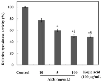
Fig. 3. Inhibitory effect of Abelmoschus esculentus extract (AEE) on tyrosinase. The tyrosinase activity assay was performed with mushroom tyrosinase. Kojic acid was used as a positive control. AEE-untreated group was used as a control. Values are presented as mean±SEM. Differences were considered statistically significant, *p<0.05 compared to the control group. §p<0.05 compared to the AEE 10μg/mL group.
Fig. 4. Anti-melanogenic effects of Abelmoschus esculentus extract (AEE). Cells were treated with 100M IBMX in presence or absence of AEE at the indicated concentration for 2 days. Untreated group (both AEE and IBMX) was used as a control. Values are presented as mean±SEM. Differences were considered statistically significant, *p<0.05, **p<0.01 compared to the AEE-untreated group, §p<0.05 compared to the AEE 10μg/mL group.
Fig. 1. Cell cytotoxicity of Abelmoschus esculentus extract (AEE) on murine melanoma B16F10 cell. AEE-untreated group was used as a control. Values are presented as mean±SEM. Differences were considered statistically significant, *p<0.05 compared to the control group, §p<0.05 compared to the AEE 10μg/mL group.
Fig. 2. Anti-oxidant effects of Abelmoschus esculentus extract (AEE). Ascorbic acid was used as a positive control. Values are presented as mean±SEM. Differences were considered statistically significant.
오크라 추출물의 tyrosinase 저해활성 Tyrosinase는 구리를 포함한 효소로서 멜라닌 형성에 중요한 역 할을 하고 있다. 이러한 멜라닌 중합체 생합성을 효과적으로 저 해하기 위한 tyrosinase 저해활성을 측정하기 위하여 오크라 추출 물에 대한 mushroom 유래의 tyrosinase 저해활성을 관찰하였다 (Fig. 3). 추출물시료 10, 50, 100 μg/mL의 경우, 각각 22.60, 40.30, 49.88%의 tyrosinase 저해활성을 나타내었다. 한편, 미백활 성의 표준시료로서 kojic acid 100 μg/mL의 경우 51.39%의 저해 활성을 나타내었다. 또한, AEE의 경우, 50% tyrosinase 저해활성 농도는 102.12 μg/mL였으며 대조군 표준시료인 kojic acid의 경우 는 97.37 μg/mL였다. 오크라 추출물시료가 대조군 kojic acid와 비 교했을 때, 50% 저해활성 농도가 거의 비슷함을 보였으며 미백 효과로서 보고된 선학초(Kim 등, 2011) 추출물의 100 μg/mL의 경 우 약 10%의 저해활성을 보이는 것과 비교할 때, 미백 기능성 소재로서의 활용 가능성이 높다고 생각된다. 오크라 추출물의 멜라닌 생성 저해활성 오크라 추출물에 대한 세포독성 실험 측정에서 정한 독성이 없 는 농도에서 멜라닌 생성 저해 활성을 측정하였다(Fig. 4). 대조 군 대비하여 10, 50, 100 μg/mL의 AEE 농도에서 각각 49.70, 62.40, 75.40%의 멜라닌 생성 억제 활성을 확인하였다. 또한, 50% 멜라닌 저해활성 농도는 17.85 μg/mL였으며 다른 천연소재 중 미 백활성 효과로서 보고된 바(Yoon 등, 2014) 있는 백합뿌리 추출 물 100 μg/mL의 경우 74% 멜라닌 생성 저해활성을 보이는 것을 고려하면 멜라닌 생성 저해 활성이 매우 우수함을 의미한다. 즉, 시험재료에 있어서 피부미백효과는 멜라닌 생성 저해에 따른 tyrosinase 저해효과가 나타나야 한다. 본 연구의 오크라 추출물의 경우도 멜라닌 저해 활성이 우수하였으며 이에 따른 tyrosinase 저해활성도 높았다. 오크라 추출물의 tyrosinase 발현 억제 효과 오크라 추출물에 의한 tyrosinase 단백질 발현에 미치는 영향을 독성이 없는 농도에서 측정하였다(Fig. 5). α-MSH 처리로 멜라 닌 생성을 유도한 후, α-MSH와 AEE 모두 처리 하지 않은 대조 군 대비하여 α-MSH와 함께 각각 무처리, 10, 50, 100 μg/mL의 AEE 농도에서 각각 133, 127, 110, 92%로 농도 의존적으로 tyro-sinase 발현이 억제되었으며, 100 μg/mL AEE 농도부터는 대조군 과 통계적으로 차이가 없이 저하되는 것을 확인하였다. 따라서 오크라 추출물의 경우, 직접적인 tyrosinase 활성저해는 단백질 발 현억제 효과에 의한 것으로 판단된다(Kim 등, 2015a). 오크라 추출물의 involucrin 발현 효과 피부장벽 보호 지표물질로서 관련된 단백질인 involucrin 발현 에 대한 오크라 추출물의 영향을 측정하였다(Fig. 6). 무처리 대 조군 및 양성대조군인 1.2 mM CaCl2와 함께 10, 50, 100 μg/mL 의 AEE 농도에서 각각 223, 242, 255%로 농도 의존적으로 involucrin 발현이 증가되었으며 1.2 mM CaCl2 291%에 비해서는 다소 낮았다. 특히. 10 μg/mL 농도에서부터 높은 involucrin 발현 증가를 보였다. 이는 피부장벽 효과로 보고된 루꼴라(Kim 등, 2014) 추출물의 경우 20 μg/mL 농도에서부터 높은 involucrin 발 현증가를 보이는 것과 비교할 때, 오크라 추출물은 피부장벽 보 호 역할이 더 우수한 것으로 보인다.
Fig. 6. Effect of Abelmoschus esculentus extract (AEE) on expression of involucrin. Each signal was quantified by scanning densitometry. β-Actin was used as an internal standard. 1.2 mM CaCl2 was used as a positive control and no treatment was used as a negative control. Values were nomalized to β-actin before calculating changes and presented as mean±SEM. Differences were considered statistically significant, **p<0.01 compared to the control group.
Fig. 5. Effect of Abelmoschus esculentus extract (AEE) on expression of tyrosinase. Western blot analysis on expression of tyrosinase. Cells were treated with 5 nM α-MSH in presence or absence of AEE at the indicated concentration for 2 days. β-Actin was used as an internal standard. Untreated group (both AEE and α-MSH) was used as a control. Values were nomalized to β-actin before calculating changes and presented as mean±SEM. Differences were considered statistically significant, *p<0.05 compared to the AEE-untreated group.
요
약
본 연구에서 오크라(Abelmoschus esculentus)에 대한 다양한 생 리활성을 조사하여 기능성소재 응용가능성을 검토하였다. 오크라 추출물은 멜라노마 세포에 대하여 낮은 세포독성을 나타냈다. 세 포독성이 거의 없는 농도에서 오크라 추출물 처리 시, 산화방지 활성(ID50, 5.24μg/mL), tyrosinase 활성저해(ID50, 102.12μg/mL)
및 멜라닌 함량 저하(ID50, 17.85μg/mL)를 보여주었다. 오크라 추 출물 농도 의존적으로 tyrosinase 발현이 억제되었으며, 이는 오 크라 추출물이 직접적인 tyrosinase 활성저해 및 세포 내 tyrosinase 발현을 억제시킴으로써 멜라닌 합성을 저해하는 것으로 판단된 다. 또한, 오크라 추출물이 피부장벽 보호 지표물질로서 관련된 단백질인 involucrin 발현을 증가시키는 것을 확인하였다. 이와 같 은 결과로 미루어 볼 때 오크라 추출물은 피부미백 소재 등 피 부장벽 개선 효과를 지닌 기능성 화장품에 활용하기 위한 매우 효과적인 재료가 될 수 있다고 판단된다.
감사의 글
본 연구는 2017년도 교육부와 한국연구재단(NRF)의 이공분야 기초연구지원사업의 지원(2017R1D1A1B03031402)에 의해 수행되 었습니다.References
Briganti S, Camera E, Picardo M. Chemical and instrumental approaches to treat hyperpigmentation. Pigment Cell Res. 16: 101-110 (2003)
Erfani Majd N, Tabandeh MR, Shahriari A, Soleimani Z. Okra (Abelmoschus esculentus) improved islets structure, and down-regulated PPARs gene expression in pancreas of high-fat diet and streptozotocin-induced diabetic rats. Cell J. 20: 31-40 (2018) Hill HZ, Li W, Xin P, Michell DL. Melanin: a two edged sword?.
Pigment Cell Res. 10: 158-161 (1997)
Hosoi J, Abe E, Suda T, Kuroki T. Regulation of melanin synthesis of B16 mouse melanoma cells by 1 alpha, 25-dihydroxyvitamin D3 and retinoic acid. Cancer Res. 45: 1474-1478 (1985)
Jeon YS, Jung YJ, Youm JK, Kim YK, Kim SN. Inhibitory effect of Endarachne binghamiae extract on melanin synthesis. Korean J. Plant Res. 26: 526-532 (2013)
Kim DH, An BJ, Lee JY. Whitening activities of the Agrimonia pilosa L. extracts. J. Appl. Biol. Chem. 54: 284-289 (2011) Kim B, Choi YE, Kim HS. Eruca sativa and its flavonoid
compo-nents, quercetin and isorhamnetin, improve skin barrier function by activation of peroxisome proliferator-activated receptor (PPAR)-α and suppression of inflammatory cytokines. Phytother. Res. 28: 1359-1366 (2014)
Kim B, Lee SH, Choi KY, Kim HS. N-nicotinoyl tyramine, a novel niacinamide derivative, inhibits melanogenesis by suppressing MITF gene expression. Eur. J. Pharmacol. 764: 1-8 (2015a) Kim YS, Lee YH, Lee JY, Yi YS. Inhibitory effect of Ginkgo biloba
extract on melanin biosynthesis. J. Soc. Cosmet. Sci. Korea 41: 383-389 (2015b)
Kim TH, You JK, Kim JM, Baek JM, Kim HS, Park JH, Choe M. Antioxidant and whitening effects of Sorbus commixta HEDL cortex extract. J. Korean Soc. Food Sci. Nutr. 39: 1418-1424 (2010)
Kobayashi T, Urabe K. Winder AJ, Jimenez-Cervantes C. Imokawa G, Brewington T, Solano F, Garcia-Borron JC, Hearing VJ. Tyro-sinase related protein-1 (TRP-1) function as a DHICA oxidase in melanin biosynthesis. EMBO J. 3: 5818-5825 (1994)
Masamoto Y, Ando H, Murata Y, Shimoishi Y, Tada M, Takahata K. Mushroom tyrosinase inhibitory activity of esculetin isolated from seeds of Euphorbia lathyris L. Biosci. Biotechnol. Biochem. 67: 631-634 (2003)
Miliauskas G, Venskutonis PR, van Beek TA. Screening of radical scavenging activity of some medicinal and aromatic plant extracts. Food Chem. 85: 231-237 (2004)
Petropoulos S, Fernandes A, Barros L, Ciric A, Sokovic M, Ferreira ICFR. The chemical composition, nutritional value and antimicro-bial properties of Abelmoschus esculentus seeds. Food Funct. 8: 4733-4743 (2017)
Ranke J, Mlter K, Stock F, Bottin-Weber U, Poczobutt J, Hoffmann J, Ondruschka B, Filser J, Jastorff B. Biological effects of imida-zolium ionic liquids with varying chain lengths in acute Vibrio fischeri and WST-1 cell viability assays. Ecotoxicol. Environ. Saf. 58: 396-404 (2004)
Sohn ES, Kim SW, Kang JS, Lee SP. Technology trend patent infor-mation analysis of cosmetic materials derived from natural prod-ucts. Appl. Chem. 8: 466-469 (2004)
Song HS, Moon HJ, Park BE, Choi BS, Lee DJ, Lee JY, Kim CJ, Sim SS. Anti-oxidant activity and whitening activity of bamboo extracts. J. Pharm. Soc. 51: 500-507 (2007)
Yoon HS, Yang KW, Kim JE, Kim JM, Lee NH, Hyun CG. Hypop-igmenting effects of extracts from bulbs of Lilium otriental hybrid ‘Siberia’ in murine B16/F10 melanoma cells. J. Korean Soc. Food Sci. Nutr. 43: 705-711 (2014)