• 검색 결과가 없습니다.

Effect of Sophora flavescens Extract on Reinforcing Skin Barrier and Alleviating Inflammation

N/A
N/A
Protected

Academic year: 2021

Share "Effect of Sophora flavescens Extract on Reinforcing Skin Barrier and Alleviating Inflammation"

Copied!
9
0
0

로드 중.... (전체 텍스트 보기)

전체 글

(1)

주 저자 (e-mail: [email protected]) call: 070-5117-0034

Vol. 46, No. 4, December 2020, 361-369 http://dx.doi.org/10.15230/SCSK.2020.46.4.361

고삼 추출물의 피부장벽 강화와 염증완화 효과

노 경 백

ㆍ신 승 우ㆍ윤 소 현ㆍ원 진 배ㆍ오 세 영ㆍ김 준 오

*

ㆍ박 덕 훈ㆍ정 은 선 바이오스펙트럼(주) 생명과학연구소

*

(주)신세계인터내셔날

(2020년 9월 1일 접수, 2020년 11월 16일 수정, 2020년 12월 10일 채택)

Effect of Sophora flavescens Extract on Reinforcing Skin Barrier and Alleviating Inflammation

Kyung-Baeg Roh , Seoungwoo Shin, Sohyun Yoon, Jin Bae Weon, Se-young Oh, Junoh Kim*, Deokhoon Park, and Eunsun Jung

Biospectrum Life Science Institute, 767 Sinsu-ro, Yongin-city, Gyeonggi-Do 16827, Korea

*

Shinsegae International

(Received September 1, 2020; Revised November 16, 2020; Accepted December 10, 2020)

5)

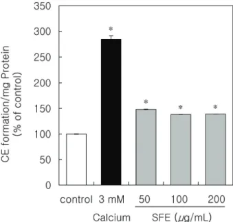
요 약: 아토피성 피부염은 피부장벽 기능장애, 염증 및 만성 소양증을 특징으로 하는 다인성의 염증성 피부질환 이다. 아토피성 피부염은 유전적, 면역학적, 환경적 요인 등의 복합적인 요인으로 피부장벽 기능과 면역기능의 장애를 유발한다고 알려져 있다. 고삼 추출물은 중국전통의학에서 사용되고 있으나, 이의 항아토피 효능에 대한 연구는 거의 진행되지 않았다. 본 연구에서는 아토피성 피부염의 주요 증상인 피부장벽 기능과 면역이상 개선에 대한 고삼추출물의 효과를 평가하였다. 고삼추출물은 피부장벽 기능에서 중요한 역할을 하는 각질세포 막의 형성을 강화하는 결과를 나타내었다. 또한 피부의 보습작용에 있어서 중요한 히알루론산의 발현을 증가 시키는 결과를 나타내었다. 아토피성 피부염 병변에서 특이적으로 증가하는 황색포도상구균에 대한 고삼추출 물의 효능도 확인하였으며, 고삼추출물이 황색포도상구균으로부터 유도된 전염증성사이토카인의 생성을 억 제함을 확인하였다. 또한 피부 스트레스 등으로 부터 생성되는 신경전달 물질인 substance P에 의해 유도된 전염증성사이토카인의 발현도 억제하는 것을 확인하였다. 이러한 결과들은 고삼추출물이 피부장벽기능과 면역반응 개선을 통해 아토피 피부염 치료에 사용될 수 있는 잠재적 후보물질임을 제시한다.

Abstract: Atopic dermatitis (AD) is a common and multifactorial inflammatory skin disease that is characterized by skin barrier dysfunction, inflammation, and chronic pruritus. AD has a complex etiology that includes genetic, immunological, and environmental factors that cause skin barrier abnormalities and immune dysfunctions. Sophora flavescens (SF) has been used in traditional Chinese medicine, but little research has been conducted on its anti-AD efficacy. In this study, we evaluated the effect of SF extract (SFE) on improving skin barrier function and immune abnormalities, which are the main symptoms of AD. SFE has the capacity to enhance the formation of cornified envelope (CE) that plays an important role in the skin barrier function. In addition, it was confirmed that SFE increased the expression of hyaluronic acid related to skin moisture. The effect of SFE against Staphylococcus aureus (S. aureus), which increases specifically in AD lesions, confirmed that SFE inhibited the production of pro-inflammatory cytokines induced by S. aureus. Furthermore, SFE was shown to inhibit the expression of pro-inflammatory cytokines induced by substance P (SP), the cause of skin neurogenic inflammation. These results demonstrate that SFE could be one of potential candidate agent for the treatment of AD by improving the skin barrier function and immune responses.

Keywords: atopic dermatitis, Sophora flavescens, skin barrier, hyaluronic acid, substance P

(2)

1. Introduction

Atopic dermatitis (AD) is a common chronic inflammatory skin disease that is characterized by xerosis, pruritus, and erythematous lesions with increased transepidermal water loss (TEWL). Although the etiology of AD is not fully elucidated, it is considered a multifactorial disorder with genetic, immunological, environmental factors, and microbiome effects that cause altered skin barrier and immune dysregulations[1].

The skin barrier function relies on the stratum corneum (SC) and tight junction with the formation of cornified layer, which are generated by epidermal differentiation. The cornified SC is composed of flattened multilayered corneocytes surrounded by an insoluble protein structure, which is the primary mediator of the epidermal permeability barrier[2]. Therefore, epidermal homeostasis plays an essential role in maintaining its remarkable ability to defend the body against irritants and dehydration.

Current evidence strongly points to the primary pathological drivers in AD being the disruption of the integrity of the skin barrier and subsequent immune dysregulation in predisposed individuals, with resultant damage to the skin barrier[3].

Immune dysregulation of the innate and adaptive immunity plays a major role in the pathogenesis of AD[4]. The microbiota of the skin is important in maintaining immune homeostasis, and preventing the growth of pathogens[5]. In contrast to healthy skin, normal microbiota is disrupted, and the diversity of microorganism in AD skin is reduced.

Sub stance P (SP) is the main neuropeptide identified in skin nerve endings, which has multiple bioactivities that include neurotransmission, proliferation of fibroblasts and keratinocytes, and mast cell degranulation[6–9]. It is considered as a major mediator of neurogenic inflammation and pruritus, and a contributor to the pathogenesis of AD[10–

12]. AD is frequently exacerbated by physical and mental stress, scratching, and sweating, which may be the result of neurogenic inflammation[13–15]. The role of SP in AD skin appears to be predominantly related to the induction of inflammation, nerve growth factor (NGF) expression, and pruritus[16,17].

Sophora flavescens (SF) is a species of plant in the genus Sophora of the family Fabaceae, which is distributed

throughout East Asia, mainly in China, Korea, and Japan. It is one of the oldest herbs used in traditional Chinese medicine (TCM) to clear heart, dry dampness, purge fire, remove toxicity, eliminate parasites, and induce diuresis. A recent study revealed that SF has a broad range of biological activities, such as anti-diabetic, anti-arthritic, anti-inflammatory, anti-bacterial, and antitumor activities[18–22]. However, little is known about the anti-AD effect of SF.

In the present study, we investigate the effects of SF on improving the skin barrier function and skin hydration. In addition, we demonstrate that SF inhibits the expression of pro-inflammatory mediators induced by Staphylococcus aureus (S. aureus)-secreted virulence factor and SP.

2. Materials and Methods

2.1. Preparation of SF Extract

The root of SF was obtained from the Samhong Herb -Medicine Co. (Korea). The dried and pulverized raw material was extracted for 3 h with purified water at 95 ℃ and concentrated by vacuum evaporation (Rotary evaporator N-1000, EYELA, Japan). The powdered extract was obtained by spray dryer (TF-S2L Mini Spray Dryer, Tefic Biotech Co., Limited, China). The extract was lyophilized to yield SF extract (SFE). The lyophilized powder was dissolved in dimethyl sulfoxide (DMSO, Sigma-Aldrich, USA) at 1000-fold higher concentration of final concentration in medium.

2.2. Cell Culture and Reagents

HaCaT (CLS Cell Line Service Gmb H, Germany), a spontaneously immortalized human keratinocyte cell line were maintained in Dulbeccoʼs Modified Eagleʼs Medium (DMEM, Welgene, Korea), containing 10% fetal bovine serum (FBS, Gibco, USA) and 1% penicillin/streptomycin (Invitrogen, USA), at 37 ℃, under 5% CO 2 . THP-1 (ATCC ® TIB-202™, ATCC, USA), a human monocytic leukemia cell line were maintained in Roswell Park Memorial Institute (RPMI) 1640 Medium (Welgene, Korea), containing 10% FBS and 1%

penicillin/streptomycin (Invitrogen, USA), at 37 ℃, under 5%

CO 2 . SP and all-trans-retinoic acid (RA) were purchased from

Sigma-Aldrich (USA).

(3)

2.3. Cornified Envelope (CE) Formation Assay CE content in HaCaT cells was quantitated as previously described with some modifications[23]. HaCaT cells were subjected to SFE for six days in FBS-free medium, when the cells reached at 90% confluency, then harvested with a cell scraper and homogenized in 2% sodium dodecyl sulfate (SDS, Sigma-Aldrich, USA). Proteins were determined according to the bicinchoninic acid (BCA) protein assay (Pierce™ BCA Protein Assay Kit, Thermo Fisher Scientific, USA) with bovine serum albumin as standard. After centrifugation at 12,000× g for 15 min, the precipitates were boiled in 2%

SDS and 20 mM dithiothreitol (DTT, Thermo Scientific™, USA) for 1 h. The amounts of cross-linked envelopes were monitored by the absorbance at 310 nm. CE content was normalized to protein concentration.

2.4. Hyaluronic Acid Assay

HaCaT cells were cultured in DMEM with 10% FBS and seeded in 24 well plate (1.0 × 10 5 cells/well). HaCaT cells at 80% confluency were treated with 10 μM RA and various concentrations (25, 50, and 100 μg/mL) of SFE in serum-free DMEM for 24 h, respectively. Cell culture supernatant was collected and centrifuged at 1000× g for 20 min. Hyaluronic acid in HaCaT cultured media was determined by Hyaluronan Enzyme-Linked Immunosorbent Assay kit (K-1200, Echelon Biosciences, USA) according to the manufacturerʼs instructions.

The absorbance was measured at 405 nm using microplate spectrophotometer (Infinite 200 ® PRO, Tecan, Switzerland).

2.5. Preparation of S. aureus Culture Supernatant (CS) S. aureus subsp. aureus Rosenbach (ATCC ® 12600 , ATCC, USA) was utilized throughout these experiments. S.

aureus was cultured aerobically on Brain heart infusion broth (Difco, USA) for 24 h. The CS was collected and centrifuged at 300× g at 4 ℃ for 10 min. Supernatant was collected and filtered through 0.22 μm filters (Merck Millipore, USA) to remove cell debris. The obtained S. aureus CS was used for further experiments.

2.6. Enzyme-linked Immunosorbent Assay (ELISA) Proinflammatory cytokines were quantified by using a

commercially available ELISA kit (R&D Systems, USA). Cell CS was colleted 24 h after treatment with S. aurues CS and SP, and assayed for proinflammatory cytokines (IL-1β, IL-8, and TNF-α). The standard curve was linearized and subjected to regression analysis. The proinflammatory cytokines were determined using a standard curve.

2.7. Statistical Analysis

Statistical significance of data was determined by a Studentʼ s t-test. All results were expressed as the means ± standard deviation (N = 3). * p < 0.05 was considered to be significant.

3. Results and Discussions

3.1. SFE Enhances the Skin Barrier Function through the Reinforcement of CE Formation

SC generated by epidermal differentiation plays a crucial role in the skin barrier function. In this process, desquamation and the formation of CE, an insoluble protein structure are required for the maintenance of skin homeostasis[2]. Many of the unique proteins required for keratinocytes differentiation,

Figure 1. SFE increases CE formation in HaCaT. The relative

CE content was expressed as the absorbance at 310 nm/mg

protein (% of control). The results are expressed as mean ± SD

(N = 3). * p < 0.001 versus the non-treated control.

(4)

including involucrin, loricrin, filaggrin, and keratins, are regulated by calcium[24]. To determine whether SFE improves skin barrier functions, we evaluated the effect of enhancing CE formation by SFE. Calcium was used as a positive control as a well-known modulator of keratinocyte differentiation[24].

When keratinocytes were treated with various concentrations (10, 100, and 200 μg/mL) of SFE, the levels of the CE formation were enhanced to 48, 38, and 39% compared to untreated cells, respectively (Figure 1). These results indicate that SFE has potentiating activity for CE formation in HaCaT.

3.2. SFE Improves Skin Hydration by Improving Hyaluronic Acid in HaCaT

In AD patients, skin barrier dysfunction manifests as an increase in TEWL and increased penetration of allergens and infectious agents, leading to inflammation and intense pruritus[25]. Dry skin caused by dysfunction of the skin barrier is a very common symptom in almost everyone

suffering from AD, which can worsen to b ecome itch and rash. Therefore, current AD management guidelines recommend moisturizer use as a key and basic step in the treatment of AD[26,27]. Regular use of moisturizers helps maintain skin hydration and improves barrier dysfunction.

Glycosaminoglycan, hyaluronic acid is a key molecule involved in skin moisture and has a unique ability to bind and retain water molecules[28]. Recent studies have shown that hyaluronic acid foam leads to statistically significant improvement in the severity score of AD[3]. Also, moisturization of the SC is highly important to maintain its flexibility and desquamation. The SC has an hyaluronic acid content equal to nearly half that of the epidermis and hyaluronic acid is a factor involved in the moisturization of SC[29]. To evaluate the effect of SFE on skin hydration, we conducted hyaluronic acid assay in HaCaT. RA was used as a positive control for the assay, since it increases hyaluronic acid synthesis by influencing epidermal differentiation associated with hyaluronic acid metabolism in keratinocytes[30]. Figure 2 shows that SFE significantly increases the expression of hyaluronic acid in a dose- dependent manner, with 45% increase at a SFE concentration of 100 μg/mL. These results suggest that SFE has the ab ility to enhance hyaluronic acid synthesis, so it is expected to be useful as a treatment for improving dry skin caused by epidermal barrier disruption.

3.3. S. aureus-induced Inflammation is Inhibited by SFE The skin is a barrier between the body and the environment that maintains homeostasis by recognizing invading pathogens from the outside, and its microbiome plays an important role in the maturation and homeostasis of skin immunity[31]. When the skin barrier collapses, or the balance between symbiotic and pathogens is broken, skin diseases or systemic diseases can occur[32]. Patients with AD have dysbiosis of the skin microbiome with S. aureus as the dominant species[33,34]. Colonization caused by S. aureus can be a major cause of inflammation, so it is associated with the severity of AD[35–39]. Since S. aureus secretes various virulence factors as inflammatory mediators, the inhibitory effect of SFE on the production of pro-inflammatory cytokines Figure 2. SFE increases hyaluronic acid expression in HaCaT.

After 24 h treatment of (25, 50, and 100) μg/mL SFE, HaCaT cultured media was collected and the amount of hyaluronic acid was analyzed by ELISA. RA (10 μM) was used as a positive control. The results are expressed as mean ± SD (N = 3). * p <

0.01 versus the non-treated control; ** p < 0.05 versus the

non-treated control.

(5)

induced by S. aureus was investigated. S. aureus CS was used to induce inflammation. The CS of S. aureus induced the production of inflammatory cytokines, such as tumor necrosis factor-α (TNF-α) and interleukin-1β (IL-1β) in THP-1 macrophages (Figure 3A and B). SFE showed an inhibitory effect of up to 22% at a concentration of 200 μg/mL against IL-1β induced by S. aureus CS, which showing a relatively weak inhibitory effect against TNF-α (Figure 3A and B). Based on these results, it seems necessary to study the suppression of the expression of more diverse inflammatory mediators through additional experiments.

3.4. SFE Inhibits Pro-inflammatory Cytokines Induced by SP

Interaction between the nervous and immune systems contributes greatly to inflammatory disease. The neurogenic components of atopy and allergy are clear and have potentially critical pathogenic relevance[40]. Neuropeptide, in particular SP, is a key mediator of acute stress through neurogenic inflammation, and alter cytokine balance and

immune cell functions[41]. Recent studies have shown that plasma levels of SP are increased in AD patients[42–44], and released from the nerve fibers stimulates keratinocytes to produce proinflammatory cytokines such as IL-1β, IL-6, IL-8 and TNF-α[45,46]. Therefore, clinical occurrence of AD is often associated with psychological stress. To determine whether SFE regulates inflammatory response, we evaluated the effects of SFE on the production of proinflammatory cytokines induced by SP. Figures 4A and B, show that SP elicited proinflammatory cytokines (TNF-α, IL-8) in THP-1 macrophages, which is significantly inhibited by SFE. It is expected that SFE can be effectively applied to the inflammatory response caused by neuropeptide SP in AD patients with psychological stress.

In a recent study, various compounds have been isolated

from SF, of which alkaloids are considered the major active

components[47]. Modern pharmacological studies have shown

that matrine and oxymatrine, the most active of these

alkaloids, has multi-pharmacological effects, including anti-

tumour, antioxidant, anti-pruritic, and anti-inflammatory

Figure 3. SFE decreases the production of pro-inflammatory cytokines induced by S. aureus-cultured media. THP-1 was

pretreated with (10, 50, 100, and 200) μg/mL SFE for 1 h, and then treated with S. aureus-cultured media for 24 h. THP-1

cultured media was collected, and the expression of IL-1β (A) and TNF-α (B) was analyzed by ELISA. The results are

expressed as mean ± SD (N = 3). *p < 0.01 versus S. aureus CS untreated control; **p < 0.001 versus S. aureus CS untreated

control; ***p < 0.05 versus S. aureus CS treated control; ****p < 0.01 versus S. aureus CS treated control.

(6)

effects[48–51]. Therefore, the anti-inflammatory effects ob served in this study may b e due to the presence of matrine or oxymatrine, quinolizidine alkaloids in SFE. The action of matrine or oxymatrine, the major components of SFE, on the anti-inflammatory and skin barrier-strengthening effects of this model should be confirmed through further studies.

4. Conclusion

In this study, we demonstrated the effect of SFE on reinforcing skin barrier and alleviating inflammation that play an important role in the pathophysiology of AD. Therefore, SFE is expected to improve AD symptoms by modulating various underlying causes. This study showed the potential of SFE as a therapeutic agent for AD, and further studies will be needed to clarify its clinical efficacy.

Figure 4. SFE decreases the production of pro-inflammatory cytokines induced by SP. THP-1 was pretreated with (10, 50, and 100) μg/mL SFE for 1 h, and then treated with SP for 24 h. THP-1 cultured media was collected, and the expression of IL-8 (A) and TNF-α (B) was analyzed by ELISA. The results are expressed as mean ± SD (N = 3). * p < 0.01 versus SP untreated control; ** p < 0.05 versus SP treated control; *** p < 0.01 versus SP treated control.

AD Atopic Dermatitis

TEWL Transepidermal Water Loss

SC Stratum Corneum

SP Substance P

SF Sophora flavescens

TCM Ttraditional Chinese Medicine S. aureus Staphylococcus aureus SFE Sophora flavescens Extract

DMSO Dimethyl Sulfoxide

DMEM Dulbeccoʼs Modified Eagleʼs Medium RPMI Roswell Park Memorial Institute

RA Retinoic Acid

CE Cornified Envelope

SDS Sodium Dodecyl Sulfate

BCA Bicinchoninic Acid

DTT Dithiothreitol

CS Culture Supernatant

ELISA Enzyme-linked Immunosorbent Assay TNF-α Tumor Necrosis Factor-α

IL-1β Interleukin-1β

List of Abbreviations

(7)

Reference

1. G. Yang, J. K. Seok, H. C. Kang, Y. Y. Cho, H. S.

Lee, and J. Y. Lee, Skin barrier abnormalities and immune dysfunction in atopic dermatitis, Int. J. Mol.

Sci., 21(8), 2867 (2020).

2. J. van Smeden and J. A. Bouwstra, Stratum corneum lipids: Their role for the skin barrier function in healthy subjects and atopic dermatitis patients, Curr.

Probl. Dermatol., 49, 8 (2016).

3. O. Pacha, B. L. Sambrano, and A. A. Hebert, Skin barrier repair in eczema: A review of current understanding of pathophysiology and treatment, Curr.

Dermatol. Rep., 1, 115 (2012).

4. T. Gavrilova, Immune dysregulation in the pathogenesis of atopic dermatitis, Dermatitis, 29(2), 57 (2018).

5. Y. Belkaid and T. Hand, Role of the Microbiota in Immunity and inflammation, Cell, 157(1), 121 (2014).

6. T. Andoh and Y. Kuraishi, Nitric oxide enhances substance P‐induced itch‐associated responses in mice, Br. J. Pharmacol., 138(1), 202 (2003).

7. M. S. Steinhoff, B. von Mentzer, P. Geppetti, C.

Pothoulakis, and N. W. Bunnett, Tachykinins and their receptors: contributions to physiological control and the mechanisms of disease, Physiol. Rev., 94(1), 265 (2014).

8. E. M. Peters, M. E. Ericson, J. Hosoi, K. Seiffert, M.

K. Hordinsky, J. C. Ansel, R. Paus, and T. E.

Scholzen, Neuropeptide control mechanisms in cutaneous biology: physiological and clinical significance, J.

Invest. Dermatol., 126(9), 1937 (2006).

9. D. Roosterman, T. Goerge, S. W. Schneider, N. W.

Bunnett, and M. Steinhoff, Neuronal control of skin function: the skin as a neuroimmunoendocrine organ, Physiol. Rev., 86(4), 1309 (2006).

10. U. Pereira, N. Boulais, N. Lebonvallet, L. Lefeuvre, A. Gougerot, and L. Misery, Development of an in vitro coculture of primary sensitive pig neurons and keratinocytes for the study of cutaneous neurogenic inflammation, Exp. Dermatol., 19(10), 931 (2010).

11. C. Hosokawa, S. Takeuchi, and M. Furue, Severity

scores, itch scores and plasma substance P levels in atopic dermatitis treated with standard topical therapy with oral olopatadine hydrochloride, J. Dermatol., 36(4), 185 (2009).

12. L. Misery, Atopic dermatitis and the nervous system, Clin. Rev. Allergy Immunol., 41(3), 259 (2011).

13. U. Raap, S. Ständer, and M. Metz, Pathophysiology of itch and new treatments, Curr. Opin. Allergy Clin.

Immunol., 11(5), 420 (2011).

14. S. Pavlovic, M. Daniltchenko, D. J. Tobin, E. Hagen, S. P. Hunt, B. F. Klapp, P. C. Arck, and E. M. J.

Peters, Further exploring the brain-skin connection:

stress worsens dermatitis via substance P-dependent neurogenic inflammation in mice, J. Invest. Dermatol., 128(2), 434 (2008).

15. H. Murota and I. Katayama, Exacerbating factors of itch in atopic dermatitis, Allergol. Int., 66(1), 8 (2017).

16. S. Ständer and T. A. Luger, NK‐1 Antagonists and itch, Handb. Exp. Pharmacol., 226, 237 (2015).

17. C. Zeidler and S. Ständer, The pathogenesis of prurigo nodularis–’Super-Itch’ in exploration, Eur. J.

Pain., 20(1), 37 (2016).

18. H. A. Jung, S. E. Jin, J. S. Park, and J. S. Choi, Antidiabetic complications and anti-alzheimer activities of sophoflavescenol, a prenylated flavonol from Sophora flavescens, and its structure-activity relationship, Phytother. Res., 25(5), 709 (2011).

19. J. H. Jin, J. S. Kim, S. S. Kang, K. H. Son, H. W.

Chang, and H. P. Kim, Anti-inflammatory and anti-arthritic activity of total flavonoids of the roots of Sophora flavescens, J. Ethnopharmacol., 127(3), 589 (2010).

20. H. P. Zhou, H. Lutterodt, Z. H. Cheng, and L. L.

Yu, Anti-inflammatory and antiproliferative activities of trifolirhizin, a flavonoid from Sophora flavescens roots, J. Agr. Food. Chem., 57(11), 4580 (2009).

21. C. S. Kim, S. N. Park, S. J. Ahn, Y. W. Seo, Y. J.

Lee, Y. K. Lim, M. O. Freire, E. Cho, and J. K.

Kook, Antimicrobial effect of sophoraflavanone G

isolated from Sophora flavescens against mutans

(8)

streptococci, Anaerobe, 19, 17 (2013).

22. M. Sun, H. Cao, L. Sun, S. Dong, Y. Bian, J. Han, L. Zhang, S. Ren, Y. Hu, C. Liu, L. Xu, and P. Liu, Antitumor activities of kushen: literature review, Evid.

Based. Complement. Alternat. Med., 2012, 373219 (2012).

23. T. Hasegawa, H. Shimada, T. Uchiyama, O. Ueda, M. Nakashima, and Y. Matsuoka, Dietary glucosylceramide enhances cornified envelope formation via transglutaminase expression and involucrin production, Lipids, 46(6), 529 (2011).

24. F. Elsholz C. Harteneck, W. Muller, and K.

Friedland, Calcium-a central regulator of keratinocyte differentiation in health and disease, Eur. J.

Dermatol., 24(6), 650 (2014).

25. L. Baumann, Skin ageing and its treatment, J.

Pathol., 211(2), 241 (2007).

26. J. Ring, A. Alomar, T. Bieb er, M. Deleuran, A.

Fink-Wagner, C. Gelmetti, U. Gieler, J. Lipozencic, T. Luger, A. P. Oranje, T. Schäfer, T. Schwennesen, S. Seidenari, D. Simon, S. Ständer, G. Stingl, S.

Szalai, J. C. Szepietowski, A. Taïeb, T. Werfel, A.

Wollenberg, and U. Darsow, European dermatology forum (EDF), European academy of dermatology and venereology (EADV), European federation of allergy (EFA), European task force on atopic dermatitis (ETFAD), European society of pediatric dermatology (ESPD), and global allergy and asthma European network (GA2LEN), guidelines for treatment of atopic eczema (atopic dermatitis) part I, J. Eur. Acad.

Dermatol. Venereol., 26(8), 1045 (2012).

27. C. Lynde, K. Barber, J. Claveau, D. Gratton, V. Ho, B. Krafchik, R. Langley, D. Marcoux, E. Murray, and N. Shear, Canadian practical guide for the treatment and management of atopic dermatitis, J. Cutan. Med.

Surg., 8(supple 5), 1 (2005).

28. G. M. Turino and J. O. Cantor, Hyaluronan in respiratory injury and repair, Am. J. Respir. Crit.

Care. Med., 167(9), 1169 (2003).

29. S. Sakai, R. Yasuda, T. Sayo, O. Ishikawa, and S.

Inoue, Hyaluronan exists in the normal stratum

corneum, J. Invest. Dermatol., 114(6), 1184 (2000).

30. R. Tammi, J. A. Ripellino, R. U. Margolis, H. I.

Maibach, and M. Tammi, Hyaluronate accumulation in human epidermis treated with retinoic acid in skin organ culture, J. Invest. Dermatol., 92(3), 326 (1989).

31. A. L. Byrd, Y. Belkaid and J. A. Segre, The human skin microbiome, Nat. Rev. Microbiol., 16(3), 143 (2018).

32. T. Nakatsuji and R. L. Gallo, The role of the skin microbiome in atopic dermatitis, Ann. Allergy Asthma Immunol., 122(3), 263 (2019).

33. S. Higaki, M. Morohashi, T. Yamagishi, and Y.

Hasegawa, Comparative study of Staphylococci from the skin of atopic dermatitis patients and from healthy subjects, Int. J. Dermatol., 38(4), 265 (1999).

34. M. Tauber, S. Balica, C. Y. Hsu, C. Jean-Decoster, C. Lauze, D. Redoules, C. Viodé, A. M. Schmitt, G.

Serre, M. Simon, and C. F. Paul, Staphylococcus aureus density on lesional and nonlesional skin is strongly associated with disease severity in atopic dermatitis, J. Allergy Clin. Immunol., 137(4), 1272 (2016).

35. T. J. Guzik, M. Bzowska, A. Kasprowicz, G.

Czerniawska-Mysik, K. Wójcik, D. Szmyd, T.

Adamek-Guzik, and J. Pryjma, Persistent skin colonization with Staphylococcus aureus in atopic dermatitis: relationship to clinical and immunological parameters, Clin. Exp. Allergy, 35(4), 448 (2005).

36. H. Y Park, C. R. Kim, I. S. Huh, M. Y. Jung, E. Y.

Seo, J. H. Park, D. Y. Lee, and J. M. Yang, Staphylococcus aureus colonization in acute and chronic skin lesions of patients with atopic dermatitis, Ann. Dermatol., 25(4), 410 (2013).

37. M. R. Williams and R. L. Gallo, Evidence that human skin microbiome dysbiosis promotes atopic dermatitis, J. Invest. Dermatol., 137(12), 2460 (2017).

38. P. Meylan, C. Lang, S. Mermoud, A. Johannsen, S.

Norrenberg, D. Hohl, Y. Vial, G. Prod’hom, G.

Greub, M. Kypriotou, and S. Christen-Zaech, Skin

colonization by Staphylococcus aureus precedes the

clinical diagnosis of atopic dermatitis in infancy, J.

(9)

Invest. Dermatol., 137(12), 2497 (2017).

39. K. Iwamoto, M. Moriwaki, R. Miyake, and M. Hide, Staphylococcus aureus in atopic dermatitis:

Strain-specific cell wall proteins and skin immunity, Allergol. Int., 68(3), 315 (2019).

40. S. Pavlovic, M. Daniltchenko, D. J. Tobin, E. Hagen, S. P. Hunt, B. F Klapp, P. C. Arck, and E. M. J, Peters, Further exploring the brain-skin connection:

stress worsens dermatitis via substance P-dependent neurogenic inflammation in mice, J. Invest. Dermatol., 128(2), 434 (2008).

41. S. Pavlovic, C. Liezmann, S. M. Blois, R. Joachim, J. Kruse, N. Romani, B. F. Klapp and E. M. J.

Peters, Substance P is a key mediator of stress-induced protection from allergic sensitization via modified antigen presentation, J. Immunol., 186(2), 848 (2011).

42. J. Salomon and E. Baran, The role of selected neuropeptides in pathogenesis of atopic dermatitis, J.

Eur. Acad. Dermatol. Venereol., 22(2), 223 (2008).

43. K. H. Kim, K. C. Park, J. H. Chung, and H. R.

Choi, The effect of substance P on peripheral blood mononuclear cells in patients with atopic dermatitis, J.

Dermatol. Sci., 32(2), 115 (2003).

44. D. J. Gordon, L. S. Ostlere, and C. A. Holden, Neuropeptide modulation of Th1 and Th2 cytokines in peripheral blood mononuclear leucocytes in atopic dermatitis and non-atopic controls, Br. J. Dermatol., 137(6), 921 (1997).

45. X. Shi, L. Wang, J. D. Clark, and W. S. Kingerya, Keratinocytes express cytokines and nerve growth factor in response to neuropeptide activation of the

ERK1/2 and JNK MAPK transcription pathways, Regul. Pept., 186, 92 (2013).

46. J. Viac, A. Gueniche, J. D. Doutremepuich, U.

Reichert, A. Claudy, and D. Schmitt, Substance P and keratinocyte activation markers: an in vitro approach, Arch. Dermatol. Res., 288(2), 85 (1996).

47. X. He, J. Fang, L. Huang, J. Wang, and X. Huang, Sophora flavescens Ait.: Traditional usage, phytochemistry and pharmacology of an important traditional Chinese medicine, J. Ethnopharmacol., 172, 10 (2015).

48. J. L. Chang, S. P. Hu, W. Y. Wang, Y. M. Li, W.

L. Zhi, S. Lu, Q. Shi, Y. Q. Wang, and Y. P. Yang, Matrine inhibits prostate cancer via activation of the unfolded protein response/endoplasmic reticulum stress signaling and reversal of epithelial to mesenchymal transition, Mol. Med. Rep., 18(1), 945 (2018).

49. P. Zhao, R. Zhou, X. Y. Zhu, Y. J. Hao, N, Li, J.

Wang, Y. Niu, T. Sun, Y. X. Li, and J. Q. Yu, Matrine attenuates focal cerebral ischemic injury by improving antioxidant activity and inhibiting apoptosis in mice. Int. J. Mol. Med., 36(3), 633 (2015).

50. G. Xiao, S. Hao, Y. Fan, D. Han, Q. Linnan, G.

Leying, W. Guanyi, Z. Chan, Y. Yan, W.

Changming, Z. Yuan, Y, Guang, L. Qin, D.

Xinzhong, Y. Lei, and T. Zongxiang, Matrine inhibits itching by lowering the activity of calcium channel, Sci. Rep., 8(1), 11328 (2018).

51. Y. J. Niu, Q. M. Dong, and R. H. Li, Matrine regulates Th1/Th2 cytokine responses in rheumatoid arthritis by attenuating the NF-κB signaling, Cell Bio.

Int., 41(6), 611 (2017).

수치

Figure  1.  SFE  increases  CE  formation  in  HaCaT.  The  relative  CE  content  was  expressed  as  the  absorbance  at  310  nm/mg  protein  (%  of  control)
Figure  4.  SFE  decreases  the  production  of  pro-inflammatory  cytokines  induced  by  SP

참조

관련 문서

Study of Thermal Effects and Structural Deformation Induced by Pulsed Laser Absorption in Human Skin.. By Jae-Young Kim Advisor

Abstract: This study was aimed to investigate the adhesion control standards of pain relieving patch (PRP) drugs and to survey it′s adverse effects on the skin of patients

Skin transcriptomes analysis from atopic dermatitis group showed 14 keratinocyte differentiation related genes, 10 negative regulation of peptidase related

In other words, pomegranate extract has an effect of inhibiting the progression of alveolar bone loss due to periodontal inflammation by reducing the expression of COX-1 and

The results of the study on how Boswellia affects the hair cycle of the inflammatory skin(? scalp) caused by DNCB in mice are as follows, Experimental method : 5 week-old

Mean Value and Skin Improvement Effect that Prepare of 50 year-old Korean by Black Soybean Powder E xtract Applied to the Human Skin ……… 32 Fig.. Correlation

Pro-allergic cytokines were important mediators of allergic inflammation, cell recruitment and allergenic response decided to further investigate the

GDP impact of COVID-19 spread, public health response, and economic policies. Virus spread and public