ABSTRACT
Purpose: Percutaneous endoscopic gastrostomy (PEG) is a safe method to feed patients with feeding difficulty. This study aimed to compare the outcomes of conventional PEG and laparoscopic-assisted PEG (L-PEG) placement in high-risk pediatric patients.
Methods: In our tertiary pediatric department, 90 PEG insertions were performed between 2014 and 2019. Children with severe thoracoabdominal deformity (TAD), previous abdominal surgery, ventriculoperitoneal (VP) shunt, and abdominal tumors were considered as high-risk patients. Age, sex, diagnosis, operative time, complications, and mortality were compared among patients who underwent conventional PEG placement (first group) and those who underwent L-PEG placement (second group).
Results: We analyzed the outcomes of conventional PEG placement (first group, n=15; patients with severe TAD [n=7], abdominal tumor [n=6], and VP shunts [n=2]) and L-PEG placement (second group, n=10; patients with VP shunts [n=5], previous abdominal surgery [n=4], and severe TAD [n=1]). Regarding minor complications, 1 (6.6%) patient in the first group underwent unplanned PEG removal and 1 (10%) patient in the second group had peristomal granuloma. We observed three major complications: colon perforation (6.6%) in a patient with VP shunt, gastrocolic fistula (6.6%) in a patient with Fallot-tetralogy and severe TAD, and pneumoperitoneum (6.6%) caused by early tube dislodgement in an autistic patient with severe TAD. All the three complications occurred in the first group (20%). No major complications occurred in the second group.
Conclusion: In high-risk patients, L-PEG may be safer than conventional PEG. Thus, L-PEG is recommended for high-risk patients.
Keywords: Percutaneous; Gastrostomy; Laparoscopy; Child; Complications
INTRODUCTION
According to the European Society for Clinical Nutrition and Metabolism guidelines, gastrostomy placement is indicated in all patients requiring supplementary feeding for >2–3 weeks. Enteral tube feeding aids in avoiding further body weight loss, correcting nutritional deficiencies, promoting growth in children, and improving patients' quality of life [1].
Original Article
Received: Oct 25, 2020 Revised: Nov 23, 2020 Accepted: Dec 13, 2020 Correspondence to Brigitta BaloghDivision of Surgery, Department of Pediatrics, University of Szeged, Korányi fasor 14-15, Szeged 6725, Hungary.
E-mail: dr.balogh.brigitta@hotmail.hu Copyright © 2021 by The Korean Society of Pediatric Gastroenterology, Hepatology and Nutrition
This is an open-access article distributed under the terms of the Creative Commons Attribution Non-Commercial License (https:// creativecommons.org/licenses/by-nc/4.0/) which permits unrestricted non-commercial use, distribution, and reproduction in any medium, provided the original work is properly cited. ORCID iDs Brigitta Balogh https://orcid.org/0000-0001-9653-8347 Dániel Szűcs https://orcid.org/0000-0003-2680-9484 Gabriella Gavallér https://orcid.org/0000-0002-5966-6208 Anna Rieth https://orcid.org/0000-0002-9294-4556 Tamás Kovács https://orcid.org/0000-0002-1288-1592 Conflict of Interest
The authors have no financial conflicts of interest.
Brigitta Balogh ,1 Dániel Szűcs ,2 Gabriella Gavallér ,2 Anna Rieth ,1 and
Tamás Kovács 1
1Division of Surgery, Department of Pediatrics, University of Szeged, Szeged, Hungary
2Division of Gastroenterology, Department of Pediatrics, University of Szeged, Szeged, Hungary
Laparoscopic-Assisted Percutaneous
Endoscopic Gastrostomy Reduces
Major Complications in High-Risk
Pediatric Patients
Percutaneous endoscopic gastrostomy (PEG) was first described in 1980 by Gauderer [2]. Currently, PEG is widely used worldwide; however, the rate of adverse effects is not low [3]. In the past decades, various technical modifications have been proposed to reduce complications. Techniques such as image-guided gastrostomy, introducer PEG, and single-stage PEG buttons or tubes have the advantage of avoiding the oropharynx and esophagus and thus, prevent the carriage of microorganisms to the peristomal site [3]. These variants of the push technique are useful in the case of esophageal tumors or surgery and can be performed even in smaller children when the internal fixation plate of the PEG is extremely large. A second intervention or anesthesia is not required to replace the tube in the push technique. Laparoscopic guidance is useful in patients with severe TAD, hepatomegaly, or previous abdominal surgery, because the site of the puncture is under visuall control, and thus hepatic or colonic interposition, and vascular injuries are avoidable and adhesions can be released easily [4]. In laparoscopic-assisted gastrostomy (LAG), a gastrostomy tube is inserted laparoscopically by a surgeon. This technique is popular and can be used during laparoscopic fundoplication. In laparoscopic-assisted PEG (L-PEG), the original pull-through technique is performed under laparoscopic and endoscopic guidance. In L-PEG the laparoscopy provides an intra-abdominal view to the endoscopist. This help is crucial in high-risk patients, although transillumination of the abdominal wall is inappropriate.
This study aimed to analyze the outcomes of conventional PEG and L-PEG in high-risk patients in our tertiary pediatric center.
MATERIALS AND METHODS
A total of 90 PEG insertions were performed between January 2014 and December 2019 in our tertiary pediatric gastroenterological and surgical centers. Patients who underwent open, LAG, and one-step gastrostomy placements were excluded from the study. We retrospectively analyzed 25 of 85 high-risk patients (patients with severe thoracoabdominal deformity [TAD], previous abdominal surgery or abdominal tumor, and ventriculoperitoneal [VP] shunt) with respect to age, sex, diagnosis, indication for surgery, operative time, minor and major complications (intraoperative/postoperative), and mortality.
This study was conducted in accordance with the Declaration of Helsinki and the recommendations of the 2015 World Health Organization (WHO) guidelines. The study protocol was approved by the Human Investigation Review Board of the University of Szeged, Albert Szent-Györgyi Clinical Center (Approval No. WHO 4015). Written informed consent was obtained from all patients.
Operative techniques
1. Original pull technique
All PEG procedures were performed under general anesthesia using a flexible gastroscope (Fujinon EG-530WR [outer diameter: 9.4 mm] or Fujinon EG-530N [outer diameter: 5.9 mm]; Fujinon, Wayne, NJ, USA). The stomach was insufflated. After transillumination, a 5-mm skin incision was made by the surgeon at the appropriate site of the anterior abdominal wall. After puncture and air aspiration, a guidewire was passed through the cannula sheath into the stomach and was grasped and pulled out through the oropharynx along with the gastroscope. The loop of the gastrostomy tube was fixed to the guidewire and pulled back
through the esophagus into the stomach and out through the puncture site until the internal fixation plate was adjacent to the anterior gastric wall.
2. L-PEG
An open (Hasson) technique was used to gain infraumbilical access to establish
pneumoperitoneum by insufflating carbon dioxide at 1–3 L/min until an intra-abdominal pressure of 8–12 mmHg was achieved. A 5-mm port and 30° optic device were placed and abdominal exploration was performed. If the abdominal cavity was adhesion-free, the conventional PEG procedure was performed under gastroscopic and laparoscopic visual control. However, in the case of adhesions, adhesions were released using 3-mm instruments introduced through separate working ports and thereafter, the gastrostomy tube was inserted using the original pull technique.
RESULTS
A total of 25 high-risk patients underwent PEG tube placement between January 2014 and December 2019. Patients who underwent open, one-step, and LAG were not included in the analysis. This retrospective study included 15 (60%) boys and 10 (40%) girls with a mean age of 70 months (range: 2.5 months to 17.5 years).
These 25 high-risk patients were divided into two groups. The first group comprised 15 (60%) patients who underwent conventional PEG placement with the pull technique only under endoscopic guidance. The second group comprised 10 (40%) patients who underwent L-PEG placement under both endoscopic and laparoscopic guidance.
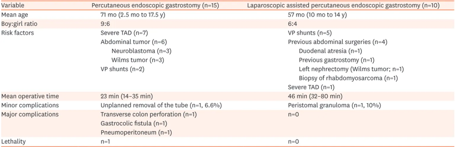
In the first group, the mean age of the patients was 71 months (range: 2.5 months to 17.5 years) and the boy:girl ratio was 9:6. In the second group, the mean age of the patients was 57 months (range: 10 months to 14 years) and the boy:girl ratio was 6:4 (Table 1).
Indications for gastrostomy in all cases were feeding difficulties or malnutrition.
Risk factors in the first group were severe TAD (n=7), abdominal tumor (n=6; neuroblastoma [n=3] and Wilms tumor [n=3]), and VP shunts (n=2), and those in the second group were VP
Table 1. Comparison of conventional and laparoscopic-assisted percutaneous endoscopic gastrostomies
Variable Percutaneous endoscopic gastrostomy (n=15) Laparoscopic assisted percutaneous endoscopic gastrostomy (n=10)
Mean age 71 mo (2.5 mo to 17.5 y) 57 mo (10 mo to 14 y)
Boy:girl ratio 9:6 6:4
Risk factors Severe TAD (n=7) VP shunts (n=5)
Abdominal tumor (n=6) Previous abdominal surgeries (n=4)
Neuroblastoma (n=3) Duodenal atresia (n=1)
Wilms tumor (n=3) Previous gastrostomy (n=1)
VP shunts (n=2) Left nephrectomy (Wilms tumor; n=1)
Biopsy of rhabdomyosarcoma (n=1) Severe TAD (n=1)
Mean operative time 23 min (14–35 min) 46 min (32–80 min)
Minor complications Unplanned removal of the tube (n=1, 6.6%) Peristomal granuloma (n=1, 10%) Major complications Transverse colon perforation (n=1) n=0
Gastrocolic fistula (n=1) Pneumoperitoneum (n=1)
Lethality n=1 n=0
shunts (n=5), previous abdominal surgeries (n=4; duodenal atresia, previous gastrostomy, left nephrectomy because of Wilms tumor, and tumor biopsy of rhabdomyosarcoma), and severe TAD (n=1). Adhesions were found in three (30%) patients, and they were released laparoscopically. There was no need for a conversion.
The mean operative time for the PEG procedure was 23 minutes (range: 14–35 minutes), whereas that for the L-PEG procedure was 46 minutes (range: 32–80 minutes) in the first group. The Welch's two-sample t-test revealed a significant difference between the length of the two procedures. The mean operative time of L-PEG was significantly (p=0.001) longer
than that of the conventional PEG, especially if adhesiolysis was required (60–80 minutes). After PEG placement, refeeding was started with water at 8 hours followed by formula at 24 hours in both the groups. The refeeding time did not significantly differ between the two groups. Hospital stay depended on refeeding time and underlying diseases and not on the operative technique.
Adverse effects were classified as minor or major according to the European Society for Pediatric Gastroenterology, Hepatic and Nutrition guidelines [5]. Minor complications occurred in two (8%) patients. In the first group, one (6.6%) patient underwent unplanned removal of the tube. The skin opening was closed immediately after unplanned removal and the internal fixation plate was emptied with a stool. In the second group, the occurrence of peristomal granuloma was noted in one (10%) patient.
We observed three major complications: transverse colon perforation, gastrocolic fistula, and pneumoperitoneum. All the three complications occurred in the first group (20%). No major complications (0%) were observed in the second group.
Regarding lethal outcome, one patient in the first group with severe comorbidities died because of severe outcomes of his general condition long after the postoperative period. However, no association was found between the fatal outcome and the operation.
DISCUSSION
Tube feeding is the method of choice when enteral nutrition is recommended and oral intake is insufficient. Previously, open gastrostomies were performed by surgeons through laparotomy. A Pezzer catheter was inserted into the stomach and fixed with a double-layer purse-string suture. Thereafter, the tube was brought out through a stab incision in the abdominal wall [2].
After PEG was first described by Gauderer [2] in 1980, this minimally invasive technique became the gold standard. The advantages of PEG are less scarring, shorter operative time, fewer infections, less postoperative pain, and shorter hospital stay [2]. In most cases, when the esophagus is patent and transillumination of the stomach through the abdominal wall is achievable, PEG tube placement is safe. The three principles of safe PEG placement are endoscopic gastric distension, endoscopically visible focal finger invagination, and transillumination [3,4]. However, these criteria are not considered in children with distorted anatomy because of severe scoliosis or intra-abdominal adhesions due to VP shunts, peritoneal dialysis, or previous operations. In these patients, a high risk of bowel or hepatic
injury exists. Laparoscopy offers better and direct visualization of the stomach, and any adhesions can be released with this minimally invasive method.
According to a literature review on the complications of PEG insertions, the most common major complications after the conventional PEG procedure are systemic infections (3.5%) and peritonitis, sepsis, or wound dehiscence (1.5%). Pneumoperitoneum occurs in 0.7% of the patients. Asymptomatic pneumoperitoneum can occur without intestinal perforation because of the procedure; however, esophagus or bowel perforations occur in 0.3% of the patients. Gastrocolic fistulas because of the interposition of the splenic flexure between the anterior abdominal and gastric walls occurs in 0.45% of the patients. Buried bumper, intra-abdominal bleeding, and ileus are detected in 1% of the patients [3]. Impaired coagulation, severe ascites, peritonitis, and local esophageal and general gastrointestinal obstructions are considered absolute contraindications for PEG placement [6]. Severe kyphoscoliosis with interposed organs and distorted anatomy are relative contraindications [6]. Vervloessem et al. [7] analyzed the potential risk factors for major complications in 449 patients and found that only VP shunts were associated with a significantly high major complication rate. Although PD catheters, hepatomegaly, esophageal stenosis, and coagulopathy had high complication rates, the difference between the two rates was not significant.
In our institute, L-PEG was started in 2014 after a major complication in a patient with a VP shunt. Thereafter, all patients at high risk for intestinal injury (patients with VP shunt, PD catheter, previous abdominal surgery, severe thoracoabdominal deformities, hepatomegaly, or intra-abdominal masses) underwent L-PEG placement. Before selection of patients,
conventional PEG placement was performed in 15 high-risk patients, that is patients with severe TAD (n=7), abdominal tumor (n=6), and VP shunts (n=2). Three major complications, namely colon perforation (n=1), gastrocolic fistula (n=1), and pneumoperitoneum (n=1), occurred. Colonic perforation was found in a patient with a 2-year-old VP-shunt. The patient developed peritonitis on the first postoperative day. Laparotomy was performed, and two perforation openings were found in the transverse colon, which were closed with a double-layer suture. The distal catheter of the VP shunt was temporarily externalized. The PEG was transferred to a gastrostomy tube. A gastrocolic fistula was observed in a 3-year-old boy with Fallot-tetralogy, severe TAD, and somatomental retardation. The internal bumper was removed endoscopically and the chronic fistula was planned to be closed; however, the patient was lost to follow-up and the chronic fistula was closed surgically. Pneumoperitoneum because of early dislodgement of the tube in the early postoperative period by an autistic patient with severe TAD was observed. Gastropexy was performed laparoscopically. This complication was independent of the surgical technique as well as patient's high-risk status.
After selection of high-risk patients, 10 L-PEG placements were performed and the indications for laparoscopic guidance were VP shunts (n=5), previous abdominal surgeries (n=4; duodenal atresia, previous gastrostomy, left nephrectomy because of Wilms tumor, and tumor biopsy from rhabdomyosarcoma), and severe TAD (n=1). Adhesions were found in three (30%) patients, of which two had a VP shunt and one had a previous gastrostomy. The advantage of L-PEG is that surgeons and endoscopists perform the same procedures, and therefore, there is no requirement for learning a new technique. The endoscopist performs the original pull technique and the surgeon attains umbilical access as in any laparoscopic procedure for a 5-mm camera port. We recommend the open (Hasson) technique over the Veress needle technique to prevent vessel, hepatic, or bowel injury. Any adhesions can be
released laparoscopically. In the case of no adhesions in the stomach, the conventional PEG procedure can be performed under double visual control. Although the laparoscopic procedure is longer, it is safer than the endoscopic procedure not only for high-risk patients but also for all patients.
This study has limitations owing to its retrospective nature and small sample size. However, L-PEG is not widespread in the literature.
Our results revealed that the major complication rate of L-PEG was lower than that of conventional PEG in high-risk patients; however, the operative time of L-PEG was significantly longer, especially if adhesiolysis was required.
Laparoscopic guidance provides a clear intra-abdominal view and offers the possibility to release adhesions and therefore, adjacent bowel or hepatic injuries can be avoided. L-PEG is recommended for children with distorted anatomy, VP shunts, or previous abdominal surgeries. L-PEG can be an emergency aid if transillumination of the gastric wall is
inappropriate during gastroscopy. PEG placement in high-risk patients is advised in centers with pediatric surgical departments, where laparoscopy is in everyday use.
REFERENCES
1. Löser C, Aschl G, Hébuterne X, Mathus-Vliegen EM, Muscaritoli M, Niv Y, et al. ESPEN guidelines on artificial enteral nutrition--percutaneous endoscopic gastrostomy (PEG). Clin Nutr 2005;24:848-61. PUBMED | CROSSREF
2. Gauderer MW, Ponsky JL, Izant RJ Jr. Gastrostomy without laparotomy: a percutaneous endoscopic technique. J Pediatr Surg 1980;15:872-5.
PUBMED | CROSSREF
3. Balogh B, Kovács T, Saxena AK. Complications in children with percutaneous endoscopic gastrostomy (PEG) placement. World J Pediatr 2019;15:12-6.
PUBMED | CROSSREF
4. Zamakhshary M, Jamal M, Blair GK, Murphy JJ, Webber EM, Skarsgard ED. Laparoscopic vs percutaneous endoscopic gastrostomy tube insertion: a new pediatric gold standard? J Pediatr Surg 2005;40:859-62. PUBMED | CROSSREF
5. Heuschkel RB, Gottrand F, Devarajan K, Poole H, Callan J, Dias JA, et al. ESPGHAN position paper on management of percutaneous endoscopic gastrostomy in children and adolescents. J Pediatr Gastroenterol Nutr 2015;60:131-41.
PUBMED | CROSSREF
6. El-Matary W. Percutaneous endoscopic gastrostomy in children. Can J Gastroenterol 2008;22:993-8. PUBMED | CROSSREF
7. Vervloessem D, van Leersum F, Boer D, Hop WC, Escher JC, Madern GC, et al. Percutaneous endoscopic gastrostomy (PEG) in children is not a minor procedure: risk factors for major complications. Semin Pediatr Surg 2009;18:93-7.