INTRODUCTION
With the development of implantology, installation of dental implants and rehabilitation in severely atro-phic bone area became more routine procedures at dental clinics. However,the need for enough bone support and sufficient bone architecture still exists in most of clinical cases. Among the available materials used for bone reconstruction, autogenous bone is cur-rently the gold-standard because it is a source of
osseous matrix, cells, and growth modulating mole-cules1). However, the use of autogenous bone is lim-ited by donor site morbidity, the quantity of donor bone, frequent infections and unpredictable resorption that may compromise aesthetics and function. To overcome the limits of autogenous bone, various sub-stitutive biomaterials are proposed. Materials of hu-man and animal origin have the potential risk of cross contamination and the disadvantages of limited sup-ply2,3). As a consequence, synthetic products as the biphasic calcium phosphate (BCP) which is a mixture of hydroxyapatite (HA) and β-tricalcium phosphate (β-TCP) were introduced4-7).
Previous reports have shown that HA particles did not elicit an inflammatory response and they provided a good scaffold for the new bone to grow in. However,
Bone regeneration capacity of two different macroporous biphasic calcium
materials in rabbit calvarial defect
Jung-Chul Park
1, Hyun-Chang Lim
1, Joo-Yeon Sohn
1, Jeong-Ho Yun
2, Ui-Won Jung
1,
Chang-Sung Kim
1, Kyoo-Sung Cho
1, Chong-Kwan Kim
1, Seong-Ho Choi
1*1. Department of Periodontology, Research Institute for Periodontal Regeneration, College of Dentistry, Yonsei University 2. Department of Dentistry, College of Medicine, Kwandong University, Myongji Hospital
ABSTRACT
Purpose: Synthetic bone products such as biphasic calcium phosphate (BCP) are mixtures of hydroxyapatite (HA) and β-tricalcium phosphate (β- TCP). In periodontal therapies and implant treatments, BCP provides to be a good bone reconstructive material since it has a similar chemical composition to biological bone apatites. The purpose of this study was to compare bone regeneration capacity of two commercially available BCP.
Methods: Calvarial defects were prepared in sixteen 9-20 months old New Zealand White male rabbits. BCP with HA and β- TCP (70:30) and BCP with Silicon-substituted hydroxyapatite (Si-HA) and β-TCP (60:40) particles were filled in each defect. Control defects were filled with only blood clots. Animals were sacrificed at 4 and 8 week postoperatively. Histomorphometric analysis was performed.
Results: BCP with HAand β- TCP 8 weeks group and BCP with Si-HA and β- TCP 4 and 8 weeks groups showed statistically significant in crease (P <0.05) in augmented area than control group. Newly formed bone area after 4 and 8 weeks was similar among all the groups. Residual materials were slightly more evident in BCP with HA and β- TCP 8 weeks group.
Conclusions: Based on histological results, BCP with HA and β- TCP and BCP with Si-HA and β- TCP appears to demonstrate acceptable space maintaining capacity and elicit significant new bone formation when compared to natural bone healing in 4 and 8 week periods. (J Korean Acad Periodontol 2009;39:223-230)
KEY WORDS: bone substitutes; hydroxyapatite wound healing.
Correspondence: Dr. Seong-Ho, Choi
Department of Periodontology, School of Dentistry, Yonsei University, Shinchon-dong, Seodaemoon-gu, Seoul, 120-752, Korea.
E-mail : [email protected], Tel : 82-2-2228-3190, Fax : 82-2-392-0398 Received: Jun 4, 2009; Accepted: Jul 24, 2009
This study was supported by a faculty research grant of Yonsei University College of Dentistry for 2008(6-2008-0071).
HA particles failed to show evidence of new perio-dontal tissue attachment, osteogenesis, or cemento-genesis in the treatment of periodontal osseous de-fects8-10). It was rather suspected that the material produced a response like a well-tolerated foreign body within the host connective tissue. On the contrary, β -TCP was reported to resorb unpredictably in biologic fields and not to provide a predictable scaffold for new bone to grow in11-13). However the combination of HA and β-TCP and the development of two-phased calcium phosphate or biphasic calcium phosphate ce-ramic made it possible to control the resorbability of the material and at the same time maintain its osteo-conductive property14).
In many cases of bone grafts in implant surgeries, BCP provides good bone reconstruction since it has similar chemical composition to biological bone apatites. Also it has been proved of its efficacy as a bone substitute material in many human clinical ap-plications3,15,16). There has been lots of trials to find ideal combination of HA/β-TCP ratio to maximize bone regeneration capacity. Early studies have shown that approximately 60% of HA and 40% of β-TCP seemed to provide a reasonable bone conductive prop-erty3,10,14,17). However the optimal ratio is still not determined. Consequently, many manufactures have come up with their unique combinations of HA/β-TCP.
The Si-substituted macroporous biphasic calcium phosphate (Bonemedik-DM◯R, META bio med Co. Ltd.,
Chungwongun, Korea) is a mixture of HA and β-TCP with a ratio of 60 : 40. The material is manufactured by substituting silicon (Si) ions of HA into phosphate (P) sites and mixing β-TCP granules with HA par-ticles to enhance both physical and chemical factors18). The presence of silicon in HA is knownto play an im-portant part on the formation of bone19). The overall granule sizes are from 0.25 to 1 mm. The other prod-uct on the market (Osteon◯R, Genoss. Co. Ltd., Suwon,
Korea) is a mixture of HA and β-TCP with a ratio of
70 : 30. The granule sizes are from 0.5 to 2 mm. The purpose of this study was to evaluate bone re-generation capacity of two commercially available bi-phasic calcium phosphate in rabbit calvarial defect.
MATERIALS AND METHODS
1. Animals
Sixteen 9-20 months old New Zealand White male rabbits (Oryctolagus cuniculus) were used. The mals were housed in cages and fed with standard ani-mal food. This study has been reviewed and approved by the Institutional Animals Care and Use Committee, Yonsei Medical Center, Seoul, Korea.
2. Surgical procedures
All surgeries were performed under sterile conditions. All rabbits were anesthetized with an in-tramuscular (IM) injection of a solution of 91% ket-amine hydrochloride (Ketalar◯R, Yuhan Co., Seoul,
Korea) and 9% xylazine(Rumpun◯R, Bayer Korea Ltd.,
Seoul, Korea). Surgical area of cranium was anes-thetized with 2% lidocaine and the scalps were shaved and disinfected. The calvariae were exposed through a mid-line skin incision. The periosteum was retracted laterally and bilaterally, and 8-mm-diameter defects were made in the parietal bones by means of a stand-ardized trephine cutting bur under physiological saline solution irrgation20).
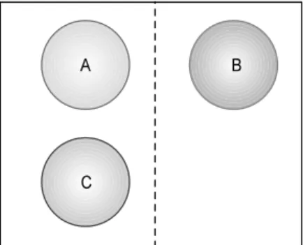
Three defects were made per one animal in triangu-lar shape with an inter defect distance of 3 mm to exclude any influence to different groups. To create the same environments, sagittal suture was avoided when creating defects. The defects were filled with the respective materials, with one cavity destined to the control group containing blood clot. The defect allocation was randomly assigned in each rabbit (Fig.
1). The graft materials were wetted in sterilized saline and gently packed into the defects. Particle size was 1.0~2.0 mm of Osteon◯R and Bonemedik-DM◯R.
A B
C
Figure 1. Schematicdrawing of designated material filled in defects. Dotted line represents sagittal suture of cranium; A: Control, B: Bonemedik-DM◯R, C: Osteon◯R.
The periosteum was drawn over the defects and su-tured by resorbable suture material (5-0 Vicryl◯R,
Ethicon, Somerville, NJ, USA). Subcutaneous mucosa were adapted and sutured with resorbable suture ma-terial (4-0 Vicryl◯R, Ethicon, Somerville, NJ, USA).
Finally the skin was closed with 4-0 absorbable mon-ofilament suture (Monosyn◯R, Braun, Tuttlingen,
Germany). The sutures were removed after 10 days after the confirmation of intact closure of skin. The rabbits were sacrificed after 4 and 8 weeks.
3. Histological preparation
The experimental specimens were obtained by using number 702 bur under physiological saline irrigation. The specimens were fixed in 10% formaldehyde sol-ution for 10 days and they were processed to the rou-tine procedures of slide preparation with 8 µm sec-tions stained with Haematoxylin-Eosin (H-E) to be analyzed under an optical microscope (Olympus BX50, Olympus Optical Co., Tokyo, Japan).
4. Statistical analysis
Numerical data was presented as mean plus one standard deviation. One way analysis of variance
(ANOVA) with Holm-Sidak method was used for mul-tiple comparisons to compare with the control. The probability level of P < 0.05 was regarded as statisti-cally significant.
RESULTS
1. Histological observation
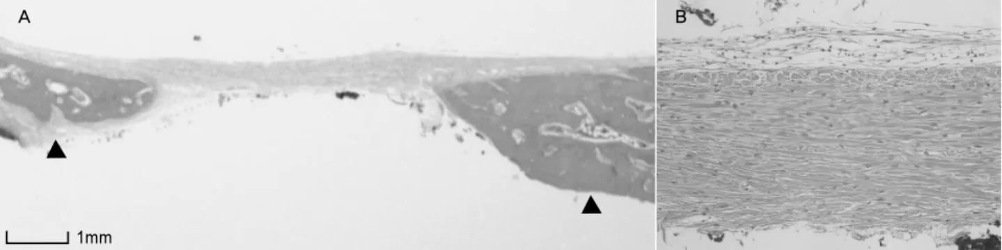
All treatment and control sites healed uneventfully with no clinical evidence of inflammatory response to the graft material. Also the distribution of residual graft materials was homogeneous on the surface of the bone defect, regardless of group (Osteon, Bonemedik-DM). For the control group, the perforated areas were filled with loose fibrous tissue and a few newly formed bone was observed. A slight decrease in bone ingrowth towards the center of the defect could be observed compared to test groups (Fig. 2, 3).
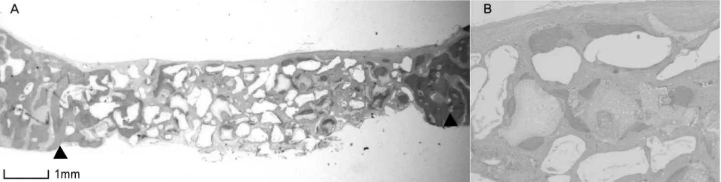
For the Osteon group, lamellar bone was apposed in close contact at the surface of the granules and con-sequent bone ingrowth was observed in both 4 and 8 weeks. However, relatively large particles of Osteon◯R
were evident in 4 weeks which were in the middle of resorption and substitution into new bone. On the contrary, particle size and total area were reduced in 8 weeks. Also the density of newly formed bone was higher compared to 4 week. Fibrous tissues were still observed between the granules (Fig. 4, 5).
For the Bonemedik-DM group, particles were in-corporated in mature new bone and were resorbed during the remodeling process in 4 weeks. The in-dividual particles were clearly identifiable and they were surrounded by varying amounts of newly formed bone without being encapsulated by loose fibrous con-nective tissues. The amount of bone ingrowth was greater than for the Osteon group. Small particles surrounded by new bone were present between the larger ones (Fig. 6, 7).
A B
Figure 2. Light micrographs of control group at 4 weeks postoperatively. Thick fibrous tissue is covering the defect ×6 (A), ×200 (B).
A B
Figure 3. Light micrographs of control group at 8 weeks postoperatively. Mature bone tissue is observed among connective tissue ×16 (A), ×200 (B).
A B
Figure 4. Light micrographs of Osteon group at 4 weeks postoperatively. Relatively large particle of residual materials are surrounded obsteoblasts and newly formed bone ×16 (A), ×200 (B).
A B
Figure 5. Light micrographs of Osteon group at 8 weeks postoperatively. Relatively small particles are scattered and newly formed bone are surrounding ×16 (A), ×200 (B).
1mm 1mm
1mm
Relatively irregular inner surface of unresorbed ce-ramic particles of Bonemedik-DM group indicates that resorption process took place before deposition of mineralized bone matrix. On the contrary, relatively smooth appearing inner surface was found in Osteon group.
2. Histomorphometric analysis
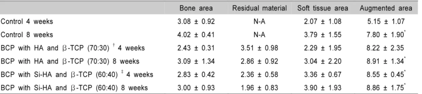
Bone substitutes are recognized through bone growth from the host bone to the graft, and pro-gressing from the outer part to the core by osteoconduction. There were osteoblastic and osteoid layers at the border of the defect and around grafted particles in Osteon and Bonemedik-DM groups. The total area of augmentation, bone area, residual mate-rial area and soft tissue area were measured.
Total augmented area including bone area, residual materials and soft tissue area was higher in Osteon 4
and 8 week groups and Bonemedik-DM 4 and 8 week groups than the control group (Table 1, Fig. 2, 3). These results were statistically significant (P< 0.05). Bone area at 4 week was slightly higher in Bonemedick- DM group which means early bone ingrowth. On the contrary, Osteon group showed more bone area than Bonemedik-DM group. Residual materials were de-creased in both Osteon and Bonemedik- DM groups with no statistical significance. Soft tissue area was dominantly wider in Bonemedik-DM groups regardless of weeks. In grafted groups, total amount of bone formation and residual materials exceed or is equal to control groups.
DISCUSSION
In the scope of bone regeneration, the augmentation of bone defects using autogenous bone to be the gold
A B
Figure 6. Light micrographs of Bonemedik-DM at 4 weeks postoperatively. Active resorption process is observed ×16 (A), ×200 (B). A B
Figure 7. Light micrographs of Bonemedik-DM at 8 weeks postoperatively. Mature bone tissue is apposed interspace of re-sidual materials ×16 (A), ×200 (B).
1mm 1mm
standard. However, autogenous bone is not always available in sufficient volume according to clinical situations. Therefore various methods such as graft materials and growth factors are proposed to replace autogenous bone. Currently various growth factors such as TGF-β, PDGF, and VEGF are studied to stim-ulate regeneration of bone tissue in several ways21-23). Also rhBM P-2, recombinant human osteogenic pro-tein-1 (rhOP-1/rhBMP-7), and recombinant human growth/differentiation factor-5 (rhGDF-5) are being pursued as therapies for reconstruction and repair of induced and congenital skeletal defects24,25). However these growth factors are not well documented and commercially unavailable. Therefore, most of clinicians are prone to choose various grafting materials to overcome bone defects in implantology or periodontal therapies in most cases.
Calcium phosphate ceramics have long been inves-tigated as biologically compatible material to used in the treatment of periodontal osseous defects and im-plant surgery. Especially two-phased calcium phos-phate or biphasic calcium phosphos-phate ceramic was de-veloped to control the resorbability of the material and at the same time maintain its osteoconductive property. The in vivo and in vitro dissolution of cal-cium phosphate ceramics was found to be dependent on the composition, crystallinity, and pH of the sol-ution26). Kwon et al27) reported that biphasic HA/β -TCP composite powders showed average solubility
between HA and β-TCP and concluded that the dis-solution rate of the calcium phosphate powders was strongly dependent on the β-TCP content. It is well known that early resorption of materials cannot maintain appropriatespace to regenerate bone tissue. On the other hand, the delayed resorption of materials inhibits new bone formation. Therefore, appropriate timing of dissolution of BCP and choosing right ratio of HA/β-TCP is crucial to maximize bone regeneration.
Gauthier et al28) used 60 % of HA and 40% of β -TCP in extraction sockets in canine models to report that well formed cortical bone over the materials was present and it inhibited the resorption of alveolar bone after 3 months. Nery et al14) used different ratio of HA/β-TCP in canine models to prove that higher HA ratio showed accelerated new bone formation and new attachment levels.
The results of this study indicated that the bone filling of calvarial defect realized with micro macro-porous biphasic calcium phosphate granules after 4 to 8 weeks have moderate bone ingrowth capacity and it appears to demonstrate a pattern that is higher HA ratio to β-TCP tends to show greater bone re-generation as shown histologically. However, statisti-cally significant different was not noted between Osteon and Bonemedik-DM.
Total residual materials were much more abundant in Osteon 8 weeks group. This may be due to the low dissolution rate of porous HA17) and easy resorbability
Table 1. Histomophometric Results at 4 and 8 Weeks. All Parameters Are Expressed As mm2 (Group Mean ± SD)
Bone area Residual material Soft tissue area Augmented area
Control 4 weeks 3.08 ± 0.92 N-A 2.07 ± 1.08 5.15 ± 1.07
Control 8 weeks 4.02 ± 0.41 N-A 3.79 ± 1.55 7.80 ± 1.90*
BCP with HA and β -TCP (70:30) † 4 weeks 2.43 ± 0.31 3.51 ± 0.98 2.29 ± 1.95 8.22 ± 2.35
BCP with HA and β -TCP (70:30) 8 weeks 3.09 ± 1.34 2.86 ± 0.92 3.04 ± 2.20 8.91 ± 1.34*
BCP with Si-HA and β -TCP (60:40) ‡ 4 weeks 2.83 ± 0.42 2.36 ± 0.58 3.36 ± 0.67 8.55 ± 0.45*
BCP with Si-HA and β -TCP (60:40) 8 weeks 3.00 ± 0.93 1.96 ± 0.83 3.90 ± 1.93 8.86 ± 1.75*
* : Significant statistical difference compared to corresponding control group at 4 weeks (P<0.05) †: Osteon®, Genoss. Co. Ltd., Suwon, Korea
of β-TCP which were proven in vitro29) and in vivo12,30). Therefore the bone quality might be influ-enced inferiorly by remaining particles. However, these residual materials may stay long enough for cell differentiation, maturation, and revascularization31). Longer period of observation would be required to fully evaluate these results.
The results from this study confirm the resorb-ability on time of Osteon◯R and Bonemedik-DM◯R andthe
scaffold effect of the HA content and high osteo-conduction property. These two crucial properties in-volved a balance of resorption and bone ingrowth at the expense of the micro-macropours bioceramics.
Although this study showed that the amount of bone regeneration of control sites was higher than graft sites, the BCP grafts also showed reasonable bone ingrowth and can be a good candidate to replace natural bone regeneration and to maintain space for longer period of time. Despite the limitation of this study because of small sample size, we could conclude that the higher portion of HA particles produced lon-ger resorption period and maintained better bone structure. Also, graft of Osteon◯R and Bonemedik-DM◯R
in the rabbit calvarial defects model was shown to be potentially beneficial at early bone healing.
REFERENCES
1. Barboza EP. Clinical and histologic evaluation of the dem-ineralized freeze-dried bone membrane used for ridge augmentation. Int J Periodontics Restorative Dent 1999; 19:601-607.
2. Daculsi G, Laboux O, Malard O, Weiss P. Current state of the art of biphasic calcium phosphate bioceramics. J Mater Sci Mater Med 2003;14:195-200.
3. Daculsi G, Passuti N, Martin S et al. Macroporous calcium phosphate ceramic for long bone surgery in humans and dogs. Clinical and histological study. J Biomed Mater Res 1990;24:379-396.
4. LeGeros RZ, Parsons JR, Daculsi G et al. Significance of
the porosity and physical chemistry of calcium phosphate ceramics. Biodegradation-bioresorption. Ann N Y Acad Sci 1988;523:268-271.
5. Levin MP, Getter L, Adrian J, Cutright DE. Healing of pe-riodontal defects with ceramic implants. J Clin Periodontol 1974;1:197-205.
6. Levin MP, Getter L, Cutright DE, Bhaskar SN. Biodegradable ceramic in periodontal defects. Oral Surg Oral Med Oral Pathol 1974;38:344-351.
7. Yukna RA, Cassingham RJ, Caudill RF et al. Six month evaluation of Calcitite (hydroxyapatite ceramic) in perio-dontal osseous defects. Int J Periodontics Restorative Dent 1986;6:34-45.
8. Froum SJ, Kushner L, Scopp IW, Stahl SS. Human clinical and histologic responses to Durapatite implants in intra-osseous lesions. Case reports. J Periodontol 1982;53: 719-725.
9. Moskow BS, Lubarr A. Histological assessment of human periodontal defect after durapatite ceramic implant. Report of a case. J Periodontol 1983;54:455-462.
10. Ellinger RF, Nery EB, Lynch KL. Histological assessment of periodontal osseous defects following implantation of hydroxyapatite and biphasic calcium phosphate ceramics: a case report. Int J Periodontics Restorative Dent 1986; 6:22-33.
11. Karabuda C, Ozdemir O, Tosun T, Anil A, Olgac V. Histological and clinical evaluation of 3 different grafting materials for sinus lifting procedure based on 8 cases. J Periodontol 2001;72:1436-1442.
12. Jarcho M. Calcium phosphate ceramics as hard tissue prosthetics. Clin Orthop Relat Res 1981:259-278.
13. Jarcho M. Biomaterial aspects of calcium phosphates. Properties and applications. Dent Clin North Am 1986; 30:25-47.
14. Nery EB, LeGeros RZ, Lynch KL, Lee K. Tissue response to biphasic calcium phosphate ceramic with different ratios of HA/beta TCP in periodontal osseous defects. J Periodontol 1992;63:729-735.
15. Block MS, Kent JN. A comparison of particulate and solid root forms of hydroxylapatite in dog extraction sites. J Oral Maxillofac Surg 1986;44:89-93.
16. Lee J, Jung U, Kim C, Choi S, Cho K. Maxillary sinus augmentation using macroporous biphasic calcium phos-phate (MBCPTM) : Three case report with histologic
evaluation. J Korean Acad Periodontol 2006;36:567-577. 17. Daculsi G, LeGeros RZ, NeryE, Lynch K, Kerebel B.
Transformation of biphasic calcium phosphate ceramics in vivo: ultrastructural and physicochemical characterization. J Biomed Mater Res 1989;23:883-894.
18. Bertoni E, Bigi A, Cojazzi G et al. Nanocrystals of magne-sium and fluoride substituted hydroxyapatite. J Inorg Biochem 1998;72:29-35.
19. Skrtic D, Antonucci JM, Eanes ED, Brunworth RT. Silica- and zirconia-hybridized amorphous calcium phosphate: ef-fect on transformation to hydroxyapatite. J Biomed Mater Res 2002;59:597-604.
20. Hollinger JO, Kleinschmidt JC. The critical size defect as an experimental model to test bone repair materials. J Craniofac Surg 1990;1:60-68.
21. Marx RE, Carlson ER, Eichstaedt RM et al. Platelet-rich plasma: Growth factor enhancement for bone grafts. Oral Surg Oral Med Oral Pathol Oral Radiol Endod 1998; 85:638-646.
22. Sandy J, Davies M, Prime S, Farndale R. Signal pathways that transduce growth factor-stimulated mitogenesis in bone cells. Bone 1998;23:17-26.
23. Horner A, Bord S, Kemp P, Grainger D, Compston JE. Distribution of platelet-derived growth factor (PDGF) A chain mRNA, protein, and PDGF-alpha receptor in rapidly forming human bone. Bone 1996;19:353-362.
24. Lee YJ, Jung SW, Chae GJ, Cho KS, Kim CS. The effect of recombinant human bone morphogenetic
protein-2/mac-roporous biphasic calcium phosphate block system on bone formation in rat calvarial defects. J Korean Acad Periodontol 2007;37:397-407.
25. Wikesjo UM, Huang YH, Polimeni G, Qahash M. Bone morphogenetic proteins: a realistic alternative to bone graft-ing for alveolar reconstruction. Oral Maxillofac Surg Clin North Am 2007;19:535-551, vi-vii.
26. Klein CP, Driessen AA, de Groot K, van den Hooff A. Biodegradation behavior of various calcium phosphate ma-terials in bone tissue. J Biomed Mater Res 1983;17: 769-784.
27. Kwon S, Jun Y, Hong S. Synthesis and dissolution behav-ior of β-TCP and HA/β-TCP composite powders J. Euro. Ceram. Soc. 2003;23:1039-1045.
28. Gauthier O, Bouler JM, Aguado E, Pilet P, Daculsi G. Macroporous biphasic calcium phosphate ceramics: influ-ence of macropore diameter and macroporosity percentage on bone ingrowth. Biomaterials 1998;19:133-139.
29. LeGeros RZ. Calcium phosphate materials in restorative dentistry: a review. Adv Dent Res 1988;2:164-180. 30. Metsger DS, Driskell TD, Paulsrud JR. Tricalcium
phos-phate ceramic--a resorbable bone implant: review and cur-rent status. J Am Dent Assoc 1982;105:1035-1038. 31. Um YJ, Hong JY, Kim ST et al. Bone formation of newly
developed biphasic calcium phosphate in rabbit calvarial defect model : A pilot study. J Korean Acad Periodontol 2008;38:163-170.