저작자표시-비영리-변경금지 2.0 대한민국 이용자는 아래의 조건을 따르는 경우에 한하여 자유롭게 l 이 저작물을 복제, 배포, 전송, 전시, 공연 및 방송할 수 있습니다. 다음과 같은 조건을 따라야 합니다: l 귀하는, 이 저작물의 재이용이나 배포의 경우, 이 저작물에 적용된 이용허락조건 을 명확하게 나타내어야 합니다. l 저작권자로부터 별도의 허가를 받으면 이러한 조건들은 적용되지 않습니다. 저작권법에 따른 이용자의 권리는 위의 내용에 의하여 영향을 받지 않습니다. 이것은 이용허락규약(Legal Code)을 이해하기 쉽게 요약한 것입니다. Disclaimer 저작자표시. 귀하는 원저작자를 표시하여야 합니다. 비영리. 귀하는 이 저작물을 영리 목적으로 이용할 수 없습니다. 변경금지. 귀하는 이 저작물을 개작, 변형 또는 가공할 수 없습니다.
Three Dimensional CT based Virtual Patellar
Resection in Female Patients undergoing Total
Knee Replacement
by
Do Young Park
Major in Medicine
Department of Medical Sciences
The Graduate School, Ajou University
Three Dimensional CT based Virtual Patellar
Resection in Female Patients undergoing Total
Knee Replacement: A Comparison between
Tendon and Subchondral Method
by
Do Young Park
A Dissertation Submitted to The Graduate School of
Ajou University in Partial Fulfillment of the Requirements
for the Degree of
Master of Medicine
Supervised by
Ye Yeon Won, M.D.
Major in Medicine
Department of Medical Sciences
The Graduate School, Ajou University
This certifies that the dissertation
of Do Young Park is approved.
SUPERVISORY COMMITTEE
Ye Yeon Won
Kyeong Jin Han
Jae-Ho Cho
The Graduate School, Ajou University
December 20th, 2011
- ABSTRACT -
Three Dimensional CT based Virtual Patellar Resection in Female
Patients undergoing Total Knee Replacement; A Comparison
between Tendon and Subchondral Method
Background: Though rotational alignment and antero-posterior positioning of the femoral
component influences patello-femoral mechanics, it is also significantly influenced by the accuracy of the patellar resection, with the resultant thickness and symmetry of the patella-implant composite
Methods: We performed virtual patellar resection with digital software using 3D-CT scans
of knees from 49 patients who underwent primary TKR at our hospital, comparing two commonly used resection methods, i.e., the tendon method (TM), and the subchondral method (SCM), to determine an ideal resection plane with respect to the symmetry and thickness of the patellar remnant
.
Results: The TM gave a thicker resected patella, and a less oval cut surface shape which
Conclusions: Though TM appears statistically better with respect to the thickness and cut
surface shape, only further intra-operative studies with long-term clinical follow up may provide us with the most appropriate patellar resection method.
Key words: patella, 3D CT, patellar resurfacing
TABLE OF CONTENTS
ABSTRACT ··· ⅰ
TABLE OF CONTENTS ··· iii
LIST OF FIGURES ··· iv
LIST OF TABLES ··· v
Ⅰ. INTRODUCTION ··· 1
Ⅱ. MATERIALS AND METHODS ··· 3
A. MATERIALS ··· 3 1. SUBJECTS ··· 3 B. METHODS ··· 5 1. SETTING ··· 5 2. STATISTICS ··· 11 III. RESULTS ··· 12 IV. DISCUSSION ··· 14 Ⅴ. CONCLUSION ··· 20 REFERENCES ··· 21 국문요약 ··· 25
LIST OF FIGURES
Fig. 1.
Reconstruction of 3-D CT images of the knee using specialized computer
software
··· 4Fig. 2.
Thickness of patella in the 3D image
··· 6Fig. 3.
Subchondral method
··· 7Fig. 4.
Anatomic landmarks chosen for resection at the fat fascia-tendon junctions
for the tendon method of resection
··· 8Fig. 5.
Measurement of ML symmetry and SI symmetry
··· 10LIST OF TABLE
Table 1. Comparison of results after virtual resection with subchondral and tendon methods ··· 13
Acknowledgements
The authors gratefully acknowledge the financial support of the Korea Healthcare Technology R&D Project, Ministry for health, welfare and family affairs, Republic of Korea (A 084120).
I.
INTRODUCTION
Though patellofemoral complications contribute as one of the main factors responsible for reoperation after total knee replacement (TKR), patellar resurfacing is not generally given as much importance as the femoral or tibial resections by surgeons during TKR. (Baldini, 2007; Gomes, 1998; Kim, 2009) Its small size, variable shape and lack of distinct anatomical landmarks especially in osteoarthritic knees, make a precise patellar resection all the more difficult. Though rotational alignment and antero-posterior positioning of the femoral component influences patello-femoral mechanics, it is also significantly influenced by the accuracy of the patellar resection, with the resultant thickness and symmetry of the patella-implant composite. (Pagnano, 2000; Baldwin, 2005; Bengs, 2006; Hsu, 1996; Kurosaka, 2002)
Asymmetric resurfacing results in a higher incidence of anterior knee pain and abnormal patellar tracking, in comparison to knees with symmetric patellar profiles. (Pagnano 2000; Anglin, 2009) The depth of bone resection and the bone prosthesis composite thickness significantly influence knee function following TKR. An over-resection resulting in a thin patella can increase the risk of stress fracture and anteroposterior instability. (Hsu, 1996; Kurosaka, 2002; Levai, 2001) An under-resection on the other hand, causes overstuffing of the joint, thereby increasing contact forces and may reduce knee flexion. (Baldwin, 2005; Hsu 1996) The shape of the resected surface
of the patella also has a relation to the coverage obtained with the prosthesis. (Kim, 2009; Baldwin, 2005; Ledger 2005)
Anglin et al (Anglin, 2009), in an effort to improve the symmetry of patellar resection, had compared different radiographic definitions of desired resection lines. The fact that their study was based on two dimensional radiographs, however, has been quoted by them as a limitation of their study. Iranpour et al (Iranpour, 2008), using three dimensional computerized tomography (3D CT) analysis, found that the width thickness ratio of patella can be used to assess the premorbid patellar thickness, which they believed, has to be restored by the bone-prosthesis composite. But they were uncertain as to whether restoring the premorbid thickness may result in overstuffing the joint in osteoarthritic patellae. They did their study on relatively normal patellae and have not assessed the symmetry of resection or the shape of the resected surface of the patella. This prompted us to do a 3D-CT study on osteoarthritic patellae, assessing all these morphological parameters to compare two commonly used methods of patellar resection, ie, the tendon method (TM) (Ledger, 2005; Lee, 1998; Lombardi, 1998), and the subchondral method (SCM) (Iranpour, 2008; Rand, 1991; Insall, 1993). Such a quantitative comparison using 3D CT on diseased patellae has not been reported so far. The purpose of our study was to compare the morphology of resected patellae between TM and SCM using 3D CT based virtual resection and assess the usefulness of these data as a guide for the surgeon in determining an ideal resection plane during patellar resurfacing.
II.
MATERIALS AND METHODS
A. Material
1. Subjects
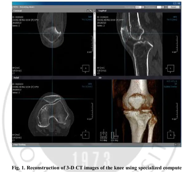
Fifty 3D-CT scans of knees from 49 female patients who underwent primary TKR for osteoarthritis at our hospital between March 2008 and October 2008 were evaluated. Exclusion criteria included any other pathologic condition other than primary osteoarthritis, as well as valgus aligned knees. All the patients had a preoperative CT scan of their diseased knee (Brilliance CT 64-channel scanner, 3rd generation, Philips, Netherlands) in a high-resolution mode using the 512 x 512 pixel matrix. Patients were supine with the involved limb in the extended position during scanning. All consecutive slices were imported into a 3D-CT reconstruction program (Lucion, Infinitt, Seoul, Korea) (Fig. 1). All patients were older than 62 years (range, 62-83 years). All of the above patients had patellofemoral osteoarthritis (Skyline Kellgren and Lawrence grade ≥2) and had received patellar resurfacing during the operation. All patients with very severe osteoarthritic changes and badly deformed patellae, where the bony landmarks were grossly obscured in the 3D CT scans were excluded from the study. The morphological parameters that were assessed were the pre-resection thickness of the whole patella, the post resection thickness, the width and height of the resected surface of the patella and the symmetry of resection, with both the TM and SCM.
Fig. 1. Reconstruction of 3-D CT images of the knee using specialized computer software. Note the corresponding coronal, sagittal and axial views(2-D images) together with the 3-D knee image.
B. Methods
1. Setting
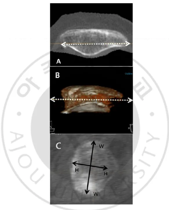
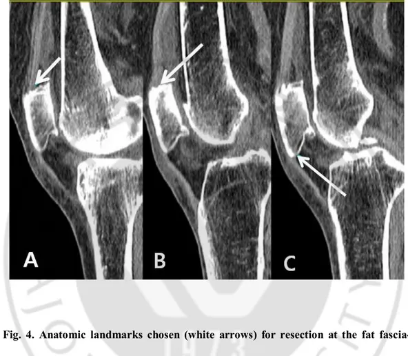
The TM involves resection through a plane that passes through the posterior limits of the quadriceps tendon medially and laterally and the patellar tendon inferiorly. (Ledger, 2005; Lee, 1998; Lombardi, 1998) The SCM involves resecting the patella at the level of the subchondral bone of the lateral facet. (Iranpour, 2008; Rand, 1991; Insall, 1993) After setting up the Lucion software, the patella was aligned in the sagittal plane with the median ridge inferiorly and the anterior surface superiorly. The thickness of the patella was then measured as the vertical distance from the midpoint of the median ridge to the dorsal surface (Fig. 2). A virtual resection in the axial plane was made at this level. This image and the 3D sagittal image were used to define a plane parallel to the anterior surface and just down to the subchondral bone of the lateral facet to virtually reproduce the SCM of resection (Fig. 3). To reproduce the TM, we used the 3D axial and sagittal images and the 2D sagittal image to select three points, one each at the posterior limits of quadriceps tendon medially and laterally and one at the posterior limit of patellar tendon inferiorly, where the Hounsfield units changed from fat (negative) to fascia-tendon (positive), just outside the bone. These three points defined the plane of resection for the TM (Fig. 4). After the 3D virtual resection was made, thickness of the resected patella was measured at the midpoint of the cut surface height in the sagittal plane. The width and height of the cut surface were measured in the 2D image as shown in Fig 3C.
Fig. 3. A-C Subchondral method showing the plane of resection(white dotted line) in the 2D axial(A) and 3D sagittal(B) images, and the cut surface in the 2D
Fig. 4. Anatomic landmarks chosen (white arrows) for resection at the fat fascia-tendon junctions for the fascia-tendon method of resection. Posterior limits of quadriceps tendon medially(A) and laterally(B) and one at the posterior limit(C) of patellar tendon inferiorly, where the Hounsfield units changed from fat (negative) to fascia-tendon (positive), just outside the bone. These three points defined the plane of resection for the TM
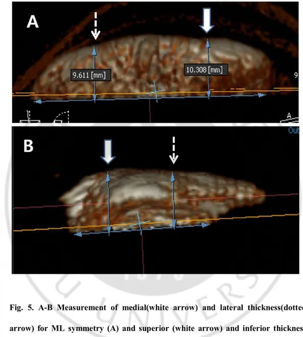
Gomes et al (Gomes, 1988) had described a method of assessing the mediolateral (ML) symmetry of the resected patella. Measuring the thickness 15mm from the medial and lateral margins of the patella, the resection is considered asymmetric when the difference between the two levels is >2mm, and as a major asymmetry when > 4mm. (Baldini, 2007) However, since these two points were too close to the center of the patella in most of our patients, we chose to assess the ML symmetry by measuring the thickness of the resected patella at the midpoints of the medial and lateral halves of the patella. (Fig. 5A) The superoinferior (SI) symmetry had not been quantitatively assessed in any previous reported studies. We assessed the SI symmetry similar to the way the ML symmetry was assessed i.e., measuring the thickness at the midpoints of the superior and inferior halves of the cut surface height in the sagittal plane. (Fig. 5B)
Fig. 5. A-B Measurement of medial(white arrow) and lateral thickness(dotted arrow) for ML symmetry (A) and superior (white arrow) and inferior thickness (dotted arrow) for SI symmetry(B).
2. Statistics
All statistical analyses were done using SPSS software (Version 13, Chicago, IL, USA) and p values less than 0.05 were considered significant. The student t test was employed to compare independent data. In order to achieve precision of the measurements, all measurements were done independently by a research fellow (SGN) and a 4th year resident (PDY), on two separate occasions with a gap of one week between the measurements. The intraclass correlation coefficients for intra and inter-observer agreement were assessed. The intraclass correlation coefficient was found to be higher than 0.818 for inter-observer agreement, and was 0.967 for intra-observer agreement. Every measurement showed significantly high values for both intra and inter-observer reliability.
III.
RESULTS
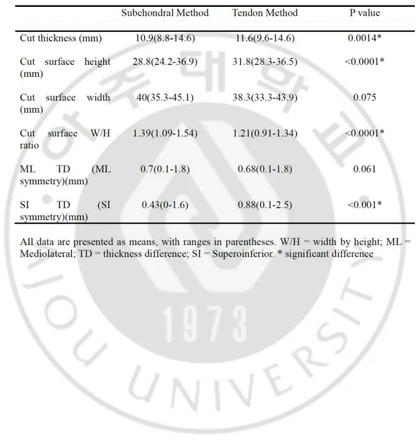
There were 27 left knees and 23 right knees in our study. The mean thickness of the patella before virtual resection with the 3D CT was 21mm (18.3mm~23.6mm, SD 1.24), and the mean width was 40.9mm (34.5mm~48.8mm, SD 3.17). The mean post-resection thickness with TM was 11.6mm (9.4mm~14.5mm, SD 0.93) and with SCM, 10.9mm (8.6mm~14.6mm, SD 1.2) the mean difference being 0.7mm (p=0.0014). The cut surface height was 28.8mm (24.2mm~36.9mm) for SCM and 31.8mm (28.3mm~36.5mm) for TM and the cut surface width was 40mm(35.3mm~45.1mm) and 38.3mm (33.3mm~43.9mm), respectively. The mean width to height (W/H) ratio of the cut surface was 1.21 with TM and 1.39 with the SCM (p<0.0001). The mean difference between the medial and lateral thicknesses (representing the ML symmetry) of the resected patella was 0.68 (0.1mm~1.8mm, SD 0.55) with TM, and 0.7mm (0.1mm~1.8mm, SD 0.52) with SCM. The mean difference between the superior and inferior thicknesses (representing the SI symmetry) of the resected patella was 0.88mm (0.1mm~2.5mm SD 0.63) with the TM and 0.43mm (0mm~1.6mm SD 0.32) with SCM(p<0.001). Thus both methods gave a symmetric patellar resection mediolaterally as well as superoinferiorly i.e., the ML and SI thickness difference being less than 2mm. The differences in various measurements after virtual resection with both methods are summarized in table 1.
Table 1. Comparison of results after virtural rescction with subchondral and tendon methos.
IV.
DISCUSSION
Patellar resurfacing was not included as a part of TKR during earlier days and the incidence of anterior knee pain was reported to be as high as 50%. (Healy, 1995; Koh, 2002) Later studies have shown that, resurfacing the patella has a role in reducing anterior knee pain. (Lombardi, 1998; Parvizi, 2005) Patellar resurfacing, however, is associated with complications which account for up to half of all revision TKRs. (Ledger, 2005; Komistek, 2000) Precise surgical technique can go a long way in avoiding these complications. The depth and symmetry of patellar resection, which are often overlooked by surgeons, can significantly influence the occurrence of these complications. (Baldwin, 2005; Bengs, 2006; Hsu, 1996; Kurosaka, 2002; Kelly, 2001)
Asymmetric patellar resection is found to be associated with anterior knee pain, bony impingement, patellar maltracking, plastic overload, and increased wear. (Baldini, 2007; Pagnano, 2000) Both the SI and ML symmetry have an important influence on the outcome following patellar resurfacing. (Anglin, 2009; Komistek, 2000) We found that both the tendon and the subchondral methods gave a symmetric resection in the ML plane since the mean ML thickness difference was 0.68mm (0.1~1.8mm) with TM and 0.7mm (0.1~1.8mm) with SCM, both values being less than 2mm. We could not come across any previous study assessing the SI symmetry of patellar resection. Both methods gave a symmetric resection superoinferiorly with a mean SI thickness difference of 0.88mm (0.1~2.5mm) with TM and 0.43mm (0~1.6mm) with SCM, both values being
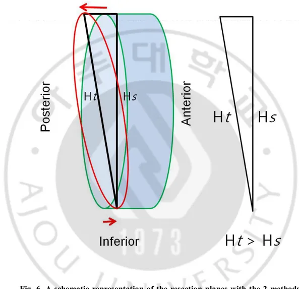
less than 2mm. However, the SI thickness difference was higher with the TM compared to the SCM (p value<0.001), the inferior thickness being lesser in all the cases. This is explained by the fact that the patellar tendon has a more anterior insertion on the patella compared to the quadriceps tendon. So, the TM gives a slightly more oblique resection compared to the SCM, where, the resection is done almost parallel to the anterior surface of the patella. (Fig. 6)
Fig. 6. A schematic representation of the resection planes with the 2 methods. The SCM gives a resection surface almost parallel with the anterior surface(green oval) while the tendon method gives a more oblique resection plane(red oval), shown here slightly exaggerated(red arrows). This demonstrates why the cut surface height is more with TM(Ht) than the SCM(Hs).
When resurfacing the patella, ideally, it is desirable that the surgeon makes an attempt to restore the thickness of the bone-prosthesis composite to that of the original patella. (Levai, 2001; Kelly, 2001) There are many studies which suggest leaving a minimum patellar thickness of 13~16mm after resection. (Levai, 2001; Reuben, 1991) Kim et al (Kim, 2009) has reported a mean patellar thickness of 21.2mm and a post resection thickness of 12.5mm in 713 female Korean patients. Though there is concern that a thinner resected patella may have increased strain and higher risk of fracture, Kim et al had only one case of periprosthetic fracture in their series, in a patient who sustained it following a fall. Thus, a resected patella, which is thinner than the minimum recommended value in literature (13~16mm), may still be acceptable for Korean patients.
The mean pre-resection thickness measured with the 3D-CT scan in our study was 21mm (18.3mm~23.6mm), which was lesser than the intra-operative value of 21.62mm (20mm~24mm). This is probably explained by the fact that the articular cartilage and prepatellar soft tissue, both of which contribute to the thickness measured intra-operatively (Iranpour, 2008; Rauh, 2002), are not included in the measurements done with the 3D-CT. Rauh et al (Rauh, 2002), after a cadaver study using spiked calipers to measure patellar thickness has mentioned that the intra-operative measurements, which are usually done with a smooth mouthed caliper, can result in an overestimation of the thickness due to the prepatellar soft tissue. The mean post resection thickness in our study was 11.59mm (9.39mm~14.50mm) with the TM and 10.85mm
patella with a mean difference of 0.7mm (p=0.0014). Intraoperative post resection thickness was 12mm (9mm-15mm) for the above patients.
The W/H ratio of the resected surface gives an idea about its approximate shape, which, when round, has a value of 1, and tend to have higher values for more oval shapes. (Baldwin, 2005) The W/H ratio of the cut surface in our study was 1.21 with the TM and 1.39 with the SCM, which means that the TM gives a less oval shape compared to the subchondral method (p<0.0001). This is because the cut surface height was more (mean difference of 3mm), and the cut surface width was less (mean difference of 1.7mm), with the TM compared to the SCM. A less oval shape for the resected surface, as obtained with the TM in our study, in turn gives better coverage for the more commonly used domed prosthesis. Lee et al (Lee, 1998) suggests that, the tendon method of resection may reduce surgical variations associated with patellar resurfacing using a domed prosthesis. Ledger et al (Ledger, 2005) mentions that, in comparison to an oval patellar prosthesis, a domed prosthesis can tolerate an oblique cut better, remaining in contact with the trochlear groove even with asymmetric cuts. There are also concerns about the possibility of implanting an oval prosthesis in malrotation. (Baldwin, 2005)
Our study has limitations. First of all, this study was done on female subjects only. The differences between female and male patella dimensions have been reported, with males showing higher values for width, height, and W/H ratio. (Baldwin, 2005) The main reason for solely using female subjects was due to the lack of male patients during the time span in which this study was undertaken. In Korea, as well as in other Asian
countries, the ratio of female to male patients receiving TKR is even more lopsided than Western countries. According to the Korean Health Insurance Review and Assessment Service data between 2002 to 2005, of 47,961 patients who had received TKR, 43,513 (90.7%) were female. Secondly, the preoperative 3D-CT tool described in our study may not be available to all facilities. The cost effectiveness of taking a CT on TKR subjects is unknown. Most importantly, the landmarks chosen for virtual resection with 3D-CT reconstruction need not accurately coincide with the landmarks used for resection during surgery. Intra-operatively, the subchondral junction is not as reliably identifiable as the posterior limits of the quadriceps and patellar tendon attachments. Thus, the virtual resections performed with 3D-CT may be difficult to be accurately reproduced intra-operatively. We believe, however, that 3D-CT was the most practical, as well as ethical way to compare the two resection methods on the above mentioned parameters.
V.
CONCLUSION
In conclusion, our results give the surgeon an idea about the difference in resected morphology between two methods. We found that when compared to SCM, the TM gives a thicker resected patella and also a less oval shape to the cut surface, which gives better coverage for a domed prosthesis. Surgeons may choose the TM method when a thicker resected patella is needed, or vice versa. Some may want to medialize the prosthesis for optimal patella tracking, in which case the SCM may be chosen for a more oval cut surface. ML and SI symmetry was achieved with both methods, but the SI symmetry was slightly better with the SCM. Surgeons may choose SCM in order to achieve a more symmetric superior –inferior cut surface to prevent periprosthetic fractures. Though the TM has shown a statistical edge over SCM in terms of post-resection thickness and the cut surface shape, to determine its actual clinical significance, and to arrive at the most appropriate patellar resection method, further prospective intra-operative studies with long-term clinical follow up are warranted.
REFERENCES
1. Baldini A, Anderson JA, Cerulli-Mariani P, et al. Patellofemoral evaluation after total knee arthroplasty. Validation of a new weight-bearing axial radiographic view. J Bone Joint Surg Am 89:1810, 2007
2. Gomes LS, Bechtold JE, Gustilo RB. Patellar prosthesis positioning in Total knee Arthroplasty. A roentgenographic study. Clin Orthop 236:72, 1988
3. Kim TK, Chung BJ, Kang YG, et al. Clinical Implications of Anthropometric Patellar Dimensions for TKA in Asians. Clin Orthop 467:1007, 2009
4. Pagnano MW, Trousdale RT. Asymmetric patella resurfacing in total knee arthroplasty. J Knee Surg Am 13:228, 2000 .
5. Baldwin JL, House CK. Anatomic dimensions of the patella measured during total knee arthroplasty. J Arthroplasty 20:250, 2005
6. Bengs BC, Scott RD. The effect of patellar thickness on intraoperative knee flexion and patellar tracking in total knee arthroplasty. J Arthroplasty 21:650, 2006
8. Kurosaka M, Yoshiya S, Mizuno K, et al. Maximizing Flexion After Total Knee Arthroplasty. J Arthroplasty 17(4 Suppl):59, 2002
9. Anglin C, Hodgson AJ, Helmy N, et al. Finding and defining the ideal patellar resection plane in total knee arthroplasty. J Biomechanics 42:2307, 2009
10. Levai JP. Technical aspects: the patellar side. Knee Surg Sports Traumatol
Arthrosc 9(suppl 1);19, 2001
11. Ledger M, Shakespeare D, Scaddan M. Accuracy of patellar resection in total knee replacement: a study using the medial pivot knee. Knee 12:13, 2005
12. Iranpour F, Merican AM, Amis AA, et al. The Width:thickness Ratio of the Patella. Clin Orthop Relat Res 466:1198, 2008
13. Lee TQ, Kim WC. Anatomically based patellar resection criteria for total knee arthroplasty. Am J Knee Surg 11:161, 1998
14. Lombardi AV Jr, Mallory TH, Maitino PD, et al. Freehand resection of the patella in total knee arthroplasty referencing the attachments of the quadriceps tendon and patellar tendon. J Arthroplasty 13:788, 1998
15. Rand JA. Total knee arthroplasty: techniques. In: Morrey BF, ed. Joint Replacement Arthroplasty. New York, NY: Churchill Livingstone, 1002, 1991
16. Insall JA. 1993-12A Insall JN. Surgical techniques, instrumentation in total knee arthroplasty. In: Insall JN, ed. Surgery of the Knee. New York, NY: Churchill Livingstone, 767, 1993
17. Healy WL, Wasilewski SA, Takei R, et al. Patellofemoral complications following total knee arthroplasty. J Arthroplasty 10:197, 1995
18. Koh JS, Yeo SJ, Lee BP, et al. Influence of patellar thickness on results of total knee arthroplasty: does a residual bony patellar thickness of below 12 mm lead to poorer clinical outcome and increased complication rates? J Arthroplasty 17:56, 2002
19. Parvizi J, Rapuri VR, Saleh KJ, et al. Failure to Resurface the Patella during Total Knee Arthroplasty May Result in More Knee Pain and Secondary Surgery.
Clin Orthop 438:191, 2005
20. Kelly MA. Patellofemoral complications following total knee arthroplasty. Instr
Course Lect 50:403, 2001
21. Komistek RD, Dennis DA, Mabe JA, et al. An in vivo determination of patellofemoral contact positions. Clinical Biomechanics 15:29, 2000
22. Reuben JD, McDonald CL, Woodard PL, et al. Effect of patella thickness on patella strain following total knee arthroplasty. J Arthroplasty 6:251, 1991
23. Rauh MA, Bayers-Thering M, Buyea CM, et al. Reliability and validity of a new caliper for measuring patellar thickness. J Arthroplasty 17:105, 2002
- 국문요약 -
슬관절
전치환술에서 3차원 단층촬영술을 이용한
슬개골의
가상 절제
아주대학교 대학원 의학과 박 도 영 (지도교수 : 원예연) 연구 목적 : 인공슬관절 전치환술에서 슬개-관절면 치환술은 많은 합병증을 야기하는 중요한 술식이지만 슬개골 절제 기준 및 절제 기구가 다양하고 정확한 해부학적 기준도 없는 실정이어서 많은 부분을 수술 중 측정한 두께와 수술자의 경험에 의존하고 있다. 저자들은 3차원 단층 촬영술을 이용한 가상 절제를 통해 현재 쓰이고 있는 2가지 절제 방법에서 예상되는 절제면의 형태, 두께 및 대칭성을 조사 및 비교함으로써 3차원 단층촬영술을 이용한 슬개골의 가상 절제의 유용성을 밝히고자 하였다. 연구 방법 : 2008년 본원에서 퇴행성 슬관절염으로 슬관절 전치환술을 시행 받은 49명의 여자 환자(55세~83세)를 대상으로 하였다. 수술 전 3차원 단층촬영술을 시행 하여 절제 전 슬개골 두께를 계측하고 수술 중 측정한 절제 전, 후의 슬개골 두께의연골 하 방법으로 가상 절제를 시행하여 절제면의 너비와 높이 및 그 비율을 측정하였고 남은 슬개골의 두께, 절제면의 넓이 및 대칭성(Gomes 등의 방법)을 계측하였다. 결과 : 3차원 단층촬영술로 측정한 슬개골 두께는 21mm(18.3mm~23.6mm, SD 1.24)였다. 수술 중 측정한 절제전의 두께는 21.62mm(20mm~24mm, SD 1.09)였다. 두 방법으로 절제 후 남은 슬개골의 두께는 각각 10.85mm(8.56mm~14.64mm, SD 1.29)와 11.59mm(9.39mm~14.50mm, SD 0.93)이었고 유의확률은(p=0.0014)이어서 두 절제 방법은 절제 후 남은 슬개골의 두께에 있어서 의미 있는 차이를 만든다고 할 수 있었다. 두 방법의 너비/높이의 비는 각각 1.39 와 1.21 이었고 통계학적으로 의미(p<0.0001)가 있어 두 절제방법은 절제면에 의미 있는 형태학적 차이를 만든다고 할 수 있었다. Gomes 등이 제시한 방법으로 절제 후 대칭성을 측정한 결과, 두 방법 모두 절제 후 횡단면상 대칭적이었다. 결론 : 저자들의 술 전 가상절제방법은 수술 전에 절제 후 슬개골의 예상 두께, 절제된 면의 형태 및 두께의 대칭성에 대한 정보를 미리 제공 할 수 있었다. 인대 방법이 골연골 방법에 비하여 타원형 대신 더 원에 가까운 절제 면이 얻어졌으므로
수술자는 슬개골 부품 형태(dome 또는 anatomical shape)에 따른 최적의 절제방법을 선택 할 수 있었다. 이외에도 횡단면 및 시상면에서의 대칭성, 절제면에서 남아 있는 경화된 뼈의 위치 및 크기를 알 수 있었다. 얻어진 다양한 정보들은 향후 항법장치에서 슬개골 치환술을 디자인하는데 도움이 될 수 있다고 본다. 결론적으로 3D CT 를 이용한 수술 전 슬개골 가상절제는 수술자에게 수술 전에 올바른 슬개골 절제를 위한 수술 전 도구가 될 수 있다고 추정된다.