ORIGINAL INVESTIGATION
Non-alcoholic steatohepatitis
and progression of carotid atherosclerosis
in patients with type 2 diabetes: a Korean
cohort study
Hyeok‑Hee Lee
1,4†, Yongin Cho
2,4†, Young Ju Choi
3, Byung Wook Huh
3, Byung‑Wan Lee
1, Eun Seok Kang
1,
Seok Won Park
1, Bong‑Soo Cha
1, Eun Jig Lee
1, Yong‑ho Lee
1,5,6*and Kap Bum Huh
3Abstract
Background: There is increasing concern regarding cardiovascular risk in individuals with non‑alcoholic fatty liver disease. This study was conducted to evaluate whether hepatic steatosis with or without fibrosis is associated with the progression of carotid atherosclerosis in patients with type 2 diabetes.
Methods: From a longitudinal cohort, we enrolled 1120 patients with type 2 diabetes who underwent repeated carotid artery ultrasonography every 1–2 years. Ultrasonographic findings at baseline and after 6–8 years were com‑ pared. Presence of hepatic steatosis was mainly assessed by abdominal ultrasonography; patients with hepatic steato‑ sis were further evaluated for hepatic fibrosis according to fibrosis‑4 index. We investigated the association between liver status and atherosclerosis progression.
Results: Of 1120 patients, 636 (56.8%) were classified as having hepatic steatosis at baseline. After 6–8 years, 431 (38.5%) showed atherosclerosis progression. Hepatic steatosis was significantly associated with atherosclerosis progression (adjusted odds ratio[AOR]: 1.370, 95% CI 1.025–1.832; p < 0.05). Among patients with hepatic steatosis, only individuals with fibrosis showed significant association with atherosclerosis progression (AOR: 1.615, 95% CI 1.005–2.598; p < 0.05). The association between hepatic fibrosis and atherosclerosis progression was significant in all metabolic subgroups regardless of age, body mass index, presence of metabolic syndrome, or insulin sensitivity (all
p < 0.05). Furthermore, subjects with hepatic steatosis & fibrosis and ≥ 4 components of metabolic syndrome criteria
showed markedly increased risk of atherosclerosis progression (AOR: 2.430, 95% CI 1.087–5.458; p < 0.05).
Conclusions: Hepatic steatosis with fibrosis is independently associated with the progression of carotid atherosclero‑ sis in patients with type 2 diabetes.
Keywords: Atherosclerosis, Hepatic fibrosis, Metabolic syndrome, Non‑alcoholic fatty liver disease, Non‑alcoholic steatohepatitis, Type 2 diabetes
© The Author(s) 2020. This article is licensed under a Creative Commons Attribution 4.0 International License, which permits use, sharing, adaptation, distribution and reproduction in any medium or format, as long as you give appropriate credit to the original author(s) and the source, provide a link to the Creative Commons licence, and indicate if changes were made. The images or other third party material in this article are included in the article’s Creative Commons licence, unless indicated otherwise in a credit line to the material. If material is not included in the article’s Creative Commons licence and your intended use is not permitted by statutory regulation or exceeds the permitted use, you will need to obtain permission directly from the copyright holder. To view a copy of this licence, visit http://creat iveco mmons .org/licen ses/by/4.0/. The Creative Commons Public Domain Dedication waiver (http://creat iveco mmons .org/publi cdoma in/ zero/1.0/) applies to the data made available in this article, unless otherwise stated in a credit line to the data.
Background
The prevalence of non-alcoholic fatty liver disease (NAFLD) is rapidly rising relative to increased obe-sity and/or type 2 diabetes [1]. NAFLD is known to be associated with various complications, such as chronic kidney disease (CKD), cancer, heart failure, or athero-sclerosis [2], and cardiovascular complications remain
Open Access
*Correspondence: yholee@yuhs.ac
†Hyeok‑Hee Lee and Yongin Cho contributed equally to this work 5 Division of Endocrinology and Metabolism, Department of Internal Medicine, Yonsei University College of Medicine, Seoul, Korea Full list of author information is available at the end of the article
the leading cause of mortality for patients with NAFLD [3–7]. Non-alcoholic steatohepatitis (NASH), one of sev-eral categories of NAFLD which is characterized by lobu-lar inflammation and hepatocyte ballooning, produces more significant liver injury like fibrosis or cirrhosis compared to simple NAFLD [8], and patients with NASH were reported to have much higher incidence of coronary artery disease-related mortality [9–11]. In this aspect, there had been numerous previous studies investigat-ing the causal relationship between NAFLD/NASH and carotid atherosclerosis [12–15], regarding carotid ather-osclerosis as a surrogate marker of coronary atheroscle-rosis. Therefore, it might be important to assess hepatic steatosis and fibrosis to identify those at high risk of car-diovascular disease, and to optimally commence medical interventions [16, 17].
This scenario is of special concern in patients with type 2 diabetes, which is known to be associated with higher risk of NAFLD [16, 18]. While NAFLD is an independent risk factor for cardiovascular complications [19], when combined with type 2 diabetes, it further increases the risk of systemic atherosclerosis [3]. Insulin resistance, a characteristic feature of both type 2 diabetes and NAFLD, is known as the key pathophysiology linking type 2 dia-betes, NAFLD, and atherosclerosis [2, 20]. However, lit-tle is known about the longitudinal effects of NAFLD or NASH on systemic atherosclerosis in type 2 diabetes.
The aim of this study was to investigate the relationship between NAFLD with or without significant fibrosis and the risk of carotid atherosclerosis progression assessed by clinical, laboratory, and repeated imaging findings in type 2 diabetes patients.
Methods Study participants
Participants were recruited from the Seoul Metabolic Syndrome Cohort, of which total 13,296 patients were diagnosed and treated for type 2 diabetes from Novem-ber 1997 to SeptemNovem-ber 2016 at Huh Diabetes Center as previously described [3, 21]. Patients aged 19 years or older who had undergone repeated carotid artery ultra-sonography at 1–2-year intervals for up to 6-8 years were enrolled. Participants were diagnosed with type 2 diabe-tes according to the American Diabediabe-tes Association clas-sification [22]. Patients were excluded for any one of the following criteria: (1) under 19 years of age; (2) diagnosed with type 1 diabetes; (3) pregnant; (4) diagnosed with liver disease other than NAFLD, such as viral or auto-immune hepatitis; and (5) history of heavy alcohol con-sumption (> 140 g/week). Patients with baseline bilateral carotid artery plaque in whom occurrence of new-onset plaque was difficult to judge in repeat ultrasonography were also excluded. In total, we enrolled 1120 patients
with type 2 diabetes who underwent repeat carotid artery ultrasonography at 1–2-year intervals for up to 6–8 years and evaluations for the presence of hepatic steatosis or fibrosis at baseline. All participants provided written informed consent, and the Ethics Committee of the Yon-sei University College of Medicine approved this study (4-2019-0270).
Measurements and definitions of clinical and laboratory parameters
At baseline, we collected information from participants regarding their medical and family history, smoking and alcohol history/consumption, and physical activity level per week. Medication history regarding aspirin, statin, and anti-diabetic drug (insulin, sulfonylurea, metformin, thiazolidinedione) usage was also reviewed. Anthropo-metrics including weight, height, and waist circumfer-ence were obtained by trained nurses who were blinded to patients’ clinical and laboratory data, and blood sam-ples were collected from participants (a) after ≥ 8 h of fasting, and (b) 2 h after a meal. Metabolic parameters including HbA1c, lipid profiles (total cholesterol, high-density lipoprotein cholesterol (HDL-C), low-high-density lipoprotein cholesterol (LDL-C), triglyceride), blood urea nitrogen (BUN), creatinine, total bilirubin, aspar-tate/alanine aminotransferase (AST/ALT), total protein, albumin, and platelet count were measured by routine laboratory methods on fresh samples at the same day of collection.
The estimated glomerular filtration rate (eGFR) was derived from the Modification of the Diet in Renal Dis-ease equation (MDRD) [23]. Diagnosis and classification of CKD was based on the Kidney Disease: Improving Global Outcomes (KDIGO) guidelines, and patients with eGFR < 60 mL/min/1.73 m2 for > 3 months were diag-nosed as CKD stage III–V accordingly [24].
Insulin sensitivity was assessed by calculating the rate constant for plasma glucose disappearance (KITT; %/ min) in a short insulin tolerance test [25]. The test was performed at 8:00AM after an overnight fast, and venous blood samples were collected at 0, 3, 6, 9, 12, and 15 min after an intravenous bolus injection of regular insulin (Humulin; Eli Lilly, Indianapolis, IN, USA) at a dosage of 0.1 U/kg. Plasma glucose concentrations were meas-ured immediately after sampling using Beckman glucose analyzer II (Beckman Coulter Inc., Brea, CA, USA), and KITT was determined by calculating the rate of the fall in log-transformed plasma glucose between 3 and 15 min. To prevent potential hypoglycemia, 100 mL of 20% dex-trose solution was administered intravenously imme-diately after testing. Insulin resistance was defined as KITT < 2.5%/min [26].
The diagnosis of metabolic syndrome was made according to a joint interim statement of the Interna-tional Diabetes Federation Task Force on Epidemiol-ogy and Prevention; National Heart, Lung, and Blood Institute; American Heart Association; World Heart Federation; International Atherosclerosis Society; and International Association for the Study of Obesity pub-lished in 2009 [27]. Hypertension was defined as sys-tolic blood pressure (BP) ≥ 140 mmHg and/or a diassys-tolic BP ≥ 90 mmHg, or current use of antihypertensive medi-cations. Individuals who drank twice a month or more were defined as regular alcohol consumers, and partici-pants who had ever smoked more than five packs of ciga-rettes were considered ever-smokers. Regular exercise was defined as moderate to vigorous physical activity for over 30 min more than once a month. Overweight was defined as body mass index (BMI) ≥ 23 kg/m2 according to scientific statement from the World Health Organiza-tion [28].
Liver status measurements
Among 1120 participants, 1086 underwent abdominal ultrasonography (iU22; Philips Healthcare, Andover, MA, USA) with a 3.5-MHz transducer after 8 h of fast-ing. Ultrasound examinations were performed by trained radiologists who were blinded to the patients’ clinical and laboratory information. According to ultrasono-graphic findings, participants were assessed on whether or not they had hepatic steatosis. The presence of hepatic steatosis in 34 patients who did not undergo abdomi-nal ultrasonography was determined by calculating the Comprehensive Non-Alcoholic Fatty Liver Disease Score (CNS) [29], in which a score ≥ 40 indicated hepatic stea-tosis. Those with hepatic steatosis were further evaluated for the presence of independent hepatic fibrosis by calcu-lating the fibrosis-4 (FIB-4) index. Significant fibrosis was defined as FIB-4 index ≥ 1.45 in this study [30].
Carotid atherosclerosis measurements
Every participant underwent repeated carotid ultra-sonography every 1–2 years to evaluate carotid athero-sclerosis status. We compared the rate of atheroathero-sclerosis progression at baseline and at 6–8 years. Both common carotid arteries were examined by high-resolution ultra-sonography (LOGIQ7; GE Healthcare, Chicago, IL, USA) by trained technicians who were blinded to the patients’ clinical and laboratory data. The mid and distal common carotid artery was scanned by lateral longitudinal projec-tion, and carotid intima-media thickness (IMT; mm) was measured at three points: far wall of mid; distal common carotid artery; and 1 cm proximal to the carotid bulb. Carotid IMT was defined as the distance between lumen-intima interface and media-adventitia interface, of which
the mean value of three measurements on each side was used to represent carotid atherosclerosis status.
Carotid atherosclerosis progression was defined as the appearance of newly developed carotid plaque lesions on repeat ultrasonography. The presence of carotid plaque was defined as meeting any one of the following criteria: (1) carotid IMT of 1.5 mm or higher; (2) protrusion of atherosclerosis into the lumen of artery with ≥ 50% thick-ness compared to the surrounding area; and (3) presence of distinct area of hyperechogenicity [31].
Statistical analysis
Baseline characteristics of study participants were ana-lyzed according to liver status: no steatosis; steatosis only; and steatosis with fibrosis. Continuous variables were expressed as mean ± standard deviation (SD) and ana-lyzed with one-way ANOVA for intergroup comparison, followed by Bonferroni test or Dunn procedure for post hoc analysis. All categorical variables were expressed as number (proportion) and compared by Chi square test.
We performed multivariable logistic regression analy-sis to calculate odds ratio (OR) of carotid atheroscleroanaly-sis progression according to the presence of hepatic steato-sis. After subdividing patients with hepatic steatosis into steatosis only and steatosis with fibrosis, Chi square test was performed to compare the proportion of carotid ath-erosclerosis progression in each liver status subgroup (no steatosis, steatosis only, and steatosis with fibrosis).
To verify independent association between liver status and carotid atherosclerosis progression, we performed multivariable logistic regression analysis in which various confounding factors were adjusted in a stepwise man-ner: age and gender were adjusted in Model 2; duration of diabetes, HbA1c, LDL-cholesterol, HDL-cholesterol, statin use, alcohol/smoking consumption, exercise status, systolic BP, diastolic BP, KITT and CKD stage III-V were adjusted in Model 3; and BMI was adjusted in model 4. Models 5 and 6 were built by further adjusting Model 4 with waist circumference and follow-up duration, respectively.
Also, logistic regression analysis was performed to detect the association between liver status and carotid atherosclerosis progression after dividing patients into two subgroups by age (70 years), BMI (overweight status: 23.0 kg/m2), presence of metabolic syndrome, or KITT (2.5%/min). Cut-off for age was chosen according to the previous report that cytochrome P450 level declines sig-nificantly after age 70 [32], which is known to be very closely related to cholesterol homeostasis and athero-sclerosis [33–35]. Finally, study participants were divided into nine subgroups according to liver status and meta-bolic syndrome criteria, and multivariable logistic regres-sion analysis was performed to calculate OR of carotid
atherosclerosis progression in each subgroup. p val-ues < 0.05 were considered statistically significant, and all statistical analyses were performed using R version 4.0.0 (R Foundation for Statistical Computing, Vienna, Aus-tria) and IBM SPSS Statistics version 24.0 (IBM Corp., Armonk, NY, USA).
Results
Baseline characteristics of study participants
Baseline characteristics are summarized in Table 1. Of 1120 participants, 636 (56.8%) had hepatic steatosis; among them, 222 (19.8%) had significant fibrosis.
The mean age of subjects with hepatic steatosis and fibrosis was 59.8 (± 7.8) years, which was significantly higher compared to the other subgroups (p < 0.001). BMI (kg/m2) was higher in those with both hepatic steatosis and fibrosis (26.1 ± 3.2) or only steatosis (25.6 ± 3.0) com-pared to those without steatosis (23.2 ± 2.8) (p < 0.001). 282 (69.8%) participants with only hepatic steatosis and 150 (67.9%) participants with both hepatic steatosis and fibrosis had metabolic syndrome, while only 174 (37.3%) participants had metabolic syndrome among those with-out hepatic steatosis (p < 0.001). KITT (%/min) was 2.4 (± 1.0) in subjects with no hepatic steatosis, which was significantly higher compared to the subgroup with only steatosis (1.9 ± 0.8) or with both steatosis and fibrosis (1.8 ± 0.7) (p < 0.001), indicating that participants without hepatic steatosis were more insulin-sensitive than those in other two subgroups.
Participants with both steatosis and fibrosis showed lower eGFR (88.0 ± 27.4) compared to those with no steatosis (94.5 ± 30.9) (p = 0.018), but it was not signifi-cantly lower than the steatosis only group (93.0 ± 28.0) (p = 0.122). There was no significant difference by sex or statin use between the three subgroups.
The mean carotid IMTs (mm) at baseline were 0.75 ± 0.15, 0.76 ± 0.15, and 0.81 ± 0.14 in patients with no hepatic steatosis, steatosis only, and steatosis with fibrosis, respectively (p < 0.001). The proportion of par-ticipants with carotid plaque at baseline was not signifi-cantly different between the three subgroups (p = 0.365).
Association between hepatic steatosis and progression of carotid atherosclerosis
The presence of hepatic steatosis increased the risk of carotid plaque progression (OR: 1.368, 95% CI 1.071– 1.748; p = 0.012). This result persisted after adjusting for age, gender, systolic BP, diastolic BP, duration of dia-betes, HbA1c, KITT, CKD stage III-V, total cholesterol, statin use, and alcohol history (adjusted odds ratio[AOR]: 1.370, 95% CI 1.025–1.832; p = 0.034) (Fig. 1).
Presence of hepatic fibrosis and carotid atherosclerosis progression in patients with NAFLD
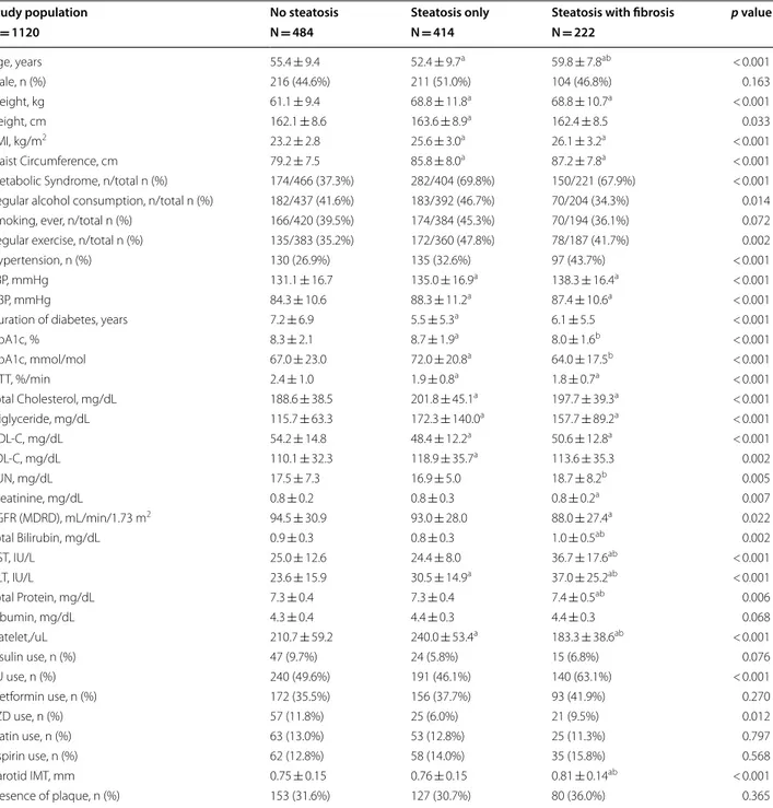
The number (%) of patients with carotid plaque progres-sion after 6–8 years was 166 (34.3%), 157 (37.9%), and 108 (48.6%), respectively, among those with no hepatic steatosis, steatosis only, and steatosis with fibrosis. The difference was statistically significant (p = 0.001) (Fig. 2).
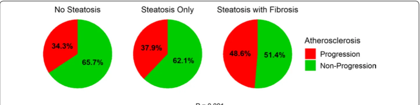
To further investigate whether presence of hepatic fibrosis is independently associated with the progression of carotid plaque in patients with NAFLD, we performed multivariable logistic regression analyses in a stepwise manner. With no adjustment (Model 1), hepatic steatosis with fibrosis was statistically significantly associated with carotid plaque progression (OR: 1.815, 95% CI 1.314– 2.507; p < 0.001), whereas steatosis only was not signifi-cant (OR: 1.170, 95% CI 0.891–1.538; p = 0.259). Steatosis with fibrosis was still significantly associated with carotid plaque progression after adjusting for age, gender (Model 2. AOR: 1.494, 95% CI 1.071–2.084; p = 0.018), duration of diabetes, HbA1c, LDL-cholesterol, HDL-cholesterol, statin use, alcohol/smoking consumption, exercise status, systolic BP, diastolic BP, KITT, CKD stage III-V (Model 3. AOR: 1.740, 95% CI: 1.111-2.723; p = 0.015), BMI (Model 4. AOR: 1.636, 95% CI 1.024–2.612; p = 0.039), and waist circumference (Model 5. AOR: 1.615, 95% CI 1.005– 2.598; p = 0.048). Further adjusting Model 4 for follow-up duration still did not alter statistical significance of the result (Model 6. AOR: 1.606, 95% CI 1.004–2.572; p = 0.048) (Fig. 3).
Risk of carotid atherosclerosis progression according to metabolic profiles
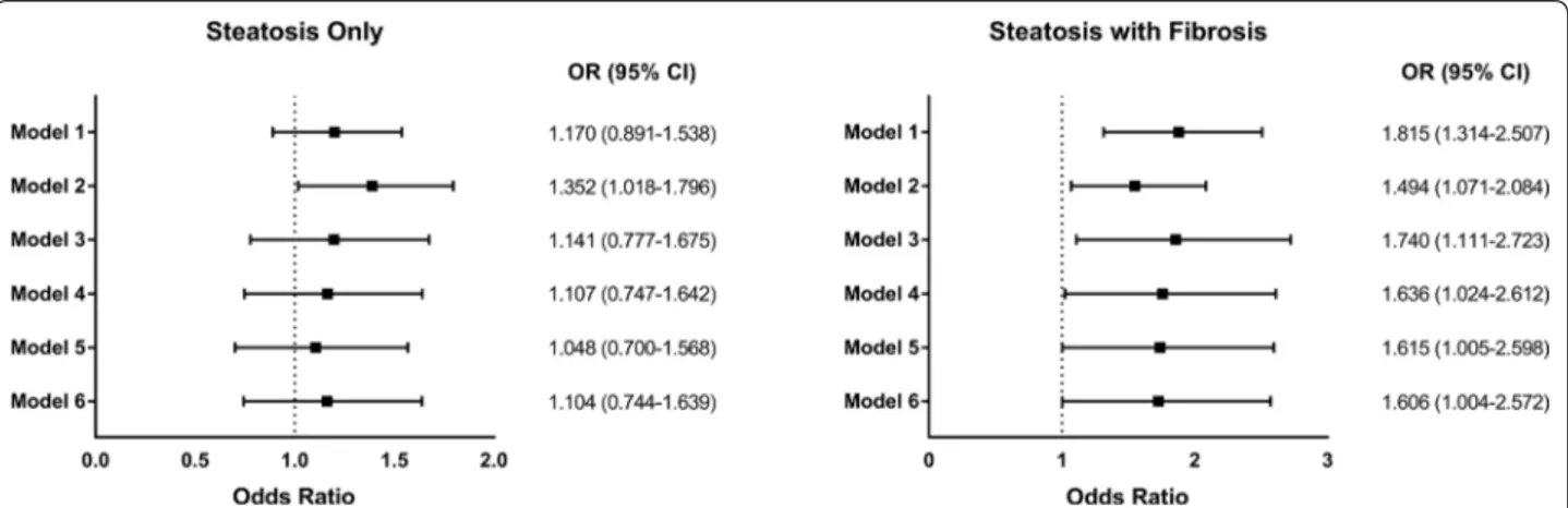
To examine the presence of potential effect modification, we analyzed the risk of carotid atherosclerosis progres-sion according to several metabolic factors. Overall, the analysis showed no difference between metabolic sub-groups, whether they were divided by age, BMI, presence of metabolic syndrome, or insulin resistance (all p inter-action > 0.05). In detail, hepatic steatosis without fibrosis was not associated with the progression of carotid ather-osclerosis in any metabolic subgroup. However, patients with combined hepatic steatosis & fibrosis showed statis-tically significantly higher risk of carotid atherosclerosis progression regardless of age (OR: 3.683, 95% CI 1.036– 13.100 in subgroup with age ≥ 70; OR: 1.653, 95% CI 1.178–2.321 in subgroup with age < 70), BMI (OR: 1.531, 95% CI 1.027–2.283 in subgroup with BMI ≥ 23; OR: 2.480, 95% CI 1.113–5.527 in subgroup with BMI < 23), presence of metabolic syndrome (OR: 1.636, 95% CI 1.051–2.548 in subgroup with metabolic syndrome; OR: 1.784, 95% CI 1.051–3.026 in subgroup without meta-bolic syndrome), or insulin sensitivity (OR: 1.712, 95% CI
1.164–2.518 in subgroup with KITT < 2.5 (insulin resist-ant); OR: 1.972, 95% CI: 1.011-3.847 in subgroup with KITT ≥ 2.5 (insulin sensitive)). There was no effect modi-fication by metabolic factors (p interaction = 0.224, 0.258,
0.815, and 0.889 for age, BMI, presence of metabolic syn-drome, and insulin sensitivity, respectively) (Fig. 4).
Consecutively, to investigate the combinatorial effects of cardiometabolic risk factors and liver status,
Table 1 Baseline Characteristics
Variables are shown as mean ± SD or n (%). ALT, alanine aminotransferase; AST, aspartate aminotransferase; BMI, body mass index; BUN, blood urea nitrogen; DBP, diastolic blood pressure; eGFR, estimated glomerular filtration rate; HDL-C, high-density lipoprotein cholesterol; IMT, intima-media thickness; KITT, rate constant for plasma glucose disappearance; LDL-C, low-density lipoprotein cholesterol; MDRD, modification of diet in renal disease equation; SBP, systolic blood pressure; SD, standard deviation; SU, sulfonylurea; TZD, thiazolidinedione
a p values < 0.05 versus no steatosis b p values < 0.05 versus steatosis only
Study population No steatosis Steatosis only Steatosis with fibrosis p value
N = 1120 N = 484 N = 414 N = 222 Age, years 55.4 ± 9.4 52.4 ± 9.7a 59.8 ± 7.8ab < 0.001 Male, n (%) 216 (44.6%) 211 (51.0%) 104 (46.8%) 0.163 Weight, kg 61.1 ± 9.4 68.8 ± 11.8a 68.8 ± 10.7a < 0.001 Height, cm 162.1 ± 8.6 163.6 ± 8.9a 162.4 ± 8.5 0.033 BMI, kg/m2 23.2 ± 2.8 25.6 ± 3.0a 26.1 ± 3.2a < 0.001 Waist Circumference, cm 79.2 ± 7.5 85.8 ± 8.0a 87.2 ± 7.8a < 0.001
Metabolic Syndrome, n/total n (%) 174/466 (37.3%) 282/404 (69.8%) 150/221 (67.9%) < 0.001 Regular alcohol consumption, n/total n (%) 182/437 (41.6%) 183/392 (46.7%) 70/204 (34.3%) 0.014 Smoking, ever, n/total n (%) 166/420 (39.5%) 174/384 (45.3%) 70/194 (36.1%) 0.072 Regular exercise, n/total n (%) 135/383 (35.2%) 172/360 (47.8%) 78/187 (41.7%) 0.002
Hypertension, n (%) 130 (26.9%) 135 (32.6%) 97 (43.7%) < 0.001
SBP, mmHg 131.1 ± 16.7 135.0 ± 16.9a 138.3 ± 16.4a < 0.001
DBP, mmHg 84.3 ± 10.6 88.3 ± 11.2a 87.4 ± 10.6a < 0.001
Duration of diabetes, years 7.2 ± 6.9 5.5 ± 5.3a 6.1 ± 5.5 < 0.001
HbA1c, % 8.3 ± 2.1 8.7 ± 1.9a 8.0 ± 1.6b < 0.001 HbA1c, mmol/mol 67.0 ± 23.0 72.0 ± 20.8a 64.0 ± 17.5b < 0.001 KITT, %/min 2.4 ± 1.0 1.9 ± 0.8a 1.8 ± 0.7a < 0.001 Total Cholesterol, mg/dL 188.6 ± 38.5 201.8 ± 45.1a 197.7 ± 39.3a < 0.001 Triglyceride, mg/dL 115.7 ± 63.3 172.3 ± 140.0a 157.7 ± 89.2a < 0.001 HDL‑C, mg/dL 54.2 ± 14.8 48.4 ± 12.2a 50.6 ± 12.8a < 0.001 LDL‑C, mg/dL 110.1 ± 32.3 118.9 ± 35.7a 113.6 ± 35.3 0.002 BUN, mg/dL 17.5 ± 7.3 16.9 ± 5.0 18.7 ± 8.2b 0.005 Creatinine, mg/dL 0.8 ± 0.2 0.8 ± 0.3 0.8 ± 0.2a 0.007 eGFR (MDRD), mL/min/1.73 m2 94.5 ± 30.9 93.0 ± 28.0 88.0 ± 27.4a 0.022 Total Bilirubin, mg/dL 0.9 ± 0.3 0.8 ± 0.3 1.0 ± 0.5ab 0.002 AST, IU/L 25.0 ± 12.6 24.4 ± 8.0 36.7 ± 17.6ab < 0.001 ALT, IU/L 23.6 ± 15.9 30.5 ± 14.9a 37.0 ± 25.2ab < 0.001 Total Protein, mg/dL 7.3 ± 0.4 7.3 ± 0.4 7.4 ± 0.5ab 0.006 Albumin, mg/dL 4.3 ± 0.4 4.4 ± 0.3 4.4 ± 0.3 0.068 Platelet,/uL 210.7 ± 59.2 240.0 ± 53.4a 183.3 ± 38.6ab < 0.001 Insulin use, n (%) 47 (9.7%) 24 (5.8%) 15 (6.8%) 0.076 SU use, n (%) 240 (49.6%) 191 (46.1%) 140 (63.1%) < 0.001 Metformin use, n (%) 172 (35.5%) 156 (37.7%) 93 (41.9%) 0.270 TZD use, n (%) 57 (11.8%) 25 (6.0%) 21 (9.5%) 0.012 Statin use, n (%) 63 (13.0%) 53 (12.8%) 25 (11.3%) 0.797 Aspirin use, n (%) 62 (12.8%) 58 (14.0%) 35 (15.8%) 0.568 Carotid IMT, mm 0.75 ± 0.15 0.76 ± 0.15 0.81 ± 0.14ab < 0.001 Presence of plaque, n (%) 153 (31.6%) 127 (30.7%) 80 (36.0%) 0.365
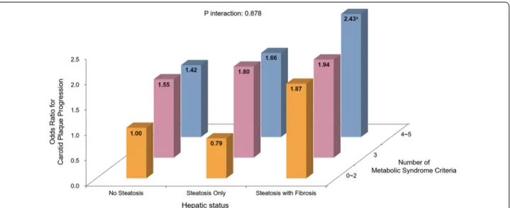
participants were divided into 9 subgroups according to liver status (no steatosis, steatosis only, steatosis with fibrosis) and the number of metabolic syndrome criteria met (0–2, 3, 4–5). In each metabolic syndrome criteria subgroup, the risk of carotid atherosclerosis progression was generally higher in subjects with hepatic steatosis, and far higher in those with both hepatic steatosis and fibrosis. Similarly, in each liver status subgroup, a higher number of metabolic syndrome criteria generally corre-lated with higher risk of carotid atherosclerosis progres-sion. Compared to those with 0–2 metabolic syndrome criteria and no hepatic steatosis, subjects with 4–5 met-abolic syndrome criteria and both steatosis and fibrosis were at significantly higher risk of carotid atherosclerosis progression (AOR: 2.430, 95% CI 1.087–5.458, p = 0.031) (Fig. 5).
Discussion
Principal findings and clinical implications
Ultrasonography is now widely accepted as a useful screening tool to detect carotid artery plaque and pre-dict cardiovascular events [31, 36]. With serial carotid ultrasonography of patients with type 2 diabetes at 1–2-year intervals for up to 6–8 1–2-years, this study demon-strated that hepatic steatosis with significant fibrosis was strongly associated with the progression of carotid artery atherosclerosis, even in relatively metabolically-healthy patients. Our results have also shown that hepatic fibro-sis and metabolic syndrome accelerate atherosclerofibro-sis progression independently of each other, delivering an additive effect when combined together.
Based on these findings, we suggest that hepatic fibro-sis can be an independent risk factor for atherogenefibro-sis acceleration, and its identification by clinical indicators may be helpful to predict the risk of atherosclerosis pro-gression. Also, patients who are already diagnosed with metabolic syndrome should especially be aware of hepatic fibrosis to blunt profoundly higher risk of ath-erosclerosis produced by the combined effect of hepatic fibrosis and metabolic syndrome.
Results in relation to other studies
NAFLD is considered a ‘hepatic manifestation of meta-bolic syndrome’. It is very closely related to type 2 diabetes or metabolic syndrome, and the main patho-physiology underlying this relationship is known to be insulin resistance [37, 38]. The association is considerably strong that NAFLD is present even in obese adolescents with dysglycemia [39]. NAFLD and metabolic syndrome can be considered to have similar effects on arteries, which accelerate atherogenesis via inflammation [40, 41], increased oxidative stress [42], atherogenic dyslipidemia [43], imbalance of adipokines [44], and hypercoagulable status [45]. As a result, NAFLD patients present lower reactive hyperemia index and higher pulse wave velocity
Fig. 1 Progression of Carotid Atherosclerosis by Presence of Hepatic
Steatosis. Odds ratio of carotid atherosclerosis progression according to the presence of hepatic steatosis. The result is adjusted for age, gender, systolic blood pressure, diastolic blood pressure, duration of diabetes, HbA1c, rate constant for plasma glucose disappearance (KITT), chronic kidney disease stage III–V, total cholesterol, statin use, and alcohol history. Levels of significance: ap = 0.012 (crude); bp = 0.034 (adjusted) (Logistic regression analysis)
Fig. 2 Progression of Carotid Atherosclerosis by Presence of Hepatic Steatosis and Fibrosis. Proportion of carotid atherosclerosis progression in
[46], and NAFLD can serve as a strong predictor of coro-nary artery calcification in metabolically healthy subjects [47].
In consequence, NAFLD was reported as an independ-ent risk factor for cardiovascular disease in the general population [7, 48]. After several efforts to prove this rela-tionship via carotid ultrasonography, it was discovered that NAFLD was significantly associated with carotid stenosis in Chinese population [49], increased carotid IMT in type 2 diabetes patients with insulin resistance [3], and higher prevalence of carotid plaque [14]. More-over, NAFLD was shown to be associated with higher cardiovascular risk in terms of carotid IMT and dyslipi-demia even in nondiabetic patients [50]. In the present study, we focused on the long-term effect of NAFLD with or without significant fibrosis on atherosclerosis by repeated carotid ultrasonography. The results showed that progression of carotid atherosclerosis after 6-8 years occurred more frequently in patients with NASH. Although it has been shown that visceral obesity is asso-ciated with cardiometabolic comorbidities of type 2 dia-betes, NAFLD, or atherosclerotic cardiovascular diseases [51], further adjustment for waist circumference along with other common metabolic factors did not alter the significance of association between hepatic fibrosis and atherosclerosis progression in our analysis.
To our knowledge, this is the first report to demon-strate that hepatic fibrosis is significantly associated with the progression of carotid artery atherosclerosis in sub-jects with type 2 diabetes. It indicates that not only pres-ence—but also severity—of metabolic liver disease can affect the risk of cardiovascular complications. Previous long-term studies showed that risk of coronary artery
disease-related mortality was much higher in patients
with NASH (12–16%) [9, 52] compared to NAFLD
(1–3%) [10, 53], and these findings were consistent with a recent meta-analysis in which increased NAFLD sever-ity produced higher risk of cardiovascular complications [11], or recent large Korean population-based cohort study which demonstrated the linear association between fatty liver index (FLI) and major adverse cardiovascular events [54]. Altered lipidomics and increased hepatic production of prothrombogenic factors, including fetuin-A in patients with fibrosing Nfetuin-ASH, can be potential con-tributors to the link between NASH and cardiovascular diseases [55].
In addition, the association between hepatic fibrosis and risk of atherosclerosis progression was significant in all metabolic subgroups regardless of age, BMI, presence of metabolic syndrome, or insulin sensitivity. It indicates that hepatic fibrosis may serve as a predictive marker for increased susceptibility to atherosclerosis progression even with less evidence of systemic metabolic alterations, implicating the possible presence of systemic profibro-genic stimuli that accelerate atherogenesis in patients with hepatic fibrosis [56].
Conversely, there was no incremental risk of ath-erosclerosis progression in hepatic steatosis without fibrosis in type 2 diabetes patients. This finding was similar to that of a previous study in which patients with hepatic steatosis and no additional feature of liver injury were found to follow a relatively benign clinical course, with mortality similar to the general population [57]. Although steatosis without fibrosis was not associated with increased risk of atherosclerosis progression in this study, repeat ultrasonography was not performed beyond
Fig. 3 Risk of Carotid Atherosclerosis Progression According to Hepatic Status. Odds ratios of carotid atherosclerosis progression according to
hepatic status. Model 1 = Crude model without any adjustment; Model 2 = Model 1 + age, gender; Model 3 = Model 2 + duration of diabetes, HbA1c, low‑density lipoprotein cholesterol, high‑density lipoprotein cholesterol, statin use, alcohol/smoking consumption, exercise status, systolic blood pressure, diastolic blood pressure, rate constant for plasma glucose disappearance (KITT), chronic kidney disease stage III‑V; Model 4 = Model 3 + body mass index; Model 5 = Model 4 + waist circumference; Model 6 = Model 4 + follow‑up duration. (Logistic regression analysis)
8 years, making it difficult to predict longer-term effect of steatosis without fibrosis on the risk of atherosclero-sis progression. Since high rates of fibroatherosclero-sis progression have been demonstrated in patients with steatosis [58], it would be important to consider its clinical significance and to manage it appropriately without overlooking the risk of cardiovascular complication.
Strengths & limits
This study has several distinguishing strengths. First, we analyzed long-term results of carotid ultrasonography in subjects with type 2 diabetes. Most previous studies using carotid ultrasonography were cross-sectional and insufficient to determine a causal relationship. In addi-tion, since this study was a hospital-based cohort study conducted at a single institution, the participants were
managed and evaluated under standardized conditions and practices.
A major limitation of this study is the fact that a bio-chemical scoring system, rather than liver biopsy, was used to evaluate hepatic fibrosis, which is currently not a gold-standard for the diagnosis. However, FIB-4 index was initially validated by comparing the results to that of liver biopsy [30], and they were shown to have fairly high accuracy to predict hepatic fibrosis [59, 60]. Sec-ondly, this study analyzed the findings of carotid deterio-ration using ultrasonography, one of the major surrogate markers of cardiovascular disease. However, our meth-ods did not allow for the investigation of cardiovascular events that could represent a direct outcome of athero-sclerosis progression. Also, antiplatelet, antihyperglyce-mic agent, or fibric acid usage were not adjusted in our models, which are potential confounding factors that
Fig. 4 Hepatic Status and Carotid Atherosclerosis Progression by Metabolic Confounders. Odds ratios of carotid atherosclerosis progression
according to hepatic status and a Age. Level of significance: p interaction = 0.224. b Body mass index. Level of significance: p interaction = 0.258.
c Presence of metabolic syndrome. Level of significance: p interaction = 0.815. d Insulin sensitivity. Level of significance: p interaction = 0.889. Risk
estimates were calculated in each subgroup using patients with no steatosis as a reference (Logistic regression analysis). BMI, body mass index; KITT, rate constant for plasma glucose disappearance
could influence our results. In addition, we did not con-sider how participants’ metabolic factors changed over time in our analyses. Finally, this study was based on a single-center cohort of Koreans with a relatively small number of participants. Therefore, further larger studies including other ethnic populations are needed to validate the results, as well as to confirm generalizability of the results.
Conclusions
In conclusion, hepatic steatosis with significant fibro-sis was independently associated with the progression of carotid atherosclerosis in patients with type 2 diabe-tes. The association was still significant in subgroups of patients who were metabolically healthy, and it became more prominent relative to criteria for metabolic syn-drome. Identification of hepatic steatosis with significant fibrosis may be helpful to predict and prevent the risk of atherosclerosis progression in individuals with type 2 diabetes.
Abbreviations
ALT: Alanine aminotransferase; AST: Aspartate aminotransferase; BMI: Body mass index; BP: Blood pressure; BUN: Blood urea nitrogen; CKD: Chronic kidney disease; CNS: Comprehensive non‑alcoholic fatty liver disease score; eGFR: Estimated glomerular filtration rate; FIB‑4: Fibrosis‑4; FLI: Fatty liver index; HDL‑ C: High‑density lipoprotein cholesterol; IMT: Intima‑media thickness; KITT: Rate constant for plasma glucose disappearance; LDL‑C: Low‑density lipoprotein cholesterol; MDRD: Modification of the Diet in Renal Disease; NAFLD: Non‑ alcoholic fatty liver disease; NASH: Non‑alcoholic steatohepatitis.
Acknowledgements
The authors would like to thank Caron Modeas, Office of Clinical Trials, The University of North Carolina at Chapel Hill, for help with preparing the manuscript.
Authors’ contributions
HL conceptualized the study, provided methodology, curated/validated data, performed analysis, and wrote the manuscript. YC conceptualized the study, provided methodology, curated/validated data, performed analysis, and wrote the manuscript. YJC curated/validated data. BWH curated/validated data. BL, ESK, SWP, BC, EJL, KBH validated data. YL conceptualized the study, provided methodology, curated/validated data, and wrote the manuscript. All authors read and approved the final manuscript.
Funding
This work was supported by the National Research Foundation of Korea (NRF) Grant funded by the Ministry of Science and ICT (Grant Number: NRF‑2016R1A5A1010764).
The study sponsor/funder was not involved in the design of the study; the col‑ lection, analysis, and interpretation of data; writing of the report; and did not impose any restrictions regarding the publication of the report.
Availability of data and materials
The datasets generated and/or analyzed during the current study are available from the corresponding author upon reasonable request.
Ethics approval and consent to participate
This study complied with the Declaration of Helsinki and was approved by the Ethics Committee of the Yonsei University College of Medicine (4‑2019‑0270). All participants provided written informed consent in this study.
Consent for publication
Not applicable.
Competing interests
The authors declare that they have no competing interests.
Fig. 5 Combinatorial Effect of Liver Status and Metabolic Syndrome Criteria on Carotid Atherosclerosis Progression. Odds ratios of carotid
atherosclerosis progression according to hepatic status and metabolic syndrome criteria. Odds ratio was calculated in each subgroup using the subgroup with 0–2 metabolic syndrome criteria and no hepatic steatosis as a reference. The result is adjusted for age, gender, duration of diabetes, HbA1c, low‑density lipoprotein cholesterol, high‑density lipoprotein cholesterol, statin use, alcohol/smoking consumption, exercise status, systolic blood pressure, diastolic blood pressure, rate constant for plasma glucose disappearance (KITT), chronic kidney disease stage III–V, body mass index, waist circumference, and follow‑up duration. Levels of significance: ap = 0.031, p interaction = 0.878 (Logistic regression analysis)
Author details
1 Department of Internal Medicine, Yonsei University College of Medicine, Seoul, Korea. 2 Department of Endocrinology and Metabolism, Inha University School of Medicine, Incheon, Korea. 3 Huh’s Diabetes Center and the 21st Cen‑ tury Diabetes and Vascular Research Institute, Seoul, Korea. 4 Graduate School, Yonsei University College of Medicine, Seoul, Korea. 5 Division of Endocrinol‑ ogy and Metabolism, Department of Internal Medicine, Yonsei University Col‑ lege of Medicine, Seoul, Korea. 6 Department of Systems Biology, Glycosylation Network Research Center, Yonsei University, Seoul, Korea.
Received: 16 April 2020 Accepted: 8 June 2020
References
1. Loomba R, Sanyal AJ. The global NAFLD epidemic. Nat Rev Gastroenterol Hepatol. 2013;10(11):686–90.
2. Vanni E, Marengo A, Mezzabotta L, Bugianesi E. Systemic complications of nonalcoholic fatty liver disease: when the liver is not an innocent bystander. Semin Liver Dis. 2015;35(3):236–49.
3. Kim SK, Choi YJ, Huh BW, Park SW, Lee EJ, Cho YW, et al. Nonalcoholic fatty liver disease is associated with increased carotid intima‑media thickness only in type 2 diabetic subjects with insulin resistance. J Clin Endocrinol Metab. 2014;99(5):1879–84.
4. Kim HC, Kim DJ, Huh KB. Association between nonalcoholic fatty liver disease and carotid intima‑media thickness according to the presence of metabolic syndrome. Atherosclerosis. 2009;204(2):521–5.
5. Fracanzani AL, Tiraboschi S, Pisano G, Consonni D, Baragetti A, Bertelli C, et al. Progression of carotid vascular damage and cardiovascular events in non‑alcoholic fatty liver disease patients compared to the general popu‑ lation during 10 years of follow‑up. Atherosclerosis. 2016;246:208–13. 6. Bhatia LS, Curzen NP, Calder PC, Byrne CD. Non‑alcoholic fatty liver
disease: a new and important cardiovascular risk factor? Eur Heart J. 2012;33(10):1190–200.
7. Han E, Lee YH. Non‑alcoholic fatty liver disease: the emerging burden in cardiometabolic and renal diseases. Diabetes Metab J. 2017;41(6):430–7. 8. Machado MV, Diehl AM. Pathogenesis of nonalcoholic steatohepatitis.
Gastroenterology. 2016;150(8):1769–77.
9. Ekstedt M, Franzen LE, Mathiesen UL, Thorelius L, Holmqvist M, Bodemar G, et al. Long‑term follow‑up of patients with NAFLD and elevated liver enzymes. Hepatology. 2006;44(4):865–73.
10. Schindhelm RK, Dekker JM, Nijpels G, Bouter LM, Stehouwer CD, Heine RJ, et al. Alanine aminotransferase predicts coronary heart disease events: a 10‑year follow‑up of the Hoorn Study. Atherosclerosis. 2007;191(2):391–6. 11. Targher G, Byrne CD, Lonardo A, Zoppini G, Barbui C. Non‑alcoholic fatty
liver disease and risk of incident cardiovascular disease: a meta‑analysis. J Hepatol. 2016;65(3):589–600.
12. Oni E, Budoff MJ, Zeb I, Li D, Veledar E, Polak JF, et al. Nonalcoholic fatty liver disease is associated with arterial distensibility and carotid intima‑ media thickness: (from the multi‑ethnic study of atherosclerosis). Am J Cardiol. 2019;124(4):534–8.
13. Madan SA, John F, Pyrsopoulos N, Pitchumoni CS. Nonalcoholic fatty liver disease and carotid artery atherosclerosis in children and adults: a meta‑ analysis. Eur J Gastroenterol Hepatol. 2015;27(11):1237–48.
14. Sookoian S, Pirola CJ. Non‑alcoholic fatty liver disease is strongly associated with carotid atherosclerosis: a systematic review. J Hepatol. 2008;49(4):600–7.
15. Sinn DH, Cho SJ, Gu S, Seong D, Kang D, Kim H, et al. Persistent nonalco‑ holic fatty liver disease increases risk for carotid atherosclerosis. Gastroen‑ terology. 2016;151(3):481–8 e1.
16. Chalasani N, Younossi Z, Lavine JE, Charlton M, Cusi K, Rinella M, et al. The diagnosis and management of nonalcoholic fatty liver disease: practice guidance from the American Association for the Study of Liver Diseases. Hepatology. 2018;67(1):328–57.
17. EASL–EASD–EASO Clinical Practice Guidelines for the management of non‑alcoholic fatty liver disease. J Hepatol. 2016;64(6):1388–402. https :// doi.org/10.1016/j.jhep.2015.11.004
18. Lee YH, Cho Y, Lee BW, Park CY, Lee DH, Cha BS, et al. Nonalcoholic fatty liver disease in diabetes. Part I: epidemiology and diagnosis. Diabetes Metab J. 2019;43(1):31–45.
19. Wagenknecht LE, Zaccaro D, Espeland MA, Karter AJ, O’Leary DH, Haffner SM. Diabetes and progression of carotid atherosclerosis: the insulin resistance atherosclerosis study. Arterioscler Thromb Vasc Biol. 2003;23(6):1035–41.
20. Baek JH, Kim H, Kim KY, Jung J. Insulin resistance and the risk of diabetes and dysglycemia in Korean general adult population. Diabetes Metab J. 2018;42(4):296–307.
21. Choe EY, Lee YH, Choi YJ, Huh BW, Lee BW, Kim SK, et al. Waist‑to‑calf circumstance ratio is an independent predictor of hepatic steatosis and fibrosis in patients with type 2 diabetes. J Gastroenterol Hepatol. 2018;33(5):1082–91.
22. Mach F, Baigent C, Catapano AL, Koskinas KC, Casula M, Badimon L, et al. ESC/EAS Guidelines for the management of dyslipidaemias: lipid modification to reduce cardiovascular risk. Eur Heart J. 2019. https ://doi. org/10.1093/eurhe artj/ehz45 5.
23. Levey AS, Bosch JP, Lewis JB, Greene T, Rogers N, Roth D. A more accurate method to estimate glomerular filtration rate from serum creatinine: a new prediction equation. Modification of Diet in Renal Disease Study Group. Ann Intern Med. 1999;130(6):461–70.
24. Chapter 1: definition and classification of CKD. Kidney Int Suppl. 2013;3(1):19–62. https ://doi.org/10.1038/kisup .2012.64.
25. Sharma R, Vikram NK, Misra A. Comparison of short insulin tolerance test with HOMA Method for assessment of insulin sensitivity in Asian Indians in north India. Diabetes Res Clin Pract. 2008;82(1):e9–12.
26. Lee MY, Koh JH, Nam SM, Jung PM, Sung JK, Kim SY, et al. Short insulin tolerance test can determine the effects of thiazolidinediones treatment in type 2 diabetes. Yonsei Med J. 2008;49(6):901–8.
27. Alberti KG, Eckel RH, Grundy SM, Zimmet PZ, Cleeman JI, Donato KA, et al. Harmonizing the metabolic syndrome: a joint interim statement of the International Diabetes Federation Task Force on Epidemiology and Prevention; National Heart, Lung, and Blood Institute; American Heart Association; World Heart Federation; International Atherosclerosis Soci‑ ety; and International Association for the Study of Obesity. Circulation. 2009;120(16):1640–5.
28. World Health Organization. Regional Office for the Western P. The Asia‑ Pacific perspective: redefining obesity and its treatment: Sydney: Health Communications Australia; 2000.
29. Lee YH, Bang H, Park YM, Bae JC, Lee BW, Kang ES, et al. Non‑laboratory‑ based self‑assessment screening score for non‑alcoholic fatty liver disease: development, validation and comparison with other scores. PLoS ONE. 2014;9(9):e107584.
30. Vallet‑Pichard A, Mallet V, Nalpas B, Verkarre V, Nalpas A, Dhalluin‑Venier V, et al. FIB‑4: an inexpensive and accurate marker of fibrosis in HCV infection. Comparison with liver biopsy and fibrotest. Hepatology. 2007;46(1):32–6.
31. Qi Y, Fan J, Liu J, Wang W, Wang M, Sun J, et al. Cholesterol‑overloaded HDL particles are independently associated with progression of carotid atherosclerosis in a cardiovascular disease‑free population: a community‑ based cohort study. J Am Coll Cardiol. 2015;65(4):355–63.
32. Sotaniemi EA, Arranto AJ, Pelkonen O, Pasanen M. Age and cytochrome P450‑linked drug metabolism in humans: an analysis of 226 subjects with equal histopathologic conditions. Clin Pharmacol Ther. 1997;61(3):331–9. 33. Elfaki I, Mir R, Almutairi FM, Duhier FMA. Cytochrome P450: polymor‑
phisms and roles in cancer, diabetes and atherosclerosis. Asian Pac J Cancer Prev. 2018;19(8):2057–70.
34. Luoma PV. Cytochrome P450–physiological key factor against cholesterol accumulation and the atherosclerotic vascular process. Ann Med. 2007;39(5):359–70.
35. Li T, Matozel M, Boehme S, Kong B, Nilsson LM, Guo G, et al. Overexpres‑ sion of cholesterol 7α‑hydroxylase promotes hepatic bile acid synthesis and secretion and maintains cholesterol homeostasis. Hepatology. 2011;53(3):996–1006.
36. Lorenz MW, Markus HS, Bots ML, Rosvall M, Sitzer M. Prediction of clinical cardiovascular events with carotid intima‑media thickness: a systematic review and meta‑analysis. Circulation. 2007;115(4):459–67.
37. Gariani K, Philippe J, Jornayvaz F. Non‑alcoholic fatty liver disease and insulin resistance: from bench to bedside. Diabetes Metab. 2013;39(1):16–26.
38. Park BH, Yoon JM, Kim JH, Moon JH, Lee YH, Jang SM, et al. Pathologic impact of insulin resistance and sensitivity on the severity of liver
•fast, convenient online submission
•
thorough peer review by experienced researchers in your field
• rapid publication on acceptance
• support for research data, including large and complex data types
•
gold Open Access which fosters wider collaboration and increased citations maximum visibility for your research: over 100M website views per year
•
At BMC, research is always in progress. Learn more biomedcentral.com/submissions
Ready to submit your research? Choose BMC and benefit from:
histopathology in pediatric non‑alcoholic steatohepatitis. Yonsei Med J. 2017;58(4):756–62.
39. Brar PC, Chun A, Fan X, Jani V, Craft M, Bhatla P, et al. Impaired myocardial deformation and ventricular vascular coupling in obese adolescents with dysglycemia. Cardiovasc Diabetol. 2019;18(1):172.
40. Al Rifai M, Silverman MG, Nasir K, Budoff MJ, Blankstein R, Szklo M, et al. The association of nonalcoholic fatty liver disease, obesity, and metabolic syndrome, with systemic inflammation and subclinical atherosclero‑ sis: the Multi‑Ethnic Study of Atherosclerosis (MESA). Atherosclerosis. 2015;239(2):629–33.
41. Fargion S, Porzio M, Fracanzani AL. Nonalcoholic fatty liver dis‑ ease and vascular disease: state‑of‑the‑art. World J Gastroenterol. 2014;20(37):13306–24.
42. Kathirvel E, Chen P, Morgan K, French SW, Morgan TR. Oxidative stress and regulation of anti‑oxidant enzymes in cytochrome P4502E1 transgenic mouse model of non‑alcoholic fatty liver. J Gastroenterol Hepatol. 2010;25(6):1136–43.
43. DeFilippis AP, Blaha MJ, Martin SS, Reed RM, Jones SR, Nasir K, et al. Nonal‑ coholic fatty liver disease and serum lipoproteins: the Multi‑Ethnic Study of Atherosclerosis. Atherosclerosis. 2013;227(2):429–36.
44. Marra F, Gastaldelli A, Svegliati Baroni G, Tell G, Tiribelli C. Molecular basis and mechanisms of progression of non‑alcoholic steatohepatitis. Trends Mol Med. 2008;14(2):72–81.
45. Targher G, Chonchol M, Miele L, Zoppini G, Pichiri I, Muggeo M. Non‑ alcoholic fatty liver disease as a contributor to hypercoagulation and thrombophilia in the metabolic syndrome. Semin Thromb Hemost. 2009;35(3):277–87.
46. Tuttolomondo A, Petta S, Casuccio A, Maida C, Corte VD, Daidone M, et al. Reactive hyperemia index (RHI) and cognitive performance indexes are associated with histologic markers of liver disease in subjects with non‑alcoholic fatty liver disease (NAFLD): a case control study. Cardiovasc Diabetol. 2018;17(1):28.
47. Gummesson A, Strömberg U, Schmidt C, Kullberg J, Angerås O, Lind‑ gren S, et al. Non‑alcoholic fatty liver disease is a strong predictor of coronary artery calcification in metabolically healthy subjects: a cross‑ sectional, population‑based study in middle‑aged subjects. PLoS ONE. 2018;13(8):e0202666.
48. Arslan U, Turkoglu S, Balcioglu S, Tavil Y, Karakan T, Cengel A. Association between nonalcoholic fatty liver disease and coronary artery disease. Coron Artery Dis. 2007;18(6):433–6.
49. Guo YC, Zhou Y, Gao X, Yao Y, Geng B, Cui QH, et al. Association between Nonalcoholic Fatty Liver Disease and Carotid Artery Disease in a Community‑Based Chinese Population: a Cross‑Sectional Study. Chin Med J (Engl). 2018;131(19):2269–76.
50. Rampally V, Biri SK, Nair IK, Vadlakonda A. Determination of association between nonalcoholic fatty liver disease and carotid artery ath‑ erosclerosis among nondiabetic individuals. J Family Med Prim Care. 2020;9(2):1182–6.
51. Fruchart JC, Santos RD, Aguilar‑Salinas C, Aikawa M, Al Rasadi K, Amarenco P, et al. The selective peroxisome proliferator‑activated recep‑ tor alpha modulator (SPPARMα) paradigm: conceptual framework and therapeutic potential: a consensus statement from the International Atherosclerosis Society (IAS) and the Residual Risk Reduction Initiative (R3i) Foundation. Cardiovasc Diabetol. 2019;18(1):71.
52. Rafiq N, Bai C, Fang Y, Srishord M, McCullough A, Gramlich T, et al. Long‑ term follow‑up of patients with nonalcoholic fatty liver. Clin Gastroenterol Hepatol. 2009;7(2):234–8.
53. Azzam H, Malnick S. Non‑alcoholic fatty liver disease—the heart of the matter. World J Hepatol. 2015;7(10):1369–76.
54. Kim JH, Moon JS, Byun SJ, Lee JH, Kang DR, Sung KC, et al. Fatty liver index and development of cardiovascular disease in Koreans without pre‑ existing myocardial infarction and ischemic stroke: a large population‑ based study. Cardiovasc Diabetol. 2020;19(1):51.
55. Lonardo A, Nascimbeni F, Mantovani A, Targher G. Hypertension, diabetes, atherosclerosis and NASH: cause or consequence? J Hepatol. 2018;68(2):335–52.
56. Villela‑Nogueira CA, Leite NC, Cardoso CR, Salles GF. NAFLD and increased aortic stiffness: parallel or common physiopathological mechanisms? Int J Mol Sci. 2016;17(4):460.
57. Dam‑Larsen S, Becker U, Franzmann MB, Larsen K, Christoffersen P, Bendt‑ sen F. Final results of a long‑term, clinical follow‑up in fatty liver patients. Scand J Gastroenterol. 2009;44(10):1236–43.
58. Farrell GC, Larter CZ. Nonalcoholic fatty liver disease: from steatosis to cirrhosis. Hepatology. 2006;43(S1):S99–112.
59. Xiao G, Zhu S, Xiao X, Yan L, Yang J, Wu G. Comparison of laboratory tests, ultrasound, or magnetic resonance elastography to detect fibrosis in patients with nonalcoholic fatty liver disease: a meta‑analysis. Hepatol‑ ogy. 2017;66(5):1486–501.
60. Wong VW, Adams LA, de Ledinghen V, Wong GL, Sookoian S. Noninvasive biomarkers in NAFLD and NASH—current progress and future promise. Nat Rev Gastroenterol Hepatol. 2018;15(8):461–78.
Publisher’s Note
Springer Nature remains neutral with regard to jurisdictional claims in pub‑ lished maps and institutional affiliations.