26
www.jkns.or.kr
Different Expression of Extracellular Matrix Genes :
Primary vs. Recurrent Disc Herniation
Sung-Uk Kuh, M.D., Young-Min Kwon, M.D., Dong-Kyu Chin, M.D., Keun-Su Kim, M.D., Byung-Ho Jin, M.D., Yong-Eun Cho, M.D. Department of Neurosurgery, Spine and Spinal Cord Institute, Yonsei University College of Medicine, Seoul, Korea
J Korean Neurosurg Soc 47 : 26-29, 2010
Objective : Recurrent lumbar disc herniation has been reported to occur in 5% to 15% of surgically treated primary lumbar disc herniation cases.
We investigated the molecular biologic characteristics of primary herniated discs and recurrent discs to see whether the recurrent discs has the similar biological features with primary herniated discs.
Methods : Primary herniated disc and recurrent disc cells were obtained by discectomy of lumbar disc patients and cells were isolated and then taken
through monolayer cultures. We compared chondrogenic and osteogenic mRNA gene expression, and western blot between the two groups.
Results : The mRNA gene expression of recurrent disc cells were increased 1.47* times for aggrecan, 1.38 times for type I collagen, 2.04 times for
type II collagen, 1.22 times for both Sox-9 and osteocalcin, and 1.31 times for alkaline phosphatase, respectively, compared with the primary herniated lumbar disc cells (*indicates p < 0.05). Western blot results for each aggrecan, type I collagen, type II collagen, Sox-9, osteocalcin, and alkaline phosphatase were similar between the primary herniated disc cells and recurrent disc cells.
Conclusion : These results indicate that the recurrent disc cells have similar chondrogenic and osteogenic gene expression compared to primary herniated disc cells. Therefore, we assumed that the regeneration of remaining discs could fill the previous discectomy space and also it could be one of the factors for disc recurrence especially in the molecular biologic field.
10.3340/jkns.2010.47.1.26
KEY WORDS : Recurrent disc˙ Aggrecan˙ Collagen˙ Sox-9˙ Osteocalcin˙ Alkaline phosphatase. Laboratory Investigation
Copyright ©2010 The Korean Neurosurgical Society
Print ISSN 2005-3711 On-line ISSN 1598-7876
INTRODUCTION
Recurrent lumbar disc herniation has been noted to occur in 5% to 15% of cases surgically treated for primary lumbar disc herniation18,19). The exact reason for disc recurrence
after discectomy has not been clearly understood, although disc recurrence after discectomy is often a result of the tech-nical problems associated with surgery such as incomplete disc removal, biomechanical factors, or disc regeneration after discectomy.
As far as we are aware, there have been no reports of the molecular biologic studies on the human recurrent discs. It was reported that the disc cells are not only degenerated but also regenerated after degeneration4,5,7-10,12-14,17). After a
lum-bar discectomy, repairing tissues substitute the part of the empty disc space or around the fenestrated annular defect11).
Our hypothesis was that there might be a regeneration of disc tissues inside the disc space after a lumbar discectomy. Thus, we studied the molecular biologic characteristics of disc materials acquired from the revision surgery for recur-rent lumbar disc herniation. All of the data were compared with the results from the primary disc herniation.
MATERIALS AND METHODS Clinical materials
We classified the patients into two groups : primary disc herniation and recurrent disc herniation, not enhanced on the Gd-DTPA contrast MRI. Disc materials of the recur-rent disc herniation group were obtained from 9 patients (6 from L4/5, 3 from L5/S1 segment). Their mean age was 47.3 (24-69) years old and the recurrent disc herniation group consisted of 4 men and 5 women. The average time interval from 1st operation and 2nd surgery for recurrent disc herniations was 5.4 years (range 5 months-11 years 11 • Received : July 16, 2009 • Revised : November 8, 2009
• Accepted : December 26, 2009
• Address for reprints : Keun-Su Kim MD., PhD.
Department of Neurosurgery, Spine and Spinal Cord Institute, Yonsei University College of Medicine, 712 Eonju-ro, Gangnam-gu, Seoul 135-720, Korea
Tel : +82-2-2019-3390, Fax : +82-2-3461-9229 E-mail : [email protected]
Different Expression of Extracellular Matrix Genes : Primary vs. Recurrent Disc Herniation| SU Kuh, et al.
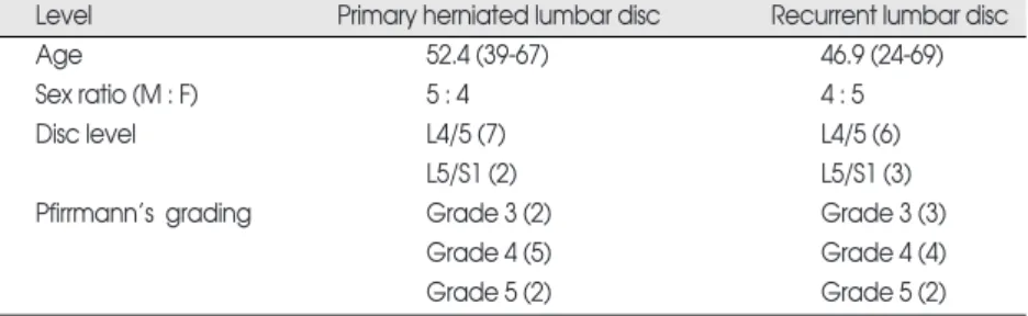
27 months). Disc materials of the primary disc herniation group were obtained from a group of 9 patients (7 from L4/5, 2 from L5/S1 segments) with a mean age of 50.6 (39-67) years old and the primary disc herniation group consisted of 5 men and 4 women. Both groups showed no statistical difference in the general demographics including Pfirrmann’s grading system of lumbar disc degeneration on MR images (Table 1).
Isolation of disc cells and culture
Unless otherwise stated, all reagents were purchased from GibcoBRL (Grand Island, NY, USA). During a lumbar discectomy, intervertebral disc materials were taken from the patients with primary and recurrent disc herniation. To make the samples homologous, disc materials were acqu-ired from nucleus pulposus not annulus. Tissue from each disc was dissected into small pieces and incubated (5% CO2, 95% room air at 37˚C) in Dulbecco’s Modified Eagle Medium and Ham’s F-12 (DMEM/F-12) media. To isolate the cells, disc tissues were digested in DMEM/F-12 media with 0.2% protease (Sigma Chemical, St. Louis, MO, USA) for 1 hour, followed by 0.025% collagenase (Sigma Chemical, St. Louis, MO, USA) for 12 hours. Cells from less than 2 passages were used
for each experiment.
Each disc cells (2 × 10⁵cells/well)
were grown as monolayer cultures for 6 days in DMEM/F-12 media with 10% fetal bovine serum (FBS) +10 U/mL penicillin + 10 g/mL strepto-mycin + 0.2 mmol/L L-glutamine. After 6 days, mRNA expression of aggrecan, type I collagen, type II col-lagen, Sox-9, osteocalcin, and alkaline phosphatase were measured with real-time PCR. And also, we compared the western blot between primary and recurrent disc herniation groups. Real-time polymerase chain reaction assay
Total RNA was extracted from each disc cells and the mRNA levels of ag-grecan, type I collagen, type II colla-gen, Sox-9, osteocalcin, and alkaline phosphatase were determined using real-time PCR. Total RNA was ext-racted with Trizol reagent (GIBCO BRL, Gaithersburg, MD, USA) and quantified by measuring absorbance
at 260 nm using a spectrophotometer (Nanodrop, Dela-ware, USA). Specific primers for aggrecan, type I collagen, type II collagen, Sox-9, osteocalcin, and alkaline phos-phatase were constructed using the complete mRNA sequ-ence from the National Center for Biotechnology Informa-tion. Forward and reverse primer sequences of aggrecan, type I collagen, type II collagen, Sox-9, osteocalcin, and alkaline phosphatase are summarized in Table 2.
We used an ABI Prism 7300 (Applied biosystems, Cali-fornia, USA) that detects SYBR Green fluorescent dye incorporated in double strand DNA. A 15 µL reaction volume included 25 ng of cDNA of RT-PCR and 5 pmole of each primer (aggrecan, type I collagen, type II collagen, Sox-9, osteocalcin, and alkaline phosphatase). Thirty-five real-time PCR cycles were performed for denaturation (95 ˚C for 30 seconds), annealing, and elongation (60˚C for 60 seconds). To confirm amplification specificity, PCR pro-ducts were subjected to a melting curve analysis. Threshold cycles (Ct) of aggrecan, type I collagen, type II collagen, Sox-9, osteocalcin, and alkaline phosphatase were standar-dized according to GAPDH. The mRNA expression of recurrent herniated disc cells were compared to primary herniated disc cells and reported as a ratio.
Table 1. Demographics of primary herniated and recurrent herniated lumbar disc patients
Level Primary herniated lumbar disc Recurrent lumbar disc
Age 52.4 (39-67) 46.9 (24-69)
Sex ratio (M : F) 5 : 4 4 : 5
Disc level L4/5 (7) L4/5 (6)
L5/S1 (2) L5/S1 (3)
Pfirrmann’s grading Grade 3 (2) Grade 3 (3)
Grade 4 (5) Grade 4 (4)
Grade 5 (2) Grade 5 (2)
Two groups have no statistical difference in the demographics
Table 2. Primer sequence for aggrecan, type I collagen, type II collagen, Sox-9, osteocalcin, and
alkaline phosphatase
Primer Sequence
Aggrecan Forward CTGCTTCCGAGGCATTTCAG Reverse CTTGGGTCACGATCCACTCC Type I collagen Forward GTCGAGGGCCAAGACGAAG
Reverse CAGATCACGTCATCGCACAAC Type II collagen Forward GGTCTTGGTGGAAACTTTGCT
Reverse GGTCCTTGCATTACTCCCAAC
Sox-9 Forward AGCGAACGCACATCAAGAC
Reverse GCTGTAGTGTGGGAGGTTGAA Osteocalcin Forward CACTCCTCGCCCTATTGGC
Reverse CCCTCCTGCTTGGACACAAAG Alkaline phosphatase Forward ATGGGATGGGTGTCTCCACA
Reverse CCACGAAGGGGAACTTGTC
GAPDH* Forward ATGGGGAAGGTGAAGGTCG
Reverse GGGGTCATTGATGGCAACAATA *GAPDH (glyceraldehyde-3-phosphate dehydrogenase) was used as a house keeping gene
J Korean Neurosurg Soc 47| January 2010
28 Statistical analysis
Student’s t-test and Fischer’s exact test were used for stati-stics. A p-value less than 0.05 was defined as statistical signi-ficance.
RESULTS
mRNA expression in the primary herniated and recurrent lumbar discs
The mRNA levels of aggrecan, type I collagen, type II collagen, and Sox-9 increased 1.47*, 1.38, 2.04, and 1.22 times, respectively, in recurrent lumbar discs. Also, the mRNA levels of osteocalcin and alkaline phosphatase were increased 1.22 and 1.31 times, respectively, in recurrent lumbar discs (Fig. 1) (* indicates p < 0.05).
Western blotting in the primary herniated and recurrent lumbar discs
There was no difference between primary herniated and recurrent lumbar discs (Fig. 2).
DISCUSSION
The main results of this experiment are that the human recurrent discs had similar molecular biologic features as the primary herniated discs on the chondrogenic and osteo-genic gene expression including western blotting results.
The recurrence of back or sciatic pain after primary dis-cectomy can be caused by a true recurrence of disc hernia-tion, new disc herniation at a different disc, epidural fibrosis, local arachnoiditis, symptomatic arthritis of the small inter-vertebral joints, secondary spinal stenosis, instability, and spondylitis or spondylodiscitis19). The recurrent disc
hernia-tion might inevitably develop in some patients, who can be determined by examining the postoperative scar using Gd-DTPA contrast-enhanced MRI study1,11). We used the
primary disc herniation and confirmed recurrent disc her-niation, not enhanced lesion on the Gd-DTPA contrast MRI to compare both discs.
It was reported that a large extruded lumbar disc resolved spontaneously without operation, and recent immunohi-stologic studies have created insights into the reabsorptive mechanisms that might account for this3,6,11,15). On the
other hand, there is another animal study which showed that the nucleus pulposus can regenerate. Three months after chymopapain injections into the intervertebral disc in the animal study, disc-space narrowing developed for 2 weeks due to the loss of proteoglycans and then the inter-vertebral disc showed an increase in height due to proteo-glycans recovering and 6 months later, the intervertebral
disc height increased more, and normal histology restored2).
The mRNA expressions of aggrecan and type 2 collagen in disc cells, chondrocytes, and bone marrow derived stem cells were increased after rhBMP-2 treatment in rabbits8).
Also, LIM Mineralization Protein (LMP)-1 could induce the BMP-2 and BMP-7 gene expression and then finally increase the protein production in both AF cells and chond-rocytes of rabbits10). But, so far, there have been no reports of
the molecular biologic studies on the human recurrent discs.
Aggrecan Collagen I Collagen II Sox-9 Osteocalcin Alkaline phosphatase 0 0.5 1 1.5 2 2.5 3 3.5
Fig. 1. mRNA expression of recurrent disc cells for aggrecan, type I colla-gen, type II collacolla-gen, Sox-9, osteocalcin, and alkaline phosphatase compared to primary herniated lumbar disc cells.
Fig. 2. Western blotting of recurrent disc cells for aggrecan, type I collagen, type II collagen, Sox-9, osteocalcin, and alkaline phosphatase compared to primary herniated lumbar disc cells.
Aggrecan Collagen I Collagen II Sox-9 Osteocalcin Alkaline phosphatase
Laus et al.11)reported that recurrent discs, which appe-ared within 1 year of surgery, were identical to that of pri-mary herniation in morphological study and so the remain-ing discs after discectomy could regenerate and then fill the resected disc space. Risbud et al.16)reported that the AF and NP derived cells also evidenced chondrogenic differentia-tion. Therefore, degenerated human discs contain skeletal progenitor cells, which suggests that these endogenous progenitors may be used to repair intervertebral discs.
There are many risk factors of recurrent lumbar disc her-niation after conventional open discectomy such as young age, male gender, smoking, and traumatic events19).
We assumed that remaining disc cells after discectomy could regenerate and proliferated during the healing process. For that reason, recurrent discs have similar chondrogenic and osteogenic gene expression. Therefore, we thought the excessive regeneration of remaining discs after discectomy could be one of the reasons for recurrent disc herniation.
CONCLUSION
These studies indicate that recurrent disc cells have similar chondrogenic and osteogenic gene expression compared to primary herniated disc cells. Therefore, we assume that regeneration of the remaining discs could fill the previous discectomy space and also it could be one of the factors for disc recurrence, especially in the molecular biologic field. �Acknowledgements
This study was supported by a faculty research grant of Yonsei University College of Medicine for 2008 (No. 6-2008-0290). References
1. Biaecki J, Lukawski S, Milecki M, Lachowicz W : Differential diag-nosis of post-surgery scars and recurrent lumbar disc herniation in MRI. Ortop Traumatol Rehabil 6 : 172-176, 2004
2. Bradford DS, Cooper KM, Oegema TR Jr : Chymopapain, chemo-nucleolysis, and nucleus pulposus regeneration. J Bone Joint Surg Am 65 : 1220-1231, 1983
3. Doita M, Kanatani T, Harada T, Mizuno K : Immunohistologic study of the ruptured intervertebral disc of the lumbar spine. Spine 21 : 235-241, 1996
4. Guehring T, Omlor GW, Lorenz H, Engelleiter K, Richter W, Carstens C, et al. : Disc distraction shows evidence of regenerative potential in degenerated intervertebral discs as evaluated by protein
expression, magnetic resonance imaging, and messenger ribonucleic acid expression analysis. Spine 31 : 1658-1665, 2006
5. Inoue G, Ohtori S, Aoki Y, Ozawa T, Doya H, Saito T, et al. : Expo-sure of the nucleus pulposus to the outside of the anulus fibrosus induces nerve injury and regeneration of the afferent fibers inner-vating the lumbar intervertebral discs in rats. Spine 31 : 1433-1438, 2006
6. Ito T, Yamada M, Ikuta F, Fukuda T, Hoshi SI, Kawaji Y, et al. : Histologic evidence of absorption of sequestration-type herniated disc. Spine 21 : 230-234, 1996
7. Johnson WE, Sivan S, Wright KT, Eisenstein SM, Maroudas A, Roberts S : Human intervertebral disc cells promote nerve growth over substrata of human intervertebral disc aggrecan. Spine 31 : 1187-1193, 2006
8. Kuh SU, Zhu Y, Li J, Tsai KJ, Fei Q, Hutton WC, et al. : A compa-rison of three cell types as potential candidates for intervertebral disc therapy : anulus fibrosus cells, chondrocytes, and bone marrow derived cells. Joint Bone Spine 76 : 70-74, 2009
9. Kuh SU, Zhu Y, Li J, Tsai KJ, Fei Q, Hutton WC, et al. : Can TGF-beta1 and rhBMP-2 act in synergy to transform bone marrow stem cells to discogenic-type cells? Acta Neurochir (Wien) 150 : 1073-1079; discussion 1079, 2008
10. Kuh SU, Zhu Y, Li J, Tsai KJ, Fei Q, Hutton WC, et al. : The AdLMP-1 transfection in two different cells; AF cells, chondrocytes as potential cell therapy candidates for disc degeneration. Acta Neurochir (Wien) 150 : 803-810, 2008
11. Laus M, Bertoni F, Bacchini P, Alfonso C, Giunti A : [Recurrent lumbar disc herniation : what recurs? (A morphological study of recurrent disc herniation).] Chir Organi Mov 78 : 147-154, 1993 12. Le Maitre CL, Richardson SM, Baird P, Freemont AJ, Hoyland JA :
Expression of receptors for putative anabolic growth factors in human intervertebral disc : implications for repair and regeneration of the disc. J Pathol 207 : 445-452, 2005
13. Lotz JC, Kim AJ : Disc regeneration : why, when, and how. Neuro-surg Clin N 16 : 657-663, 2005
14. Meisel HJ, Siodla V, Ganey T, Minkus Y, Hutton WC, Alasevic OJ : Clinical experience in cell-based therapeutics : Disc chondrocyte transplantation A treatment for degenerated or damaged interverte-bral disc. Biomol Eng 24 : 5-21, 2007
15. Reyentovich A, Abdu WA : Multiple independent, sequential, and spontaneously resolving lumbar intervertebral disc herniations : a case report. Spine 27 : 549-553, 2002
16. Risbud MV, Guttapalli A, Tsai TT, Lee JY, Danielson KG, Vaccaro AR, et al. : Evidence for skeletal progenitor cells in the degenerate human intervertebral disc. Spine 32 : 2537-2544, 2007
17. Schnake KJ, Putzier M, Haas NP, Kandziora F : Mechanical con-cepts for disc regeneration. Eur Spine J 15 Suppl 3 : S354-S360, 2006 18. Swartz KR, Trost GR : Recurrent lumbar disc herniation. Neurosurg
Focus 15 : E10, 2003
19. Suk KS, Lee HM, Moon SH, Kim NH : Recurrent lumbar disc her-niation : results of operative management. Spine 26 : 672-676, 2001
Different Expression of Extracellular Matrix Genes : Primary vs. Recurrent Disc Herniation| SU Kuh, et al.