저작자표시-비영리-변경금지 2.0 대한민국 이용자는 아래의 조건을 따르는 경우에 한하여 자유롭게 l 이 저작물을 복제, 배포, 전송, 전시, 공연 및 방송할 수 있습니다. 다음과 같은 조건을 따라야 합니다: l 귀하는, 이 저작물의 재이용이나 배포의 경우, 이 저작물에 적용된 이용허락조건 을 명확하게 나타내어야 합니다. l 저작권자로부터 별도의 허가를 받으면 이러한 조건들은 적용되지 않습니다. 저작권법에 따른 이용자의 권리는 위의 내용에 의하여 영향을 받지 않습니다. 이것은 이용허락규약(Legal Code)을 이해하기 쉽게 요약한 것입니다. Disclaimer 저작자표시. 귀하는 원저작자를 표시하여야 합니다. 비영리. 귀하는 이 저작물을 영리 목적으로 이용할 수 없습니다. 변경금지. 귀하는 이 저작물을 개작, 변형 또는 가공할 수 없습니다.
Implication of white matter
hyperintensity on the long-term
mortality in patients with cryptogenic
stroke
Seong Ho Jeong
Department of Medicine
Implication of white matter
hyperintensity on the long-term
mortality in patients with cryptogenic
stroke
Directed by Professor Hyo Suk Nam
The Master's Thesis
submitted to the Department of Medicine
the Graduate School of Yonsei University
in partial fulfillment of the requirements for the degree
of Master of Medical Science
This certifies that the Master's Thesis of
Seong Ho Jeong is approved.
Thesis Supervisor : Hyo Suk Nam
---Thesis Committee Member#1 : Ji Hoe Heo
---Thesis Committee Member#2 : Sung Soo Ahn
The Graduate School
Yonsei University
ACKNOWLEDGEMENTS
I would like to express my gratitude to all those who gave me
the possibility to complete this thesis. I am deeply indebted to
my supervisor Prof. Hyo Suk Nam for suggestions and
encouragement in all the time of research, working and writing
this thesis. Additionally, I would like to thank Prof. Ji Hoe Heo
and Prof. Sung Soo Ahn and for comments and advice that
greatly improved manuscript. Minyoul Baik, my fellow in the
department of neurology, supported me in my work. I want to
thank him for all his help, support, interest and hints. At last, I
owe more than thanks to my parents and girlfriend for their
constant love and support to complete this work.
<TABLE OF CONTENTS>
ABSTRACT ···1
I. INTRODUCTION ···3
II. MATERIALS AND METHODS ···5
1. Patients and evaluation ···5
2. Stroke subtype classification···6
3. Risk factors···6
4. Image acquisition and scales for the severity of white matter
hyperintensity ···7
5. Short-term functional outcome and long-term mortality···10
6. Statistic analysis ···10
III. RESULTS ···12
1. Subject characteristics ···12
A. Patients enrollments ···12
B. Demographic characteristics ···14
C. Association between deep white matter hyperintensity and
periventricular white matter hyperintensity···16
2. Functional outcome at 3 months ···18
3. Cumulative death rates ···20
4. Univariable and multivariable analyses of long-term mortality ··22
5. Subgroup analysis of long-term mortality according to age···25
IV. DISCUSSION ···29
V. CONCLUSION ···33
REFERENCES ···34
LIST OF FIGURES
Figure 1. Patient selection ···13
Figure 2. The rates of cumulative death and fatal stroke
according to the severity of white matter hyperintensity ····21
Figure 3. Kaplan–Meier survival curve and Cox regression
survival curve according to the severity of white matter
hyperintensity ···24
Figure 4. Kaplan-Meier survival curve according to age ····27
LIST OF TABLES
Table 1. The rating scale for white matter hyperintensity on
magnetic resonance imaging by Fazekas et al.···9
Table 2. Demographic characteristics according to the severity
of white matter hyperintensity···15
Table 3. Association between the severity of deep white matter
hyperintensity and periventricular white matter hyperintensity
···17
Table 4. Univariable and multivariable analysis for poor
outcome at 3 months (modified Rankin scale score 3–6) ···19
Table 5. Cox regression analysis of long-term mortality ···23
Table 6. Demographic characteristics according to age ···26
Table 7. Cox regression analysis of long-term mortality in
subgroup of younger patients ···28
ABSTRACT
Implication of white matter hyperintensity on the long-term
mortality in patients with cryptogenic stroke
Seong Ho Jeong
Department of Medicine
The Graduate School, Yonsei University
(Directed by Professor Hyo Suk Nam)
Background: Etiology of a quarter to fifth of ischemic strokes is
unknown despite extensive evaluations. Although mortality rate is
substantially high in cryptogenic strokes (CS), little is known regarding
the factors associated with long-term mortality in patients with CS. The
white matter hyperintensity (WMH) is prevalent in ischemic stroke.
The risk of death is increased when the patients have WMH. We
investigated the association of WMH with the long-term mortality in
patients with CS.
Methods: During a 6.5-year period (January 2001-June 2007),
consecutive patients with CS were enrolled and followed up until
December 2013. Long-term mortality and causes of death were identified
using death certificates from the Korean National Statistical Office,
medical records, or interview. The presence of WMH was determined in
the fluid attenuation inversion recovery magnetic resonance imaging
(MRI) by two independent investigators who were blinded to the clinical
information. The WMH was categorized into three groups according to
Fazekas’ scale; Fazekas 0 (no WMH), Fazekas 1 to 2 (mild WMH), and 3
to 6 (severe WMH). The total Fazekas scale was calculated by adding the
periventricular WMH and deep WMH scores.
Results: During the study period, 2732 consecutive patients with
first-ever ischemic stroke were registered to the Yonsei Stroke Registry.
Among them, 599 (21.9%) patients were classified as CS. After
excluding 104 patients who underwent MRI in other hospitals, 495
patients were finally enrolled and followed-up for a median of 8.1 years
(IQR, 6.6-9.5). Severe WMH was found in 192 (38.8%) patients. During
the follow-up period, 158 (31.9%) patients died. The Kaplan-Meier
survival analysis showed that the CS patients with severe WMH showed
higher long-term mortality compared to those without. The Cox
regression analysis revealed that severe WMH along with age, diabetes
mellitus, initial stroke severity were independent predictors for long-term
mortality. The CS patients with severe WMH had a higher death rate
compared to those without (HR 1.62, 95% CI, 1.14-2.31). Subgroup
analysis according to age revealed the younger patients (age < 65) with
severe WMH were associated with higher death rate compared to
younger patients with no or mild WMH (HR 3.45, 95% CI, 1.82-6.55).
Conclusion: In this study, we demonstrated that severe WMH was
independently associated with long-term mortality in CS patients,
especially in younger patients
---Key words: white matter hyperintensity, cryptogenic stroke, long-term
mortality, Fazekas’ score
Implication of white matter hyperintensity on the long-term
mortality in patients with cryptogenic stroke
Seong Ho Jeong
Department of Medicine
The Graduate School, Yonsei University
(Directed by Professor Hyo Suk Nam)
I. INTRODUCTION
To reveal the etiology of stroke is important to prevent the recurrence of stroke. However, the etiology is unknown in about 20 to 25% of patients with ischemic strokes despite extensive evaluations. The unknown etiology is more common in young stroke patients (48% in age ≤55 years old).1
Cryptogenic stroke (CS) is defined as “stroke of undetermined cause that is attributable to negative evaluation” according to the Trial of ORG 10172 in Acute Stroke Treatment classification. The Cryptogenic Stroke/ESUS International Working Group suggested that CS could be caused by artery-to-artery thromboembolism, paroxysmal atrial fibrillation, patent foramen ovale, or minor-risk of cardiac structural abnormalities.1 Although CS
has substantial mortality rate and significantly high rate of recurrent stroke, the factors associated with prognosis in patients with CS have not been thoroughly determined.1
The detection of white matter hyperintensity (WMH) has been increasing in the general population as magnetic resonance imaging (MRI) has become popular. WMH is one of the features for cerebral small vessel disease and it is also called leukoaraiosis.2 WMH is the patchy or confluent lesion in the
periventricular or subcortical areas. Theses lesions are usually identified a high signal intensity in T2-weighted or fluid-attenuated inversion recovery image.2
WMH has been regarded as one of imaging biomarkers for brain frailty.3
Clinically, the patients with WMH are associated with the increased risk of stroke,4,5 dementia,6,7 gait disturbance,8 hemorrhagic transformation,9 or poor
functional outcome after stroke.10,11
The long-term prognosis of patients who have WMH has been known to be poor. Poor prognosis of WMH was reported not only in the high risk populations with a history of stroke, depression, or minor neurological symptoms4,12-17 but also in the general population.15,18-21 However, little is
known on the association between the burden of WMH and long-term prognosis in CS patients. Therefore, we aimed to investigate the clinical characteristics of CS patients with WMH and their long-term prognosis.
II. MATERIALS AND METHODS
1. Patients and evaluation
Subjects for this study were drawn from patients with acute ischemic stroke who had been registered in the Yonsei Stroke Registry. For this study, we included the consecutive patients admitted from Jan 2001 to June 2007. Consecutive patients with cerebral infarction or transient ischemic attack within 7 days after symptom onset are registered into the Yonsei Stroke Registry.22
During admission, all patients were thoroughly investigated for medical history, clinical manifestations, and the presence of vascular risk factors. Every patient was evaluated with 12-lead electrocardiography, chest x- ray, lipid profile, and standard blood tests. All registered patients underwent brain imaging studies including brain computed tomography (CT) and/or MRI. An angiographic study using CT angiography, magnetic resonance angiography, or digital subtraction angiography was the standard evaluation. For the patients younger than 45 years old, additional blood tests for coagulopathy or prothrombotic conditions were performed.23 Transesophageal echocardiography was a part of the standard
evaluation, except in patients with decreased consciousness, impending brain herniation, poor systemic conditions, inability to accept an esophageal transducer because of swallowing difficulty or tracheal intubation, or lack of informed consent. Transthoracic echocardiography, heart CT, and Holter monitoring were also performed.23-25 A stroke unit was opened in the study
hospital on December 2002. Since then, most patients have been admitted to the stroke unit and are monitored continuously with electrocardiography during their stay in the stroke unit. Since July 2006, heart CT using multi-slice CT had been performed for the evaluation of the coronary artery, aorta and heart.
Indications of heart CT were at least one of the following: (1) the presence of atherosclerosis in the intracranial or extracranial cerebral artery; (2) the presence of ≥2 risk factors for coronary artery disease such as hypertension, diabetes mellitus, dyslipidemia, cigarette smoking, and central obesity; and (3) old age (men older than 45 years, women older than 55 years).26
2. Stroke subtype classification
The stroke subtypes were determined according to the Trial of ORG 10172 in Acute Stroke Treatment classification. We defined CS as stroke of undetermined etiology attributable to negative evaluation despite extensive work-up. Stroke classification was determined in the weekly conferences based on the consensus of stroke neurologists. Data of clinical information, risk factors, findings of imaging studies, laboratory or other special evaluation were collected. Along with these data, prognosis during hospitalization and long-term outcomes were also determined. Data was entered into a web-based database.
3. Risk factors
Hypertension was defined when a patient had a resting systolic blood pressure ≥140 mm Hg or diastolic blood pressure ≥90 mm Hg on repeated measurements during hospitalization or had been taken antihypertensive medication. Diabetes mellitus was diagnosed when a patient had a fasting plasma glucose value ≥7 mmol/L or had been treated with an oral hypoglycemic agent or insulin. Hyperlipidemia was diagnosed when a patient had a fasting serum total cholesterol level ≥6.2 mmol/L, low- density lipoprotein cholesterol ≥4.1 mmol/L, or if the patient had been using a lipid- lowering drug after the
diagnosis of hyperlipidemia. A current smoker was defined as an individual who smoked at the time of stroke or had quit smoking within 1 year.27
4. Image acquisition and scales for the severity of white matter hyperintensity
All MRI examinations were performed on a 3.0T MRI machine (Achieva 3.0T; Philips Medical Systems, Best, Netherlands or MAGNETOM 3.0T Trio; Siemens, Germany). Patients underwent MRI within 3 days after admission, and the following parameters were used. For diffusion-weighted imaging: repetition time (TR)/echo time (TE) = 2,600–6,500/42–70 milliseconds, 2-mm interslice gap, field of view (FOV) = 230 X 230 mm, slice thickness = 5 mm, 6 different directions of diffusion gradient (x, y, z, xy, yz, zx), and 2 b values (0, 1,000); for fluid-attenuated inversion recovery: TR/TE = 9,000/120 milliseconds, pixel spacing = 0.449 mm/0.449 mm, FOV = 230 X 230 mm, and slice thickness = 5 mm.
WMH was determined on fluid-attenuated inversion recovery MRI in the supratentorium according to the Standards for Reporting Vascular changes on Neuroimaging criteria28 and graded according to the Fazekas scale (Table 1).
The Fazekas scale is one of subjective scales for scoring WMH lesions and this scale has been widely used for the patients with stroke.29-31The total Fazekas
scale was calculated by adding the periventricular WMH and deep WMH scores. Perivascular spaces and lacunes of presumed vascular origin were excluded. WMH was separately assessed in each hemisphere but only the score from the non-ischemic hemisphere was considered. We then dichotomized the degree of WMH to Fazekas 0 (no), Fazekas 1 to 2 (mild), and 3 to 6 (severe). Two
investigators blinded to the clinical information independently rated the severity of WMH. Inter-rater reliability for the ratings of periventricular WMH, deep WMH, and overall WMH were good (kappa values were 0.842, 0.798, and 0.873, respectively).
Table 1. Rating scale of white matter hyperintesity by Fazekas scale.32
Periventricular white matter hyperintensity
0 Absence
1 Caps or pencil thin lining
2 Smooth halo
3 Irregular white matter hyperintensity extending into deep white matter Deep white matter hyperintensity
0 Absence
1 Punctate foci
2 Beginning confluence of foci 3 Large confluent area
5. Short-term functional outcome and long-term mortality
Short-term functional outcomes at 3 months were determined based on the modified Rankin Scale (mRS). Poor outcome was defined as an mRS score ≥3. Long-term mortality and causes of death were identified using death certificates from the Korean National Statistical Office. In Korea, by law, all deaths of Koreans must be reported to the National Statistical Office. Deaths during the hospitalization or within 48 hours after discharge from a hospital are certified by the physician who examined the decedent. For deaths not certified by physicians, any vague or missing items on the death certificate are clarified by the National Statistical Office via telephone.33 The causes of death in death
statistics of the Korean National Statistical Office have been reported to be reliable.34,35Deaths among subjects from Jan 2001 to December 31, 2013, were
confirmed by matching the information in the death records and identification numbers assigned to subjects at birth.36,37
6. Statistical analysis
SPSS for Windows (version 20.0; SPSS, Chicago, IL) was used for statistical analysis. The Pearson χ2 test was used to compare frequencies. For continuous variables, the normality was examined using the Kolmogorov- Smirnov test. Provided that the data did not deviate from a normal distribution, the mean and standard deviation were calculated and independent sample t tests were used for comparisons. For data that were not normally distributed, we reported descriptive statistics as the median and interquartile range (IQR) and compared them using the Mann–Whitney U test or Kruskal-Wallis H test. Independent predictors for poor outcome at 3 months were determined using the logistic
regression analysis. To compare the long-term mortality according to the severity of WMH, we used Kaplan-Meier survival analysis to calculate the 1-year, 3-year, 5-year actuarial risks of events during follow-up, censored at death or December 31, 2013. The Cox proportional hazard regression analysis was performed to calculate crude and adjusted hazard ratios with 95% confidence interval (CI). For univariable Cox analysis, the severity of WMH and possible confounding factors including age, sex, hypertension, diabetes, current smoking, hyperlipidemia, and initial National Institutes of Health Stroke Scale (NIHSS) scores were compared. Variables with P<0.2 in the univariable analyses were entered into the multivariable Cox regression model to identify independent predictors of long-term mortality. Because age is a major determinant of WMH, age was dichotomized at the median age (65 years is the median age): <65 years and ≥65 years. The association of WMH with mortality was compared in the dichotomized age groups (younger as <65 years old vs. older as ≥65 years old) using Kaplan-Meier survival analysis.
III. RESULTS
1. Subject characteristics
A. Patients enrollments
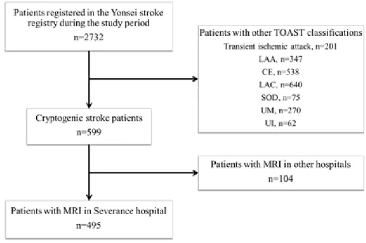
A total of 2732 patients who registered in the Yonsei stroke registry were analyzed. After excluding 2133 patients of transient ischemic attack or other stroke subtypes, 599 patients (21.9%) were categorized into CS. After excluding 104 patients (3.8%) who underwent MRI in other hospitals, 495 patients (18.1%) were included in this study (Figure 1).
Those 495 patients were followed-up for a median of 8.1 years (IQR, 6.6-9.6). Median age of the patients was 64 years (IQR, 56-71) and 62.5% were men. Overall, 172 patients (34.8%) were younger than 60 years and 158 patients (32.0%) were older than 70 years.
Figure 1. Patient selection. TOAST, Trial of ORG 10172 in Acute Stroke
Treatment; FLAIR, fluid-attenuated inversion recovery; LAA, large artery atherosclerosis; CE indicates cardioembolism; LAC, lacune; SOD, stroke of other determined etiology; UM, stroke of undetermined etiology because of multiple causes; UI, stroke of undetermined etiology because of incomplete evaluation.
B. Demographic characteristics
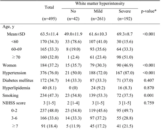
WMH was found in 453 patients (91.5%); severe WMH in 192 patients (38.8%) and mild WMH in 261 patients (52.7%). Only 42 patients (8.5%) did not have WMH.
When comparing patients with no or mild WMH, those with severe WMH were older, more likely to be women, more frequently had hypertension, and less frequently were current smokers (Table 2).
Table 2. Demographic characteristics according to the severity of white matter hyperintensity
Total White matter hyperintensity
p-value* No Mild Severe (n=495) (n=42) (n=261) (n=192) Age, y Mean±SD 63.5±11.4 49.0±11.9 61.6±10.3 69.3±8.7 <0.001 <60 170 (34.3) 33 (78.6) 107 (41.0) 30 (15.6) 60-69 165 (33.3) 8 (19.0) 93 (35.6) 64 (33.3) ≥70 160 (32.0) 1 (2.4) 61 (23.4) 98 (51.0) Women 184 (37.2) 15 (35.7) 79 (30.3) 90 (46.9) <0.001 Hypertension 376 (76.0) 21 (50.0) 188 (72.0) 167 (87.0) <0.001 Diabetes mellitus 172 (34.7) 14 (33.3) 87 (33.3) 71 (37.0) 0.407 Hyperlipidemia 40 (8.1) 0 (0) 24 (9.2) 16 (8.3) 0.870 Smoking 234 (47.3) 23 (54.8) 139 (53.3) 72 (37.5) 0.001 NIHSS score 3 [1-5] 2 [1-4] 3 [1-5] 3 [1-5] 0.759 0-2 237 (48.0) 23 (54.8) 119 (45.6) 95 (49.7) 3-6 166 (33.6) 14 (33.3) 97 (37.2) 55 (28.8) ≥7 91 (18.4) 5 (11.9) 45 (17.2) 41 (21.5)
Data are expressed as the median [interquartile range] or number (%). *p values are compared between severe WMH and others.
C. Association between deep white matter hyperintensity and periventricular white matter hyperintensity
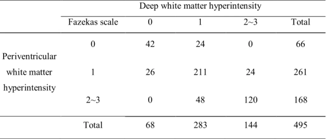
We determined the association between deep WMH and periventricular WMH. Linear-by-linear association showed strong association between the severity of deep WMH and that of periventricular WMH (P<0.001) (Table 3).
Table 3. Association between severity of deep white matter hyperintensity and periventricular white matter hyperintensity
Deep white matter hyperintensity
Fazekas scale 0 1 2~3 Total
Periventricular white matter hyperintensity 0 42 24 0 66 1 26 211 24 261 2~3 0 48 120 168 Total 68 283 144 495 Linear-by-linear association, p<0.001.
2. Functional outcome at 3 months
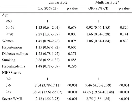
Data of mRS at 3 months were available in all patients. Univariable analysis showed that poor outcome was more frequent in patients with old age, high NIHSS score, and severe WMH. In multivariable analysis, initial stroke severity and the severity of WMH were independent predictors of poor outcome at 3 months. The CS patients with severe WMH had higher possibility of short-term poor outcome (odds ratio, 2.75; 95% CI, 1.56–4.85; Table 4).
Table 4. Univariable and multivariable analysis for poor outcome at 3 months (modified Rankin scale score 3–6)
Univariable Multivariable*
OR (95% CI) p value OR (95% CI) p value
Age <60 1 1 60-69 1.13 (0.64-2.01) 0.678 0.92 (0.46-1.85) 0.820 ≥70 2.27 (1.33-3.87) 0.003 1.66 (0.84-3.28) 0.141 Women 1.45 (0.94-2.26) 0.095 1.06 (0.61-1.84) 0.830 Hypertension 1.15 (0.68-1.92) 0.605 Diabetes mellitus 1.23 (0.78-1.92) 0.371 Smoking 0.86 (0.55-1.32) 0.485 Hyperlipidemia 1.48 (0.71-3.07) 0.296 NIHSS score 0-2 1 1 3-6 8.04 (3.78-17.11) <0.001 9.46 (4.35-20.59) <0.001 ≥7 38.70 (17.61-85.07) <0.001 44.65 (19.64-101.48) <0.001 Severe WMH 2.42 (1.56-3.75) <0.001 2.75 (1.56-4.85) <0.001
*Variables (age, sex, initial NIHSS scores, and severe WMH), which showed P<0.2 in the univariable analysis, were included in the multivariable analysis.
OR, odds ratio; CI, confidence interval; NIHSS, National Institute of Health Stroke Scale; WMH, white matter hyperintensity.
3. Cumulative death rates
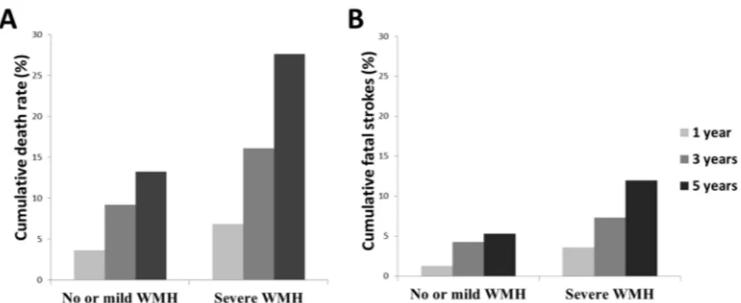
During the follow-up period, 158 (31.9%) patients died. The cumulative death rates were 4.8% within 1 year, 11.9% within 3 years, and 18.8% within 5 years. The overall cumulative death rates were higher in patients with severe WMH (6.8% within 1 year, 16.1% within 3 years, 27.6% within 5 years) than those with no or mild WMH (3.6% within 1 year, 9.2% within 3 years, 13.2% within 5 years) (Figure 2A). Cumulative fatal stroke rates were 2.2% within 1 year, 5.5% within 3 years, and 7.9% within 5 years. The cumulative fatal stroke rates were higher in patients with severe WMH (3.6% within 1 year, 7.3% within 3 years, 12.0% within 5 years) than those with no or mild WMH (1.3% within 1 year, 4.3% within 3 years, 5.3% within 5 years) (Figure 2B).
Figure 2. Cumulative death rate and cumulative fatal stroke rate according to the severity of white matter hyperintensity. Overall cumulative death rate (A) and cumulative fatal stroke rate (B) within 1 year, 3 years, and 5 years were
higher in patients with severe white matter hyperintensity than those with no or mild white matter hyperintensity. WMH, white matter hyperintensity.
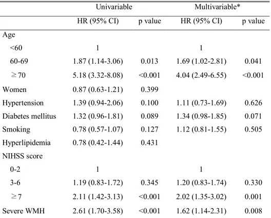
4. Univariable and multivariable analyses of long-term mortality
The Kaplan-Meier survival analysis revealed that more patients with severe WMH died during long-term follow-up compared with those with no or mild WMH (Log rank test, p<0.001) (Figure 3A). Univariable Cox regression analyses showed that older age, history of diabetes, the severity of WMH, and initial stroke severity were associated with long-term mortality. On
multivariable Cox regression analyses, older age, the severity of WMH, and initial stroke severity were independent predictors of long-term mortality. Patients with severe WMH showed higher death rate compared with those with no or mild WMH (hazard ratio 1.62; 95% CI 1.14-2.31; Table 5 and Figure 3B).
Table 5. Cox regression analysis of long-term mortality
Univariable Multivariable*
HR (95% CI) p value HR (95% CI) p value
Age <60 1 1 60-69 1.87 (1.14-3.06) 0.013 1.69 (1.02-2.81) 0.041 ≥70 5.18 (3.32-8.08) <0.001 4.04 (2.49-6.55) <0.001 Women 0.87 (0.63-1.21) 0.399 Hypertension 1.39 (0.94-2.06) 0.100 1.11 (0.73-1.69) 0.626 Diabetes mellitus 1.32 (0.96-1.81) 0.089 1.34 (0.98-1.85) 0.071 Smoking 0.78 (0.57-1.07) 0.127 1.12 (0.81-1.55) 0.505 Hyperlipidemia 0.78 (0.42-1.44) 0.431 NIHSS score 0-2 1 1 3-6 1.19 (0.83-1.72) 0.345 1.20 (0.83-1.74) 0.330 ≥7 2.11 (1.42-3.13) <0.001 2.02 (1.35-3.02) 0.001 Severe WMH 2.61 (1.70-3.58) <0.001 1.62 (1.14-2.31) 0.008
*Variables (age, hypertension, diabetes mellitus, smoking, initial NIHSS scores, and severe WMH), which showed P<0.2 in the univariable analysis, were included in the multivariable analysis.
HR, hazard ratio; CI, confidence interval; NIHSS, National Institute of Health Stroke Scale; WMH; white matter hyperintensity.
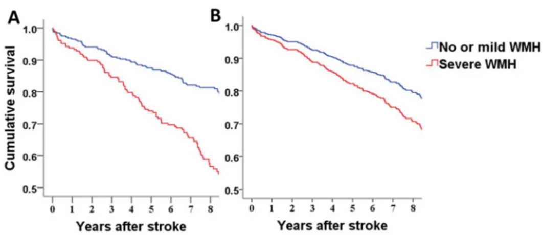
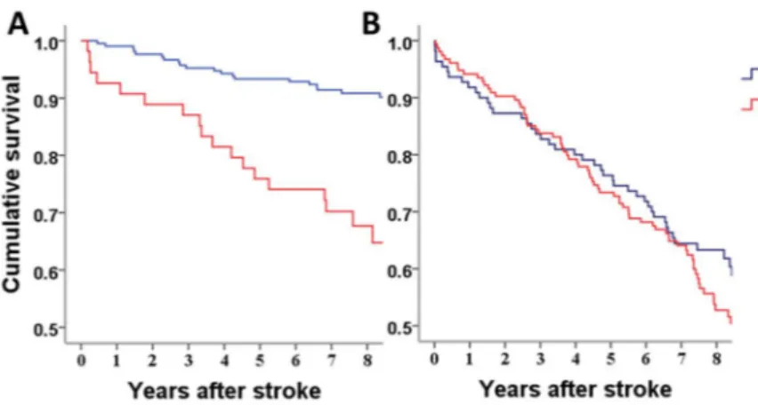
Figure 3. Kaplan–Meier survival curve and Cox regression survival curve according to the severity of white matter hyperintensity. A. Univariable
Kaplan– Meier survival analysis revealed that more patients with severe white matter hyperintensity (red) died during follow-up compared with no or mild white matter hyperintensity (blue) (P<0.001). B. Multivariable Cox regression curve shows that the risk of long-term death was higher in patients with severe white matter hyperintensity (red) compared with no or mild white matter hyperintensity (blue) after adjustment of age, hypertension, diabetes, smoking, and initial stroke severity. WMH, white matter hyperintensity.
5. Subgroup analysis for long-term mortality according to age
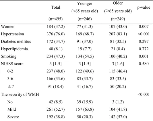
When comparing with older patients (≥65 years), younger patients (<65 years) were less likely to be women, less frequently had hypertension, more frequently were current smokers, and more frequently had severe WMH (Table 6). In younger patients, the Kaplan-Meier survival analysis showed that more patients with severe WMH died during long-term follow-up compared with those with no or mild WMH (Log rank test, p<0.001; Figure 4A). In contrast, there was no difference in survival rate according to the presence of severe WMH in older patients (Log rank test, p=0.129; Figure 4B). Multivariable Cox regression analyses revealed initial stroke severity, diabetes mellitus and the presence of severe WMH were independent predictors of long-term mortality in the subgroup of age < 65 years. The patients with severe WMH showed higher death rate compared to those with no or mild WMH in the subgroup of younger patients (hazard ratio 3.45; 95% CI, 1.82-6.55; Table 7).
Table 6. Demographic characteristics according to age
Total Younger
(<65 years old)
Older
(≥65 years old) p-value
(n=495) (n=246) (n=249) Women 184 (37.2) 77 (31.3) 107 (43.0) 0.007 Hypertension 376 (76.0) 169 (68.7) 207 (83.1) <0.001 Diabetes mellitus 172 (34.7) 91 (37.0) 81 (32.5) 0.297 Hyperlipidemia 40 (8.1) 19 (7.7) 21 (8.4) 0.772 Smoking 234 (47.3) 134 (54.5) 100 (40.2) 0.001 NIHSS score 3 [1-5] 3 [1-5] 3 [1-6] 0.580 0-2 237 (48.0) 122 (49.6) 115 (46.4) 3-6 166 (33.6) 83 (33.7) 83 (33.5) ≥7 91 (18.4) 41 (16.7) 50 (20.2) The severity of WMH <0.001 No 42 (8.5) 39 (15.9) 3 (1.2) Mild 261 (52.7) 157 (63.8) 104 (41.8) Severe 192 (38.8) 50 (20.3) 142 (57.0)
Data are expressed as the median [interquartile range] or number (%). NIHSS, National Institute of Health Stroke Scale; WMH, white matter
Figure 4. Kaplan-Meier survival curve according to age. A. In young
patients, Kaplan-Meier survival analysis showed that cryptogenic stroke patients with severe white matter hyperintensity (red) had higher mortality compared to those with no or mild white matter hyperintensity (blue) (Log rank test, p<0.001). B. Univariable Kaplan-Meier survival analysis in older patients showed no difference between two groups (Log rank test, p=0.214).WMH, white matter hyperintensity
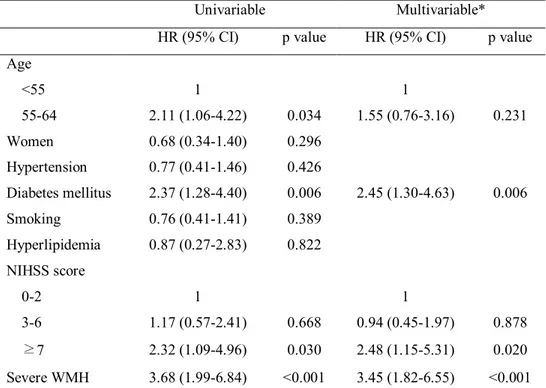
Table 7. Cox regression analysis of long-term mortality in subgroup of younger patients
Univariable Multivariable*
HR (95% CI) p value HR (95% CI) p value
Age <55 1 1 55-64 2.11 (1.06-4.22) 0.034 1.55 (0.76-3.16) 0.231 Women 0.68 (0.34-1.40) 0.296 Hypertension 0.77 (0.41-1.46) 0.426 Diabetes mellitus 2.37 (1.28-4.40) 0.006 2.45 (1.30-4.63) 0.006 Smoking 0.76 (0.41-1.41) 0.389 Hyperlipidemia 0.87 (0.27-2.83) 0.822 NIHSS score 0-2 1 1 3-6 1.17 (0.57-2.41) 0.668 0.94 (0.45-1.97) 0.878 ≥7 2.32 (1.09-4.96) 0.030 2.48 (1.15-5.31) 0.020 Severe WMH 3.68 (1.99-6.84) <0.001 3.45 (1.82-6.55) <0.001
*Variables (age, diabetes mellitus, initial NIHSS scores, and severe WMH), which showed P<0.2 in the univariable analysis, were included in the multivariable analysis. HR, hazard ratio; CI, confidence interval; NIHSS, National Institute of Health Stroke Scale; WMH; white matter hyperintensity.
IV. DISCUSSION
This study demonstrated that severe WMH was frequent in CS patients. Accompanying WMH was independently associated with long-term mortality in CS patients. Age was a major determinant of death in CS patients with WMH. Younger CS patients (age <65) with severe WMH were associated with higher death rates compared to those with no or mild WMH, whereas old patients did not show these association.
In this study, the prevalence of WMH was 91.5% and the proportion of severe WMH was 39.4%. The
p
revalence of WMH in stroke patients differs between studies, from 6.8%38 to 51.7%39. This variability can be explained bymethodological differences between the studies including different methods to assess imaging findings, and differences in the presence of risk factors between study populations.
Our study demonstrated that severe WMH was an independent predictor of short-term and long-term poor outcome in CS patients. Prior studies of WMH and stroke outcome have been inconsistent; a direct association of WMH on stroke morbidity has been demonstrated in some10,13,40-42 but not in other43,44
studies. The mechanisms underlying the influence of WMH on functional outcomes in stroke patients are uncertain. However, there are some hypotheses that explain the relationship between WMH and clinical outcome. First, it is well known that WMH regions have reduced vascular density and cerebral blood flow, which may induce infarct growth after ischemic event in the acute stage of stroke.45,46Second, the presence of dysfunctional neuronal networks in
WMH and poor outcome. An intact system of intra-hemispheric and inter-hemispheric connectivity is important for functional recovery following stroke.47 A neuropathological study has shown the correlation between
decreased neuronal connectivity and WMH,48 which is associated with the
demyelination, loss of axons and oligodendrocytes, and astrocytic gliosis in white matter.49 This may result in decreased functional connectivity of distant
cortical regions,50 which could impair plasticity and inhibit recovery. Third,
because WMH is associated with cognitive decline, gait disturbance, and post stroke depression,50-53 poor compliance with medical treatment and
rehabilitation may affect stroke outcome.52,53
Along with short-term poor prognosis, severe WMH was an independent predictor of long-term mortality in patients with CS. Meta-analysis demonstrated that the extent of WMH was associated with mortality in the general population.54 Some studies showed the relationship between long-term
mortality and WMH in stroke patients.13,20,55 However, another study failed to
show the association between long-term mortality and severe WMH.42 Other
studies showed that this association was more remarkable for cardiovascular mortality.23,25,28
The underlying mechanisms why severe WMH in CS patients are associated with long-term mortality are uncertain. There may be several speculations. First, polyvascular diseases are known to be poor prognosis in stroke patients.56,57
WMH are a surrogate marker for an important burden of vascular risk factors and a marker of diffuse vascular damage involving not only the cerebral arteries but also coronary and peripheral arteries.33,58 In this regards, atherosclerotic
patients. Second, shot-term outcomes were poor in CS patients with severe WMH. As the short-term prognosis is a major determinant of long-term outcome,59 poor short-term functions outcomes in CS patients with severe
WMH might be partly responsible for the high long-term mortality. Third, severe WMH may imply that there are hidden cardioembolic source. Long-term monitoring of heart rhythm identifies paroxysmal atrial fibrillation in up to 16% of patients with cryptogenic stroke.60,61 Other cardioembolic sources, such as
left atrial enlargement, atrial septal aneurysm and patent foramen ovale, are known to be associated with CS, too.62,63The burden of WMH was associated
with atrial fibrillation, patent foramen ovale, or atrial septal aneurysm, which suggests that cardioembolism may cause WMH in some patients.64-66 In this
regard, severe WMH may be associated with hidden cardioembolic source in some patients with CS. When considering that stroke patients with cardioembolism have the most unfavorable outcome among stroke subtypes,1
poor long-term outcome in patients with CS and severe WMH might be associated with this uncovered cardioembolism.
Of note, a substantial proportion of our young patients with CS harbored WMH in brain MRI (84.1%). In this study, young CS patients were at a 3.45-fold higher risk of death when they had severe WMH when compared with those with no or mild WMH. However, old CS patients with severe WMH did not show association between severe WMH and long-term mortality. Even though older patients have no or mild WMH, they could have multiple medical problems as well as subclinical aging process in other organs.67 Accompanied
multiple comorbidity strongly influence on long-term mortality, which may dilute the effects of severe WMH on long-term prognosis. On the other hand, severe WMH in younger CS patients might be one of marker of unhealthy brain
and other comorbidity. To confirm this finding, prospective study for younger CS patient is needed.
In consistent with our study, most previous studies have found WMH to be positively related to age and hypertension.68-73However, previous investigations
have been contradictory; some indicated higher incidence of WMH in women, others in men.70,74Studies of the effect of diabetes mellitus and hyperlipidemia
on WMH have shown conflicting results71,72,75,76 and we found no relationship
with severe WMH. We found that severe WMH was less frequent in current smoker. Previous study showed that conflicting report whether a history of current or previous smoking is associated with WMH. Rotterdam study showed significant inverse correlation between smoking and WMH in univariable analysis.50 However, Fukuda et al. showed weak positive correlation between
cigarette smoking and periventricular WMH in multivariable analysis.77. We
hypothesized that this conflicting finding may by related that age is more strongly related with WMH than smoking, which resulted in inverse correlation between WMH and current smoking. To support our hypothesis, the Pearson χ2 test showed the inverse correlation between age and current smoking status (54.9% in younger group vs. 39.0% in older group, p < 0.001, Table 6).
Our study has several limitations. First, this study was confined to the patients who had diffusion-weighted imaging and
fluid attenuation inversion
recovery
image. The patients with severe strokes who were unable to undergo MRI and patients who had contraindications for MRI were excluded. However, exclusion of these patients may affect our results, most research of WMH is underwent using MRI. Second, our patient cohort was all Asian, further study to test our finding in other ethnic groups is warranted.V. CONCLUSION
Severe WMH in CS patients was associated with poor short-term functional outcome and long-term mortality. The association between severe WMH and long-term mortality was observed in younger patients, but not in older patients. Our findings provide evidence for the usefulness of identifying WMH for predicting the prognosis of patients with CS patients.
REFERENCES
1. Li L, Yiin GS, Geraghty OC, Schulz UG, Kuker W, Mehta Z, et al. Incidence, outcome, risk factors, and long-term prognosis of cryptogenic transient ischaemic attack and ischaemic stroke: a population-based study. Lancet Neurol 2015;14:903-13.
2. Hachinski VC, Potter P, Merskey H. Leuko-araiosis. Arch Neurol 1987;44:21-3.
3. Association between brain imaging signs, early and late outcomes, and response to intravenous alteplase after acute ischaemic stroke in the third International Stroke Trial (IST-3): secondary analysis of a randomised controlled trial. Lancet Neurol 2015;14:485-96.
4. Fu JH, Lu CZ, Hong Z, Dong Q, Luo Y, Wong KS. Extent of white matter lesions is related to acute subcortical infarcts and predicts further stroke risk in patients with first ever ischaemic stroke. J Neurol Neurosurg Psychiatry 2005;76:793-6.
5. Henon H, Vroylandt P, Durieu I, Pasquier F, Leys D. Leukoaraiosis more than dementia is a predictor of stroke recurrence. Stroke 2003;34:2935-40.
6. Inzitari D, Pracucci G, Poggesi A, Carlucci G, Barkhof F, Chabriat H, et al. Changes in white matter as determinant of global functional decline in older independent outpatients: three year follow-up of LADIS (leukoaraiosis and disability) study cohort. Bmj 2009;339:b2477. 7. Skoog I, Berg S, Johansson B, Palmertz B, Andreasson LA. The
influence of white matter lesions on neuropsychological functioning in demented and non-demented 85-year-olds. Acta Neurol Scand 1996;93:142-8.
8. Briley DP, Wasay M, Sergent S, Thomas S. Cerebral white matter changes (leukoaraiosis), stroke, and gait disturbance. J Am Geriatr Soc 1997;45:1434-8.
9. Neumann-Haefelin T, Hoelig S, Berkefeld J, Fiehler J, Gass A, Humpich M, et al. Leukoaraiosis is a risk factor for symptomatic intracerebral hemorrhage after thrombolysis for acute stroke. Stroke 2006;37:2463-6.
10. Arsava EM, Rahman R, Rosand J, Lu J, Smith EE, Rost NS, et al. Severity of leukoaraiosis correlates with clinical outcome after ischemic stroke. Neurology 2009;72:1403-10.
11. Koton S, Schwammenthal Y, Merzeliak O, Philips T, Tsabari R, Orion D, et al. Cerebral leukoaraiosis in patients with stroke or TIA: clinical correlates and 1-year outcome. Eur J Neurol 2009;16:218-25.
12. Tveiten A, Ljostad U, Mygland A, Naess H. Leukoaraiosis is associated with short- and long-term mortality in patients with intracerebral hemorrhage. J Stroke Cerebrovasc Dis 2013;22:919-25.
13. Miyao S, Takano A, Teramoto J, Takahashi A. Leukoaraiosis in relation to prognosis for patients with lacunar infarction. Stroke 1992;23:1434-8.
14. Yamauchi H, Fukuda H, Oyanagi C. Significance of white matter high intensity lesions as a predictor of stroke from arteriolosclerosis. J Neurol Neurosurg Psychiatry 2002;72:576-82.
15. Ikram MA, Vernooij MW, Vrooman HA, Hofman A, Breteler MM. Brain tissue volumes and small vessel disease in relation to the risk of mortality. Neurobiol Aging 2009;30:450-6.
16. Kerber KA, Whitman GT, Brown DL, Baloh RW. Increased risk of death in community-dwelling older people with white matter
hyperintensities on MRI. J Neurol Sci 2006;250:33-8.
17. Oksala NK, Oksala A, Pohjasvaara T, Vataja R, Kaste M, Karhunen PJ, et al. Age related white matter changes predict stroke death in long term follow-up. J Neurol Neurosurg Psychiatry 2009;80:762-6.
18. Kuller LH, Arnold AM, Longstreth WT, Jr., Manolio TA, O'Leary DH, Burke GL, et al. White matter grade and ventricular volume on brain MRI as markers of longevity in the cardiovascular health study. Neurobiol Aging 2007;28:1307-15.
19. Bokura H, Kobayashi S, Yamaguchi S, Iijima K, Nagai A, Toyoda G, et al. Silent brain infarction and subcortical white matter lesions increase the risk of stroke and mortality: a prospective cohort study. J Stroke Cerebrovasc Dis 2006;15:57-63.
20. Briley DP, Haroon S, Sergent SM, Thomas S. Does leukoaraiosis predict morbidity and mortality? Neurology 2000;54:90-4.
21. Debette S, Beiser A, DeCarli C, Au R, Himali JJ, Kelly-Hayes M, et al. Association of MRI markers of vascular brain injury with incident stroke, mild cognitive impairment, dementia, and mortality: the Framingham Offspring Study. Stroke 2010;41:600-6.
22. Lee BI, Nam HS, Heo JH, Kim DI. Yonsei Stroke Registry. Analysis of 1,000 patients with acute cerebral infarctions. Cerebrovasc Dis 2001;12:145-51.
23. Cho HJ, Choi HY, Kim YD, Nam HS, Han SW, Ha JW, et al. Transoesophageal echocardiography in patients with acute stroke with sinus rhythm and no cardiac disease history. J Neurol Neurosurg Psychiatry 2010;81:412-5.
24. Nam HS, Han SW, Lee JY, Ahn SH, Ha JW, Rim SJ, et al. Association of aortic plaque with intracranial atherosclerosis in patients with stroke.
Neurology 2006;67:1184-8.
25. Han SW, Nam HS, Kim SH, Lee JY, Lee KY, Heo JH. Frequency and significance of cardiac sources of embolism in the TOAST classification. Cerebrovasc Dis 2007;24:463-8.
26. Yoo J, Yang JH, Choi BW, Kim YD, Nam HS, Choi HY, et al. The frequency and risk of preclinical coronary artery disease detected using multichannel cardiac computed tomography in patients with ischemic stroke. Cerebrovasc Dis 2012;33:286-94.
27. Song TJ, Kim J, Lee HS, Nam CM, Nam HS, Kim YD, et al. Distribution of cerebral microbleeds determines their association with impaired kidney function. J Clin Neurol 2014;10:222-8.
28. Wardlaw JM, Smith EE, Biessels GJ, Cordonnier C, Fazekas F, Frayne R, et al. Neuroimaging standards for research into small vessel disease and its contribution to ageing and neurodegeneration. Lancet Neurol 2013;12:822-38.
29. Helenius J, Goddeau RP, Jr., Moonis M, Henninger N. Impact of Leukoaraiosis Burden on Hemispheric Lateralization of the National Institutes of Health Stroke Scale Deficit in Acute Ischemic Stroke. Stroke 2016;47:24-30.
30. Helenius J, Henninger N. Leukoaraiosis Burden Significantly Modulates the Association Between Infarct Volume and National Institutes of Health Stroke Scale in Ischemic Stroke. Stroke 2015;46:1857-63.
31. Onteddu SR, Goddeau RP, Jr., Minaeian A, Henninger N. Clinical impact of leukoaraiosis burden and chronological age on neurological deficit recovery and 90-day outcome after minor ischemic stroke. J Neurol Sci 2015;359:418-23.
32. Fazekas F, Chawluk JB, Alavi A, Hurtig HI, Zimmerman RA. MR signal abnormalities at 1.5 T in Alzheimer's dementia and normal aging. AJR Am J Roentgenol 1987;149:351-6.
33. Khang YH, Lynch JW, Kaplan GA. Health inequalities in Korea: age-and sex-specific educational differences in the 10 leading causes of death. Int J Epidemiol 2004;33:299-308.
34. Kim HC, Choi DP, Ahn SV, Nam CM, Suh I. Six-year survival and causes of death among stroke patients in Korea. Neuroepidemiology 2009;32:94-100.
35. Won TY, Kang BS, Im TH, Choi HJ. The Study of Accuracy of Death Statistics. Journal of The Korean Society of Emergency Medicine 2007;18:256-62.
36. Kim YD, Song D, Kim EH, Lee KJ, Lee HS, Nam CM, et al. Long-term mortality according to the characteristics of early neurological deterioration in ischemic stroke patients. Yonsei Med J 2014;55:669-75. 37. Nam HS, Kim HC, Kim YD, Lee HS, Kim J, Lee DH, et al. Long-term
mortality in patients with stroke of undetermined etiology. Stroke 2012;43:2948-56.
38. Wiszniewska M, Devuyst G, Bogousslavsky J, Ghika J, van Melle G. WHat is the significance of leukoaraiosis in patients with acute ischemic stroke? Archives of Neurology 2000;57:967-73.
39. Lee S-J, Kim J-S, Lee K-S, An J-Y, Kim W, Kim Y-I, et al. The leukoaraiosis is more prevalent in the large artery atherosclerosis stroke subtype among Korean patients with ischemic stroke. BMC Neurology 2008;8:1-6.
40. Zhang J, Puri AS, Khan MA, Goddeau RP, Jr., Henninger N. Leukoaraiosis predicts a poor 90-day outcome after endovascular stroke
therapy. AJNR Am J Neuroradiol 2014;35:2070-5.
41. Henninger N, Lin E, Baker SP, Wakhloo AK, Takhtani D, Moonis M. Leukoaraiosis predicts poor 90-day outcome after acute large cerebral artery occlusion. Cerebrovasc Dis 2012;33:525-31.
42. Kissela B, Lindsell CJ, Kleindorfer D, Alwell K, Moomaw CJ, Woo D, et al. Clinical prediction of functional outcome after ischemic stroke: the surprising importance of periventricular white matter disease and race. Stroke 2009;40:530-6.
43. Jorgensen HS, Nakayama H, Raaschou HO, Olsen TS. Leukoaraiosis in stroke patients. The Copenhagen Stroke Study. Stroke 1995;26:588-92. 44. Palumbo V, Boulanger JM, Hill MD, Inzitari D, Buchan AM.
Leukoaraiosis and intracerebral hemorrhage after thrombolysis in acute stroke. Neurology 2007;68:1020-4.
45. Moody DM, Thore CR, Anstrom JA, Challa VR, Langefeld CD, Brown WR. Quantification of afferent vessels shows reduced brain vascular density in subjects with leukoaraiosis. Radiology 2004;233:883-90. 46. O'Sullivan M, Lythgoe DJ, Pereira AC, Summers PE, Jarosz JM,
Williams SC, et al. Patterns of cerebral blood flow reduction in patients with ischemic leukoaraiosis. Neurology 2002;59:321-6.
47. Grefkes C, Nowak DA, Eickhoff SB, Dafotakis M, Kust J, Karbe H, et al. Cortical connectivity after subcortical stroke assessed with functional magnetic resonance imaging. Ann Neurol 2008;63:236-46. 48. Yamanouchi H, Sugiura S, Tomonaga M. Decrease in nerve fibres in
cerebral white matter in progressive subcortical vascular encephalopathy of Binswanger type. An electron microscopic study. J Neurol 1989;236:382-7.
1989;236:65-79.
50. Nordahl CW, Ranganath C, Yonelinas AP, Decarli C, Fletcher E, Jagust WJ. White matter changes compromise prefrontal cortex function in healthy elderly individuals. J Cogn Neurosci 2006;18:418-29.
51. De Groot JC, De Leeuw FE, Oudkerk M, Van Gijn J, Hofman A, Jolles J, et al. Periventricular cerebral white matter lesions predict rate of cognitive decline. Ann Neurol 2002;52:335-41.
52. Sinyor D, Amato P, Kaloupek DG, Becker R, Goldenberg M, Coopersmith H. Post-stroke depression: relationships to functional impairment, coping strategies, and rehabilitation outcome. Stroke 1986;17:1102-7.
53. Firbank MJ, Teodorczuk A, van der Flier WM, Gouw AA, Wallin A, Erkinjuntti T, et al. Relationship between progression of brain white matter changes and late-life depression: 3-year results from the LADIS study. Br J Psychiatry 2012;201:40-5.
54. Debette S, Markus HS. The clinical importance of white matter hyperintensities on brain magnetic resonance imaging: systematic review and meta-analysis. Bmj 2010;341:c3666.
55. Putaala J, Haapaniemi E, Kurkinen M, Salonen O, Kaste M, Tatlisumak T. Silent brain infarcts, leukoaraiosis, and long-term prognosis in young ischemic stroke patients. Neurology 2011;76:1742-9.
56. Blanco M, Sobrino T, Montaner J, Medrano V, Jimenez C, Masjuan J, et al. Stroke with polyvascular atherothrombotic disease. Atherosclerosis 2010;208:587-92.
57. Cimminiello C, Zaninelli A, Carolei A, Sacco S, Toni D, Gensini G. Atherothrombotic burden and medium-term prognosis in patients with acute ischemic stroke: findings of the SIRIO study. Cerebrovasc Dis
2012;33:341-7.
58. Gerdes VE, Kwa VI, ten Cate H, Brandjes DP, Buller HR, Stam J. Cerebral white matter lesions predict both ischemic strokes and myocardial infarctions in patients with established atherosclerotic disease. Atherosclerosis 2006;186:166-72.
59. Eriksson M, Norrving B, Terent A, Stegmayr B. Functional outcome 3 months after stroke predicts long-term survival. Cerebrovasc Dis 2008;25:423-9.
60. Gladstone DJ, Spring M, Dorian P, Panzov V, Thorpe KE, Hall J, et al. Atrial fibrillation in patients with cryptogenic stroke. N Engl J Med 2014;370:2467-77.
61. Sanna T, Diener HC, Passman RS, Di Lazzaro V, Bernstein RA, Morillo CA, et al. Cryptogenic stroke and underlying atrial fibrillation. N Engl J Med 2014;370:2478-86.
62. Cabanes L, Mas JL, Cohen A, Amarenco P, Cabanes PA, Oubary P, et al. Atrial septal aneurysm and patent foramen ovale as risk factors for cryptogenic stroke in patients less than 55 years of age. A study using transesophageal echocardiography. Stroke 1993;24:1865-73.
63. Yaghi S, Moon YP, Mora-McLaughlin C, Willey JZ, Cheung K, Di Tullio MR, et al. Left Atrial Enlargement and Stroke Recurrence: The Northern Manhattan Stroke Study. Stroke 2015;46:1488-93.
64. de Leeuw FE, de Groot JC, Oudkerk M, Kors JA, Hofman A, van Gijn J, et al. Atrial fibrillation and the risk of cerebral white matter lesions. Neurology 2000;54:1795-801.
65. Karmarkar A, Helenius J, Henninger N, McManus DD. Abstract 17308: Leukoaraiosis is Associated With Atrial Fibrillation and Greater Disability at 90-days in Patients Hospitalized With a Cardioembolic
Stroke. Circulation 2015;132:A17308.
66. Ueno Y, Shimada Y, Tanaka R, Miyamoto N, Tanaka Y, Hattori N, et al. Patent Foramen Ovale with Atrial Septal Aneurysm May Contribute to White Matter Lesions in Stroke Patients. Cerebrovasc Dis 2010;30:15-22.
67. Karlamangla A, Tinetti M, Guralnik J, Studenski S, Wetle T, Reuben D. Comorbidity in older adults: nosology of impairment, diseases, and conditions. J Gerontol A Biol Sci Med Sci 2007;62:296-300.
68. Fernando MS, Simpson JE, Matthews F, Brayne C, Lewis CE, Barber R, et al. White matter lesions in an unselected cohort of the elderly: molecular pathology suggests origin from chronic hypoperfusion injury. Stroke 2006;37:1391-8.
69. Pantoni L, Garcia JH. The significance of cerebral white matter abnormalities 100 years after Binswanger's report. A review. Stroke 1995;26:1293-301.
70. Park K, Yasuda N, Toyonaga S, Yamada SM, Nakabayashi H, Nakasato M, et al. Significant association between leukoaraiosis and metabolic syndrome in healthy subjects. Neurology 2007;69:974-8.
71. Padovani A, Di Piero V, Bragoni M, Di Biase C, Trasimeni G, Lannili M, et al. Correlates of leukoaraiosis and ventricular enlargement on magnetic resonance imaging: a study in normal elderly and cerebrovascular patients. Eur J Neurol 1997;4:15-23.
72. Streifler JY, Eliasziw M, Benavente OR, Hachinski VC, Fox AJ, Barnett HJ. Lack of relationship between leukoaraiosis and carotid artery disease. The North American Symptomatic Carotid Endarterectomy Trial. Arch Neurol 1995;52:21-4.
1997;28:652-9.
74. Longstreth WT, Jr., Manolio TA, Arnold A, Burke GL, Bryan N, Jungreis CA, et al. Clinical correlates of white matter findings on cranial magnetic resonance imaging of 3301 elderly people. The Cardiovascular Health Study. Stroke 1996;27:1274-82.
75. van Swieten JC, Hijdra A, Koudstaal PJ, van Gijn J. Grading white matter lesions on CT and MRI: a simple scale. J Neurol Neurosurg Psychiatry 1990;53:1080-3.
76. Bogousslavsky J, Regli F, Uske A. Leukoencephalopathy in patients with ischemic stroke. Stroke 1987;18:896-9.
77. Fukuda H, Kitani M. Cigarette smoking is correlated with the periventricular hyperintensity grade of brain magnetic resonance imaging. Stroke 1996;27:645-9.
ABSTRACT