tion in peripheral sensory input or behavioral manipulation6,16). A much greater extent of structural and functional change can be observed after large-scale injuries such as SCI or limb amputa-tion11,18). Spinal lesions in primates reshape the sensory repre-sentational map in the cortex11,15). EEG and PET in SCI patients has shown reorganization related to the recovery of limb func-tions and extensive changes in cortical and subcortical activa-tion3,8,9). On the other hand, transcranial magnetic stimulation in paraplegics disclosed an enlargement of the cortical repre-sentations of non-affected muscles in the primary motor cor-tex, together with an increased excitability7). A few reports have shown the alterations in the expression of the neurotrophins (NTs), NTs receptors, and prohormone convertase in the in-jured site following SCI10). Epigenetically, histone deacetylase (HDAC) family have been reported it can be important role in
INTRODUCTION
Structural and functional changes following spinal cord injury (SCI) include both retrograde and antrograde cell degenera-tion2,24). SCI removes supraspinal input to the spinal sensory and motor networks and thus results in severe and permanent sen-sory and motor function impairment after injury site. Some de-gree of functional recovery, however, can be observed without management4). Since regeneration of injured axons is limited in mature CNS, spontaneous recovery in motor function appears to be mediated by reorganization of spared neuronal system. This compensatory remodeling occurs at multiple level of the neur-axis including spinal motor centers, descending supraspinal tracts, brainstem and brain cortex19). The sensorimotor cortex in adults retains the capability to reorganize in response to
altera-Epigenetic Regulation in the Brain after Spinal Cord
Injury : A Comparative Study
Bit-Na-Ri Park, M.D.,1 Seok Won Kim, M.D., Ph.D.,4 Sung-Rae Cho, M.D., Ph.D.,2 Ji Yong Lee, P.T., M.S.,3 Young-Hee Lee, M.D., Ph.D.,1
Sung-Hoon Kim, M.D., Ph.D.1
Department of Rehabilitation Medicine,1 Wonju Christian Hospital, Yonsei University Wonju College of Medicine, Wonju, Korea Department of Rehabilitation Medicine and Research Institute,2 Yonsei University College of Medicine, Seoul, Korea
Department of Anatomy,3 Yonsei University Wonju College of Medicine, Wonju, Korea Department of Neurosurgery,4 College of Medicine, Chosun University, Gwangju, Korea
Objective : After spinal cord injury (SCI), functional and structural reorganization occurs at multiple levels of brain including motor cortex. However, the underlying mechanism still remains unclear. The current study was performed to investigate the alterations in the expression of the main regula-tors of neuronal development, survival and death, in the brain following thoracic contusive SCI in a mouse model.
Methods : Eight-week-old female imprinting control region mice (n=60; 30-35 g) were used in this study. We analyzed the expression levels of regulators such as brain-derived neurotrophic factor (BDNF), glial cell line-derived neurotrophic factor (GDNF), nerve growth factor (NGF) and his-tone deacetylase (HDAC) 1 in the brain following thoracic contusive SCI.
Results : The expression of BDNF levels were elevated significantly compared with control group at 2 weeks after injury (p<0.05). The expression of NGF levels were elevated at 2, 4 weeks compared with control group, but these difference were not significant (p>0.05). The GDNF levels were elevated at 2 week compared with control group, but these differences were not significant (p>0.05). The difference of HDAC1 levels were not sig-nificant at 2, 4 and 8 weeks compared with control group (p>0.05).
Conclusion : These results demonstrate that the upregulation of BDNF may play on important role in brain reorganization after SCI. Key Words : Spinal cord injury · Regulators · Brain · Brain derived neurotrophic factor · Epigenetic.
Laboratory Investigation
•Received : January 8, 2013 •Revised : April 3, 2013 •Accepted : June 19, 2013 •Address for reprints : Sung-Hoon Kim, M.D., Ph.D.
Department of Rehabilitation Medicine, Wonju Christian Hospital, Yonsei University Wonju Colleage of Medicine, 20 Ilsan-ro, Wonju 220-701, Korea Tel : +82-33-741-1423, Fax : +82-33-742-1409, E-mail : kimrehab@yonsei.ac.kr
•This is an Open Access article distributed under the terms of the Creative Commons Attribution Non-Commercial License (http://creativecommons.org/licenses/by-nc/3.0)
which permits unrestricted non-commercial use, distribution, and reproduction in any medium, provided the original work is properly cited.
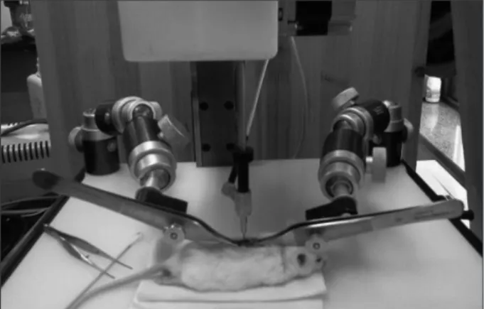
erate force of 50 kdyne. Feedback from the IH device, including the actual impact force, cord displacement, graph of time versus force, and graph of time versus displacement, were recorded for each animal (Fig. 1). After SCI, the muscles were sutured in lay-ers with 5-0 chromic gut, and skin was closed with 7-mm wound clips. After surgery, mice were allowed to recover in a warmed cage with water and food easily accessible. Gentamicin (5 mg/kg, intramuscular; Abbott Laboratories, North Chicago, IL, USA) was administered immediately post-surgery. Animals were dou-bly housed with Mouse Houses for the duration of the study. Post-operative care consisted manual bladder expression twice of a day until mice recovered some bladder expression, then once a day for the duration of the study. The mice were main-tained for 8 week after injury with all brains collected at 2, 4, 8 week for HDAC related protein, brain-derived neurotrophic fac-tor (BDNF), glial cell line-derived neurotrophic facfac-tor (GDNF) and nerve growth factor (NGF) analysis. All animal care, inter-ventions and euthanasia procedures, were in accordance with the National Institute of Health Guide for the Care and Use of Laboratory Animals and guideline approved by Animal Care and Use Committees of the Yonsei University Wonju College of Medicine. Data was analyzed and unblended by the statistician at the end of the experiment.
Immuno blotting
To compare the expression levels of neurotrophic factors, mouse brains were lysed in 500 μL of cold RIPA buffer [50 mM Tris-HCl, pH 7.5, 1% Triton X-100, 150 mM NaCl, 0.1% sodi-um dodecyl sulfate (SDS), and 1% sodisodi-um deoxycholate] with a protease inhibitor cocktail (Sigma-Aldrich, St. Louis, MO, USA). Tissue lysate was centrifuged at 13000×g for 15 minutes at 4°C. The supernatant was harvested, and protein concentration was analyzed using a Qunt-iT protein assay kit (Molecular Probes, Eugene, OR, USA). For electrophoresis, 50 μg of protein was dissolved in sample buffer (60 mM Tris-HCl, pH 6.8, 14.4 mM β-mercaptoethanol, 25% glycerol, 2% SDS, and 0.1% bromo-phenol blue), boiled for 10 minutes and separated on a 10% SDS reducing gel. Separated proteins were transferred onto polyvi-nylidene difluoride membranes (Invitrogen, Carlsbad, CA, USA) using a trans-blot system. Blots were blocked for 1 hour in Tris-buffered saline (TBS) (10 mM Tris-HCl, pH 7.5, 150 mM NaCl) containing 5% nonfat dry milk at room temperature, washed three times with TBS and incubated at 4°C overnight with an anti-mouse monoclonal BDNF (1 : 1000, Abcam, Cambridge, MA, USA), anti-mouse glial cell-derived neurotrophic factor (GDNF, 1 : 1000, Abcam) and anti-GAPDH (1 : 3000, Cell sig-naling, Boston, MA, USA) antibody in TBST (10 mM Tris, pH 7.5, 150 mM NaCl, and 0.02% Tween 20) containing 5% nonfat dry milk. On the next day, blots were washed three times with TBST and incubated for 1 hour with horseradish peroxidase-conjugated secondary antibodies (1 : 3000, SantaCruz Biotech, Santa Cruz, CA, USA) in TBST containing 3% nonfat dry milk at room temperature. After washing three times with TBST, pro-a vpro-ariety of neurodegenerpro-ative disepro-ases pro-and neurologicpro-al
condi-tions, such as stroke or traumatic brain injury1,13). Despite these ongoing studies, molecular mechanism in the brain after SCI re-mains unexplored. In this study, we have analyzed the changes in the expression of the main regulators of neuronal survival and death in the mouse model of SCI.
MATERIALS AND METHODS
Experimental animalEight-week-old female imprinting control region mice (n=60; 30-35 g) were used in this study. All animal care, interventions and euthanasia procedures, were in accordance with the National Institute of Health Guide for the Care and Use of Laboratory An-imals and guideline approved by Animal Care and Use Commit-tees of our institute. Data was analyzed and unblended by the statistician at the end of the experiment. At the beginning of the experiment animals were randomly assigned to one of two groups : naive control (n=30), SCI (n=30).
Moderate spinal cord contusion
The prescribed animals underwent a contusive impact SCI in-duced by the Infinite Horizons (IH) device (Precision Systems and Instrumentation, Lexington, NY, USA). Animals were anes-thetized with mixture of ketamine and xylazine (0.05 mL/kg) and absence of blink and withdrawal reflexes were ensured. Lac-rilube ophthalmic ointment (Allergan Pharmaceuticals, Irvine, CA, USA) was applied to the eye to prevent drying. During sur-gery, the mice were kept on homeothermic blanket system (Har-vard Apparatus, Ltd., Kent, UK) to maintain the body tempera-ture at 37±0.5°C as measured by rectal probe. Following anesthesia, a vertical incision was made along the low thoracic vertebra and the superficial muscle and skin retracted. A lami-nectomy performed at thoracic vertebra T12 exposed the dor-sal surface of the spinal cord without disrupting the duramater. Stabilization clamps were placed around the vertebrae at T9 and L2 to support the column during impact. The injury was induced by the tip of impactor probe, which transduces a
mod-Fig. 1. Schematic drawing illustrating operation of spinal cord injury
(1.16±0.11) and 8 weeks (1.01±0.09) compared with those of control group after SCI, however the differences were not sig-nificant (p>0.05) (Fig. 4).
Among the regulators of gene expression, we investigated his-tone deacetylase-related protein. The expressions of HDAC1 were elevated at 2 weeks (1.11±0.15), 4 weeks (1.15±0.21) and 8 teins were visualized with an ECL detection system (Amersham
Pharmacia Biotech, Piscataway, NJ, USA).
Statistical analysis
Statistical analysis was performed using the Student’s t-test and one-way ANOVA (Prism Graph Pad Software, San Diego, CA, USA). The data was expressed as mean±standard error of the mean. The significance level was assumed at p<0.05, unless otherwise indicated.
RESULTS
To identify the neuromodulators associated with the neuro-nal cell development, growth, survival and apoptosis, the ex-pression of four specific neurotrophic factors such as BDNF, NGF, GDNF and HDAC1 were investigated in the whole brain by Western blotting and multiplex ELISA assay.
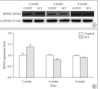
Expression of BDNF was significantly elevated in the SCI group compared with those of control group at 2 weeks after SCI (1.38±0.11, p<0.05). However, expressions of BDNF were not significant compared with those of control group at 4 weeks (0.80±0.04) and 8 weeks (0.87±0.04) after SCI (Fig. 2).
The level of NGF was elevated in the SCI group at 2 weeks (1.21±0.10) and 4 weeks (1.17±0.16), and decreased at 8 weeks (0.87±0.03) compared with those of control group after SCI, however the differences were not significant (p>0.05) (Fig. 3).
The level of GDNF was elevated in the SCI group at 2 weeks Fig. 2. Semi-quantitative immunoblot analysis of BDNF expression in
brain after contusion of the spinal cord in mice (A and B). BDNF signifi-cantly elevated compared with those of control group at 2 week after SCI (p<0.05). In this figure (B) data are plotted as mean±standard error of the mean. A sterisk indicate values significantly different from those of SCI and of control group by using a two-way repeated measures analysis of variance with repeated time factor. BDNF : brain-derived neurotrophic factor, SCI : spinal cord injury, CONT : control, GAPDH : glyceraldehyde 3-phosphate dehydrogenase.
Fig. 4. Semi-quantitative immunoblot analysis of GDNF expression in
brain after contusion of the spinal cord in mice (A and B). GDNF elevated compared with those of control group at 2 weeks, however the differ-ence is not significant (p>0.05). In this figure (B) data are plotted as mean±standard error of the mean. GDNF : glial cell line-derived neuro-trophic factor, SCI : spinal cord injury, CONT : control, GAPDH : glyceral-dehyde 3-phosphate dehydrogenase.
Fig. 3. Semi-quantitative immunoblot analysis of NGF expression in
brain after contusion of the spinal cord in mice (A and B). NGF elevated compared with those of control group at 2 and 4 weeks, however the differences were not significant (p>0.05). In this figure (B) data are plotted as mean±standard error of the mean. NGF : nerve growth factor, SCI : spi-nal cord injury, CONT : control, GAPDH : glyceraldehyde 3-phosphate de-hydrogenase. 0.0 0.0 0.0 0.5 0.5 0.5 1.0 1.0 1.5 1.0 2.0 1.5 1.5 BD N F ex pr es sio n lev el G D N F ex pr es sio n lev el N G F ex pr es sio n lev el 2 weeks 2 weeks 2 weeks Time Time Time 4 weeks 4 weeks 4 weeks 8 weeks 8 weeks 8 weeks Control SCI Control SCI Control SCI 2 weeks 2 weeks 2 weeks CONT CONT CONT CONT CONT CONT CONT CONT CONT SCI SCI SCI SCI SCI SCI SCI SCI SCI 4 weeks 4 weeks 4 weeks 8 weeks 8 weeks 8 weeks BDNF (28 kD) GDNF (24 kD) NGF (27 kD) GAPDH (37 kD) GAPDH (37 kD) GAPDH (37 kD) A A A B B B
morphology of dendritic spines underlie reorganization of syn-aptic connectivity in the motor cortex following SCI. And PSD-95, a major postsynaptic density protein, and PSA-NCAM, a marker of active synaptic remodeling increase their expression. Some evidences observed that the alteration in the expression of enzymes in the spinal cord following SCI. Mature NTs in-duce cell survival upon binding to Trk receptors, their imma-ture pro-protein counterparts promote apoptosis via activating P75NTR in the spinal cord13). In accordance with this reports the members of PC enzymes functioning within regulated secretory pathway of neurons and neuroendocrine cells are significantly down-regulated in the spinal cord after SCI21). In comparison, the expression of pro-NGF and pro-NT3 regulating enzymes are ubiquitous expressed in the cell of SCI. Pro-BDNF is primarily regulated secretory pathway of neuron in the spinal cord16,20). In the recent year, BDNF have emerged as important upstream regulators of long term potentiation in brain regions, including neocortex and hippocampus. These mean that BDNF appear to play an important role in neuronal cell development and sur-vival. One of novel finding is significant elevation of BDNF lev-el at 2 weeks. This is the first direct demonstration that BDNF signaling can be important regulation process in the brain reor-ganization following SCI. Although several HDACs are ex-pressed at high level in the brain, their role in brain function has not been fully explored. The action of HDACs is opposed by histone acetyltransferases (HATs) such as CREB-binding pro-tein and p300, which catalyze the transfer an acetyl moiety from acetyl-coenzyme A to specific lysine residues of histones14). Acet-ylation of histones relaxes the chromatin structure to a state that is transcriptionally active, while histone deacetylation trans-forms chromatin to a transcriptionally repressed state. Hence, gene expression is regulated, in part, by the balance of HDAC and HAT activities14). Recent study have reported that neuronal apoptosis by HDAC inhibitors involves stimulation of E2F-1, a transcription factor with established proapoptotic activity in neu-rons1). The expression of E2F target genes, such as those for cyclin E and Apaf-1, is induced in cerebellar granule neurons by phar-macological inhibition of HDACs1). Our data show elevated ten-dency of HDAC1 level at 2, 4 and 8 weeks but the elevation was not significant. Histone deacetylase-related protein expression is down regulated in apoptotic neurons, and this occurs prior to the time at which these neurons become irreversibly commit-ted to death. Hence it is need to evaluate expression level of HAD1 at the acute phase.
CONCLUSION
In summary, our studies reveal elevated expression of BDNF in the brain after SCI. Also it is need to explore neurorestorative effect might be induced by upregulation of BDNF, extending evidence that pharmacological means of BDNF may play an important role in brain reorganization following SCI. All to-gether, the actions of NGF, GDNF and HDAC 1 on neuronal weeks (1.09±0.13) in SCI group compared with those of control
group, but the differences were not significant (p>0.05) (Fig. 5).
DISCUSSION
The purpose of this study was to investigate the alterations in the expression of the main regulators of neuronal development, survival and death related proteins. Here, we have examined the changes in expression of BDNF, NGF, GDNF and HDAC1 in the whole brain at specific time-points after SCI, in the mouse model of spinal cord contusion. BDNF protein showed signifi-cant increase at 2 weeks. NGF and GDNF proteins showed a tendency toward increase at 2 weeks compared with control, but it was not statistically significant. In addition, protein in-volved in gene regulation by relaxing the chromatin structure to a state that is transcriptionally active, HDAC1, showed ele-vated tendency, but the alteration was not significant. Over 100 years ago, it has been shown that following injury of a peripher-al nervous system, axon distperipher-al to the injury undergo progressive retrograde degeneration, a process termed Wallerian degenera-tion.22) Also, recent studies suggest that Wallerian degeneration occur in the spinal cord of SCI patients, the topic of SCI results in anatomical changes of supraspinal central nervous system were clarified5). For example, the recent study by Wrigley et al.22) found in living human that complete SCI results in anatomical changes in the human motor cortex and in the descending path-ways from the motor cortex and anatomical changes in areas of the brain not directly involved in motor control, that is the me-dial prefrontal and anterior cingulated cortices. Furthermore, Kim et al.12) observed that dynamic changes in density and Fig. 5. Semi-quantitative immunoblot analysis of HDAC1 expression in
brain after contusion of the spinal cord in mice (A and B). The difference of HDAC1 level in SCI and control group is not significant (p>0.05). In this figure (B) data are plotted as mean±standard error of the mean. HDAC : histone deacetylase, SCI : spinal cord injury, CONT : control, GAPDH : glyceraldehyde 3-phosphate dehydrogenase.
0.0 0.5 1.0 1.5 H DA C1 ex pr es sio n lev el 2 weeks Time 4 weeks 8 weeks Control SCI 2 weeks
CONT SCI CONT4 weeksSCI CONT8 weeksSCI
HDAC1 (65 kD) GAPDH (37 kD)
A
alterations of neurotrophins, their receptors and prohormone conver-tases in a rat model of spinal cord contusion. Neurosci Lett 441 : 261-266, 2008
11. Jain N, Catania KC, Kaas JH : Deactivation and reactivation of somato-sensory cortex after dorsal spinal cord injury. Nature 386 : 495-498, 1997
12. Kim BG, Dai HN, McAtee M, Vicini S, Bregman BS : Remodeling of synaptic structures in the motor cortex following spinal cord injury. Exp
Neurol 198 : 401-415, 2006
13. Lee R, Kermani P, Teng KK, Hempstead BL : Regulation of cell survival by secreted proneurotrophins. Science 294 : 1945-1948, 2001
14. Legube G, Trouche D : Regulating histone acetyltransferases and deacetylases. EMBO Rep 4 : 944-947, 2003
15. McKinley PA, Jenkins WM, Smith JL, Merzenich MM : Age-dependent capacity for somatosensory cortex reorganization in chronic spinal cats.
Brain Res 428 : 136-139, 1987
16. Mowla SJ, Pareek S, Farhadi HF, Petrecca K, Fawcett JP, Seidah NG, et al. : Differential sorting of nerve growth factor and brain-derived neuro-trophic factor in hippocampal neurons. J Neurosci 19 : 2069-2080, 1999 17. Nudo RJ, Milliken GW, Jenkins WM, Merzenich MM : Use-dependent
alterations of movement representations in primary motor cortex of adult squirrel monkeys. J Neurosci 16 : 785-807, 1996
18. Pons TP, Garraghty PE, Ommaya AK, Kaas JH, Taub E, Mishkin M : Massive cortical reorganization after sensory deafferentation in adult macaques. Science 252 : 1857-1860, 1991
19. Raineteau O, Schwab ME : Plasticity of motor systems after incomplete spinal cord injury. Nat Rev Neurosci 2 : 263-273, 2001
20. Seidah NG, Benjannet S, Pareek S, Chrétien M, Murphy RA : Cellular processing of the neurotrophin precursors of NT3 and BDNF by the mammalian proprotein convertases. FEBS Lett 379 : 247-250, 1996 21. Seidah NG, Chrétien M : Proprotein and prohormone convertases : a
family of subtilases generating diverse bioactive polypeptides. Brain Res
848 : 45-62, 1999
22. Waller A : Experiments on the section of glossopharyngeal and hypo-glossal nerves of the frog and observations of the alternatives produced thereby in the structure of their primitive fibers. Phil Trans R Soc Lond
140 : 423, 1850
23. Wrigley PJ, Gustin SM, Macey PM, Nash PG, Gandevia SC, Macefield VG, et al. : Anatomical changes in human motor cortex and motor pathways following complete thoracic spinal cord injury. Cereb Cortex
19 : 224-232, 2009
24. Yu SH, Cho DC, Kim KT, Nam KH, Cho HJ, Sung JK : The neuropro-tective effect of treatment of valproic Acid in acute spinal cord injury. J
Korean Neurosurg Soc 51 : 191-198, 2012 changes in brain after SCI should be evaluated. Modulation of
process such as extending its temporal window or histological analysis related alteration of proteins may be a future strategy to clarify main protein in the brain after SCI.
• Acknowledgements
This study was supported by a grant of the Korea Health Technology R & D Project, Ministry of Health & Welfare, Republic of Korea (No. A100054), research grant from Yonsei University Wonju College of Medicine (YU-WCM-2011-49), and grant from the National Research Foundation (NRF-2010-0020408) funded by the Ministry of Education, Science Technology, Republic of Korea.
References
1. Boutillier AL, Trinh E, Loeffler JP : Selective E2F-dependent gene tran-scription is controlled by histone deacetylase activity during neuronal apoptosis. J Neurochem 84 : 814-828, 2003
2. Bregman BS, Goldberger ME : Infant lesion effect : II. Sparing and re-covery of function after spinal cord damage in newborn and adult cats.
Brain Res 285 : 119-135, 1983
3. Bruehlmeier M, Dietz V, Leenders KL, Roelcke U, Missimer J, Curt A : How does the human brain deal with a spinal cord injury? Eur J
Neuro-sci 10 : 3918-3922, 1998
4. Burns SP, Golding DG, Rolle WA Jr, Graziani V, Ditunno JF Jr : Recov-ery of ambulation in motor-incomplete tetraplegia. Arch Phys Med
Re-habil 78 : 1169-1172, 1997
5. Buss A, Brook GA, Kakulas B, Martin D, Franzen R, Schoenen J, et al. : Gradual loss of myelin and formation of an astrocytic scar during Wal-lerian degeneration in the human spinal cord. Brain 127 (Pt 1) : 34-44, 2004
6. Clark SA, Allard T, Jenkins WM, Merzenich MM : Receptive fields in the body-surface map in adult cortex defined by temporally correlated inputs. Nature 332 : 444-445, 1988
7. Cohen LG, Roth BJ, Wassermann EM, Topka H, Fuhr P, Schultz J, et al. : Magnetic stimulation of the human cerebral cortex, an indicator of re-organization in motor pathways in certain pathological conditions. J
Clin Neurophysiol 8 : 56-65, 1991
8. Curt A, Bruehlmeier M, Leenders KL, Roelcke U, Dietz V : Differential effect of spinal cord injury and functional impairment on human brain activation. J Neurotrauma 19 : 43-51, 2002
9. Green JB, Sora E, Bialy Y, Ricamato A, Thatcher RW : Cortical senso-rimotor reorganization after spinal cord injury : an electroencephalo-graphic study. Neurology 50 : 1115-1121, 1998