J. Microbiol. Biotechnol. (2015), 25(2), 302–306
http://dx.doi.org/10.4014/jmb.1501.01007
Research Article
jmb
Review
VvpM Induces Human Cell Death via Multifarious Modes Including
Necroptosis and Autophagy
Mi-Ae Lee
1, Jeong-A Kim
1, Mee-Young Shin
2, Jeong K. Lee
1, Soon-Jung Park
2, and Kyu-Ho Lee
1*
1Department of Life Science, Sogang University, Seoul 121-742, Republic of Korea
2Department of Environmental Medical Biology, Brain Korea 21 PLUS Project for Medical Science, Yonsei University College of Medicine,
Seoul 120-752, Republic of Korea
Vibrio vulnificus causes a fatal septicemia in humans via ingestion of V. vulnificus-enriched marine organisms or exposure to V. vulnificus in seawater [18]. This bacterium is able to destroy human blood cells and Jurkat T-cell lines in a necrotic manner, via NADH-oxidase-derived production of reactive oxygen species and a subsequent activation of MAPKs [8, 9]. One of the secreted enzymes responsible for this pathology is the elastolytic zinc-metalloprotease VvpE [6]. Another metalloprotease, VvpM, detected in the culture supernatants of V. vulnificus [11], induces apoptotic cell death via a pathway consisting of ERK activation, cytochrome c release, and activation of caspases-9 and -3 [12]. Interestingly, only 30~40% of the VvpM-treated cells demonstrated the apoptotic characteristics. Therefore, the present study has been aimed to define the other pathways involved in the VvpM-induced cell death.
Recombinant VvpM (rVvpM) was prepared from the culture supernatant of V. vulnificus as previously described [12]. A human colorectal carcinoma cell line, HCT-116 (10247, Korean Cell Line Bank), was grown at 37oC in
RPMI-1640 medium (Gibco BRL) supplemented with 10% heat-inactivated fetal bovine serum (FBS), 2 mM L-glutamine, 100 U/ml penicillin G, 100 mg/ml streptomycin, 25 mM
sodium bicarbonate, and 25 mM HEPES. The HCT-116 cells were seeded in a 6-well plate (1 × 10⁶ cells per well) and
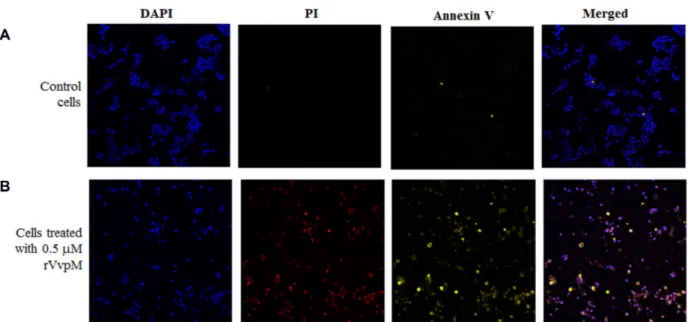
incubated for 24 h. Prior to treatment with rVvpM, HCT-116 cells were incubated for 1 h in serum-free RPMI-1640. The cells were treated with 0.5 µM rVvpM for 6 h, and stained with Annexin V and propidium iodide (PI) using a GFP-certified Apoptosis/Necrosis Detection kit (Enzo Life Science) according to the manufacturer’s instructions. The cells were stained with 4,6’-diamino-2-phenylindole (DAPI; Sigma), rinsed with phosphate-buffered saline, and mounted with anti-fade mounting medium. They were then observed under a Zeiss LSM 510 laser scanning confocal microscope. The control cells were rarely stained with PI and Annexin V (Fig. 1A). rVvpM-treated cells were stained with both Annexin V and PI, which appeared as yellow and red cells, respectively (Fig. 1B). This observation indicated that rVvpM induced cell death in both an apoptotic and a necrotic manner.
In subsequent experiments, the human cell lines were incubated for 6 h with various amounts of rVvpM and the mortality of these cells was monitored by staining with PI (Figs. 2A and 2B). A human colonic carcinoma cell line, HT-29 (HTB-38; American Type Culture Collection), was
Received: January 6, 2015 Revised: January 13, 2015 Accepted: January 14, 2015 First published online February 3, 2015 *Corresponding author Phone: +82-2-705-7963; Fax: +82-2-704-3601; E-mail: kyuholee@sogang.ac.kr pISSN 1017-7825, eISSN 1738-8872 Copyright© 2015 by
The Korean Society for Microbiology and Biotechnology
VvpM, one of the extracellular metalloproteases produced by Vibrio vulnificus, induces apoptotic cell death via a pathway consisting of ERK activation, cytochrome c release, and activation of caspases-9 and -3. VvpM-treated cells also showed necrotic cell death as stained by propidium iodide (PI). The percentage of PI-stained cells was decreased by pretreatment with Necrostatin-1, indicating that VvpM-mediated cell death occurs through necroptosis. The appearance of autophagic vesicles and lipidated form of light-chain-3B in rVvpM-treated cells suggests an involvement of autophagy in this process. Therefore, the multifarious action of VvpM might be one of the factors responsible for V. vulnificus pathogenesis.
added in this assay, which was cultured in Dulbecco’s modified Eagle’s medium (Gibco BRL) supplemented with 10% FBS, 100 U/ml penicillin G, and 100 µg/ml streptomycin. HCT-116 or HT-29 cells were seeded in a 12-well plate (5 × 10⁵ cells per well) and incubated for 24 h. After washing
with FBS-free medium, the cells were treated with various concentrations of rVvpM (from 0.01 to 0.5 µM) for 3 h. As a positive control for cell death, each cell line was treated with 10 mM H2O2 for 6 h. Cells were then stained with 0.5 µg/ml
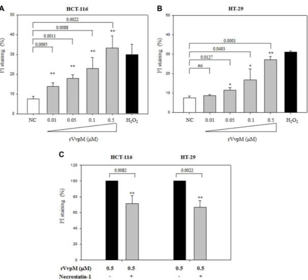
PI, and the degree of dye-binding was assessed using fluorescence-activated cell sorting (FACS) analysis (Becton Dickinson). Statistical analysis for pairwise comparison was performed using Student’s t-test (SYSTAT program, SigmaPlot ver. 9). Seven percent of HCT-116 cells were stained with PI in the absence of rVvpM, whereas 33% of the cells were stained with PI after treatment with 0.5 µM rVvpM (p = 0.0022). In the same manner, less than 8% of HT-29 cells were stained with PI without rVvpM treatment, but 27% of HT-29 cells were stained by PI after treatment with 0.5 µM rVvpM (p = 0.0001).
Apoptosis and necrosis have been considered as two representative pathways for cell death. In contrast to the apoptotic pathway involving the sequential activation of caspases [5, 24], necrotic cell death occurs drastically, showing cytoplasmic swelling due to plasma membrane permeabilization. It has been recently reported that necrosis could occur in a tightly programmed fashion named
necroptosis [4]. Necroptosis requires the interaction of receptor-interacting serine-threonine kinase 1 (RIP1) with another kinase, RIP3 [2, 5]. To investigate whether rVvpM-mediated cell death is related to necroptosis [20], necrostatin-1 (Nec-1), a chemical compound inhibiting the kinase activity of RIP-1 [3], was treated to cells prior to rVvpM-challenge (Fig. 2C). HCT-116 or HT-29 cells were seeded in a 12-well plate and incubated for 24 h. The cells were pre-treated with Nec-1 at 50 µM for 1 h, and then exposed to 0.5 µM rVvpM for 6 h. As a control, the cells were incubated with 1% DMSO for 1 h before challenging with rVvpM. Degree of cell death was monitored by PI staining and subsequent FACS analysis. Pre-treatment of HCT-116 or HT-29 with Nec-1 reduced the degree of PI staining of rVvpM-treated cells, which was estimated to be 83% (p = 0.0082) or 67% (p = 0.0022) of the control, respectively. This result indicates that RIP1-mediated necroptosis is one of the pathways involved in the rVvpM-initiated cell death. However, the degree of reduction in PI staining of cells pre-treated with necrostatin-1 was only <35%, suggesting that death of rVvpM-treated cells also occurred via RIP-1-independent mechanisms.
When HT-29 cells treated with rVvpM were observed under a transmission electron microscope, these cells showed the morphological changes indicating apoptosis, as previously described [12]. Interestingly, apparent autophagosome-like structures, in addition to the morphological changes derived
Fig. 1. Staining of the rVvpM-treated HCT-116 cells with Annexin V and PI.
HCT-116 cells were treated with 0.5 µM rVvpM at 37oC for 6 h, and then stained with Annexin V-FITC (yellow) and PI (red) using a GFP-certified
Apoptosis/Necrosis Detection kit. The cells were then reacted with DAPI (blue), mounted with anti-fade mounting medium, and observed under a laser scanning confocal microscope. As a control, HCT-116 cells not treated with rVvpM were included in this assay.
304 Lee et al.
from the apoptosis, were observed in the transmission electron micrograms of VvpM-treated cells (Fig. 3). Autophagy is a cellular mechanism by which cytoplasmic components are degraded under stress conditions [13]. Recently, autophagy has been considered as one of the host defense mechanisms to respond to bacterial invasion by delivering intracellular pathogens to lysosomes [15]. Representative markers for autophagic events include beclin-1 for the nucleation process [1], light-chain-3 (LC3) involved in autophagosome formation and elongation [22], and scaffold
protein p62 delivering ubiquitinated proteins to the autophagosome [14].
Thus, we examined whether VvpM acted to elicit a characteristic of autophagy by monitoring for LC3B [10]. LC3B involving the autophagosome formation [16] could be present in either the cytoplasmic LC3B-I or the lipidated LC3B-II associated with the autophagosomal membrane [7]. HCT-116 or HT-29 cells were seeded in a 12-well plate and incubated for 24 h. Before treatment with rVvpM, one set of cell cultures was pre-treated with 1% DMSO/2%
Fig. 2. PI staining of the rVvpM-treated HCT-116 and HT-29 cells, and the effect of Nec-1 on PI staining.
(A, B) HCT-116 or HT-29 cells (5 × 10⁵ cells per well) were treated with various concentrations of rVvpM (from 0.01 to 0.5 µM) at 37oC for 6 h. The
cells treated with 10 mM H2O2 or medium only (NC) were included as a positive and a negative control, respectively. Cells were then stained with
PI at a concentration of 0.5 µg/ml, and the degree of binding was assessed using fluorescence-activated cell sorting (FACS) analysis. FACS analyses were performed on at least 10,000 cells per sample with a FACScan (Becton Dickinson). (C) HCT-116 and HT-29 (5 × 10⁵ cells per well)
pre-treated with 1% DMSO solution containing 50 µM Necrostatin-1 for 1 h were exposed to 0.5 µM rVvpM for 6 h at 37oC. As a control, the cells
pre-treated with 1% DMSO were challenged with rVvpM. Degree of cell death was monitored by PI staining and subsequent FACS analysis as described above. Results were expressed as the averages ± standard deviations from at least three independent experiments. The p-values were derived from comparison with the control: A p-value <0.05 was considered statistically significant and the p-values are presented in the corresponding figures with a single asterisk (*) for 0.01< p <0.05 or double asterisks (**) for p < 0.01. ns, non-significant difference.
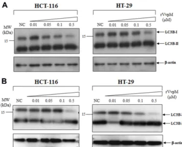
ethanol, and the other set was pre-treated with 1% DMSO/2% ethanol solution containing protease inhibitors (10 µg/ml E64d and 10 µg/ml pepstatin). After 3 h of incubation, they were added with various concentrations of rVvpM (0.01 ~ 0.5 µM) and further incubated for 6 h. Twenty micrograms of the cellular extracts were separated by 15% SDS-PAGE and probed with anti-LC3B monoclonal antibodies (Cell Signaling Technology). For a loading control of the western blot, anti-β-actin antibodies were used. In both cell lines, the levels of LC3B-I were gradually decreased as the added concentrations of rVvpM were increased, suggesting the occurrence of conversion of LC3B-I to LC3B-II. The apparent increase in the LC3B-II levels, however, were not observed in the same cells (Fig. 4A). Since it is possible that the LC3B-II formed might be degraded during the subsequent step(s) of autophagy, such as lysosomal degradation [19], the LC3B levels were also monitored after a pre-treatment with protease inhibitors. Formation of LC3B-II was evidently observed in rVvpM-treated cells incubated with E64d and pepstatin (Fig. 4B), indicating that this rVvpM-induced cell death was related to autophagy.
The co-occurrence of apoptosis and autophagy in rVvpM-treated cells [12] might indicate the presence of a connection between autophagy and apoptosis, possibly via the association of another autophagy marker, beclin-1, with
Fig. 3. Transmission electromicrographs of the control HT-29 (A) and the rVvpM-treated HT-29 cells (B, C).
HT-29 cells were cultured for 6 h in the absence (A) or presence (B, C) of 0.1 µM rVvpM. Autophagic vesicles are indicated by arrowheads. The scale bars in panels A and B are 2,000 nm, whereas the bar in panel C is 500 nm.
Fig. 4. Formation of LC3B-II in the rVvpM-treated HCT-116 and HT-29 cells.
Effects of rVvpM-treatment on the conversion to LC3B-II were examined in HCT-116 and HT-29 cells. Before treatment of various concentrations of rVvpM (from 0.01 to 0.5 µM), one set was treated with 1% DMSO/2% ethanol (A) and the other set was treated with E64d and pepstatin (each 10 µg/ml of 1% DMSO/2% ethanol) for 3 h (B). Twenty micrograms of the protein extracts separated on 15% SDS-PAGE was probed with anti-LC3B monoclonal antibodies (Cell Signaling Technology). The level of β-actin in the same samples was monitored as a loading control (the lower panels).
306 Lee et al.
a key member for the apoptotic process, such as the Bcl-2 family member, as reviewed by Wang [21]. It has been previously described that some substances, such as calcium ions and sphingolipids, lead to a co-occurrence of autophagy and apoptosis [17, 23].
This study demonstrates that rVvpM-induced cell death occurs via multiple pathways, including necroptosis and autophagy in addition to apoptosis. The implication of this multifarious action of VvpM in the pathogenesis of V. vulnificus should be evaluated.
Acknowledgments
This work was supported by a grant (14162MFDS972) from the Ministry of Food and Drug Safety, and by the Pioneer Research Center Program through NRF funded by the Ministry of Science, ICT & Future Planning (2013M3C1A3064325).
References
1. Chen Y, Lu Y, Lu C, Zhang I. 2009. Beclin-1 expression is a predictor of clinical outcome in patients with esophageal squamous cell carcinoma and correlated to hypoxia-inducible factor (HIF)-1 alpha expression. Pathol. Oncol. Res. 15: 487-493. 2. Cho YS, Challa S, Moquin D, Genga R, Ray TD, Guildford M, Chan FK. 2009. Phosphorylation-driven assembly of RIP1-RIP3 complex regulates programmed necrosis and virus-induced inflammation. Cell 137: 1112-1123.
3. Degterev A, Huang Z, Boyce M, Li Y, Jagtap P, Mizushima N, et al. 2005. Chemical inhibitor of nonapoptotic cell death with therapeutic potential for ischemic brain injury. Nat. Chem. Biol. 1: 112-119.
4. Goldstein P, Kroemer G. 2007. Cell death by necrosis: towards a molecular definition. Trends Biochem. Sci. 32: 37-43. 5. Hitomi J, Christofferson DE, Ng A, Yao J, Degterev A,
Xavier RJ, Yuan J. 2008. Identification of a molecular signaling network that regulates a cellular necrotic cell death pathway. Cell 135: 1311-1323.
6. Jeong KC, Jeong HS, Rhee JH, Lee SE, Chung SS, Starks AM, et al. 2000. Construction and phenotypic evaluation of a Vibrio vulnificus vvpE mutant for elastolytic protease. Infect. Immun. 68: 5096-5106.
7. Kabeya Y, Mizishima N, Ueno T, Yamamoto A, Kirisako T, Noda T, et al. 2000. LC3, a mammalian homologue of yeast Apg8p, is localized in autophagosome membranes after processing. EMBO J. 19: 5720-5728.
8. Kim WH, Goo SY, Lee K-H, Park S-J. 2009. Vibrio vulnificus-induced cell death of human mononuclear cells requires ROS-dependent activation of p38 and ERK 1/2 MAPKs. Immunol. Invest. 38: 31-48.
9. Kim WH, Goo SY, Shin MH, Chun S-J, Lee H, Lee K-H, Park S-J. 2008. Vibrio vulnificus-induced death of Jurkat T-cells requires activation of p38 mitogen-activated protein kinase by NADPH oxidase-derived reactive oxygen species. Cell. Immunol. 253: 81-91.
10. Kroemer G, El-Deiry WW, Golstein P, Peter ME, Vaux D, Vandenabeele P, et al. 2005. Classification of cell death: recommendations of the nomenclature committee on cell death. Cell Death Differ. 12: 1463-1467.
11. Lee H-J, Kim J-A, Lee M-A, Park S-J, Lee K-H. 2013. Regulation of haemolysin (VvhA) production by ferric uptake regulator (Fur) in Vibrio vulnificus: repression of vvhA transcription by Fur and proteolysis of VvhA by Fur-repressive exoproteases. Mol. Microbiol. 88: 813-826.
12. Lee M-A, Kim J-A, Yang YJ, Shin MY, Park S-J, Lee K-H. 2014. VvpM, an extracellular metalloprotease of Vibrio vulnificus, induces apoptotic death of human cells. J. Microbiol. 52: 1036-1043.
13. Levine B, Kroemer G. 2008. Autophagy in the pathogenesis of disease. Cell 132: 27-42.
14. Mizushima N, Yoshimori T, Levine B. 2010. Methods in mammalian autophagy research. Cell 140: 313-326.
15. Modtowy S. 2013. Autophagy and bacterial clearance: a not so clear picture. Cell. Microbiol. 15: 395-402.
16. Sivridis E, Koukourakis MI, Zois CE, Ledaki I, Ferguson DJ, Harris AL, et al. 2010. LC3A-positive light microscopy detected patterns of autophagy and prognosis in operable breast carcinomas. Am. J. Pathol. 176: 2477-2489.
17. Sm aili SS, Pereira GJ, Costa MM, Rocha KK, Rodrigues L, do Carmo LG, et al. 2013. The role of calcium stores in apoptosis and autophagy. Curr. Mol. Med. 13: 252-265. 18. Strom MS, Paranjpye RN. 2000. Epidemiology and pathogenesis
of Vibrio vulnificus. Microbes Infect. 2: 177-188.
19. Tanida I, Minematsu-Ikeguchi N, Ueno T, Kominami E. 2005. Lysosom al turnover, but not a cellular level, of endogenous LC3 is a marker for autophagy. Autophagy 1: 84-91.
20. Vandenabeele P, Galluzzi L, Vanden Berghe T, Kroemer G. 2010. Molecular mechanisms of necroptosis: an ordered cellular explosion. Nat. Rev. Mol. Cell Biol. 11: 700-714. 21. Wang J. 2008. Beclin 1 bridges autophagy, apoptosis, and
differentiation. Autophagy 4: 947-948.
22. Yoshioka A, Miyata H, Doki Y, Yamasaki M, Sohma I, Gotoh K, et al. 2008. LC3, an autophagosome marker, is highly expressed in gastrointestinal cancers. Int. J. Oncol. 33: 461-468.
23. Young MM, Kester M, Wang HG. 2013. Sphingolipids: regulators of crosstalk between apoptosis and autophagy. J. Lipid Res. 54: 5-19.
24. Zhang DW, Shao J, Lin J, Zhang N, Lu BJ, Lin SC, et al. 2009. RIP3, an energy metabolism regulator that switches TNF-induced cell death from apoptosis to necrosis. Science 325: 332-336.