INTRODUCTION
Pancreatic ductal adenocarcinoma (PDAC) is a lethal
gastroin-testinal malignant disease, with an estimated overall 5-year survival rate of less than 5%. While the most effective treatment option is margin-negative pancreatectomy, only about 15–20% of patients with a new diagnosis of PDAC can undergo surgi-cal resection, and their 5-year survival rate is around 20%.1 Most PDAC patients experience recurrence and cancer-relat-ed mortality.
In investigating the biologic behavior of PDAC, cancer-relat-ed factors and host-relatcancer-relat-ed factors necancer-relat-ed to be considercancer-relat-ed. Sev-eral cancer-related factors, such as margin status, tumor size, tumor (T) stage, nodal (N) stage, tumor differentiation, lym-phovascular invasion, perineural invasion, major vascular in-vasion, and high levels of preoperative or postoperative CA19-9, have been identified as important cancer-related prognostic
Yonsei Criteria, a Potential Linkage to Intratumoral
Foxp3
+
/CD8
+
Ratio for the Prediction of Oncologic
Outcomes in Resected Left-Sided Pancreatic Cancer
Ho Kyoung Hwang
1,2, Sung Hwan Lee
3, Hyoung-Il Kim
4, Se Hoon Kim
5,
Junjeong Choi
6, Chang Moo Kang
1,2, and Woo Jung Lee
1,21Division of Hepatobiliary and Pancreatic Surgery, Department of Surgery, Yonsei University College of Medicine, Seoul; 2Pancreatobiliary Cancer Center, Yonsei Cancer Center, Severance Hospital, Seoul;
3Division of Hepatobiliary and Pancreatic Surgery, Department of Surgery, CHA Bundang Medical Center, CHA University School of Medicine,
Seongnam;
Departments of 4Gastrointestinal Surgery and 5Pathology, Severance Hospital, Yonsei University College of Medicine, Seoul; 6Department of Pharmacy, Yonsei University College of Pharmacy, Seoul, Korea.
Purpose: This study sought to investigate associations among Yonsei criteria (tumor confined to the pancreas, intact fascia layer between the distal pancreas and the left adrenal gland and kidney, and tumor located more than 1–2 cm from the celiac axis) and tumor infiltrating lymphocytes in pancreatic cancer.
Materials and Methods: Patients who underwent curative distal pancreatectomy due to left-sided pancreatic cancer from Janu-ary 2000 to December 2011 were enrolled. Follow-up was completed September 30, 2015.
Results: Fifty patients were enrolled. Having ≥ two metastatic lymph nodes (LNs, p=0.002), intraoperative transfusion (p=0.011), low levels of tumor infiltrating CD8+ T-cells (p=0.001), and a high Foxp3+/CD8+ ratio (p=0.009) were independent risk factors for disease-free survival. Not satisfying the Yonsei criteria (p=0.021), having ≥ two metastatic LNs (p=0.032), low levels of tumor infil-trating CD8+ T-cells (p=0.040) and a high Foxp3+/CD8+ ratio (p=0.032) were associated with unfavorable overall survival. High lev-els of CA19-9 and not satisfying the Yonsei criteria were significantly associated with a high Foxp3+/CD8+ ratio [Exp(β)=3.558; 95% confidence inverval: 1.000–12.658; p=0.050].
Conclusion: Yonsei criteria may be clinically detectable biologic marker with which to predict immunologic status and survival in pancreatic cancer patients.
Key Words: Pancreatic cancer, tumor infiltrating lymphocyte, cytotoxic T lymphocyte, regulatory T lymphocyte
pISSN: 0513-5796 · eISSN: 1976-2437
Received: September 16, 2019 Revised: March 3, 2020 Accepted: March 3, 2020
Corresponding author: Chang Moo Kang, MD, PhD, Division of Hepatobiliary and
Pancreatic Surgery, Department of Surgery, Yonsei University College of Medicine, Pancreatobiliary Cancer Center, Yonsei Cancer Center, Severance Hospital, 50-1 Yon-sei-ro, Seodaemun-gu, Seoul 03722, Korea.
Tel: 82-2-2228-2135, Fax: 82-2-313-8289, E-mail: [email protected] •The authors have no potential conflicts of interest to disclose. © Copyright: Yonsei University College of Medicine 2020
This is an Open Access article distributed under the terms of the Creative Com-mons Attribution Non-Commercial License (https://creativecomCom-mons.org/licenses/ by-nc/4.0) which permits unrestricted non-commercial use, distribution, and repro-duction in any medium, provided the original work is properly cited.
Yonsei Med J 2020 Apr;61(4):291-300 https://doi.org/10.3349/ymj.2020.61.4.291
factors.1 Host-related factors, for example, physical activity2 and host immune responses to cancer, have also been reported to be important in determining the disease-related prognosis for a variety of cancers.3-6 Because tumor infiltrating lymphocytes (TILs) are thought to reflect the host immune response against tumors, TILs are considered prognostic factors.7-10
Tumor infiltrating CD8+ cytotoxic T lymphocytes and CD4+ helper T lymphocytes act as antitumor effectors and are asso-ciated with a favorable outcome.11 Activated CD8+ T-cells at-tack tumor cells presenting tumor associated antigens via the peptide/major histocompatibility complex class I on the tumor cell surfaces.12 Activated CD8+ T-cell express granzyme B on their surface.13 Unlike CD4+ and CD8+ T-cells, regulatory T lymphocytes (Treg) have been shown to have an adverse ef-fect on the prognosis of patients by suppressing efef-fector T-cells and the production of several immunosuppressive cytokines, including interleukin-10 and transforming growth factor-β.14 The forkhead/winged helix transcription factor, forkhead P3 (Foxp3), which is genetically defective in autoimmune and in-flammatory syndromes in humans and mice, is specifically ex-pressed in naturally arising CD4+ Tregs.15 In our previous study of the prognostic impact of TILs in resected left-sided PDAC,16 we demonstrated that a low tumor infiltrating Foxp3+ /gran-zyme B+ ratio was an independent factor predicting good dis-ease-free survival (DFS) and overall survival (OS) outcomes.
We have been performing minimally invasive radical pan-createctomies in well-selected left-sided PDAC.17-19 In doing so, we have applied the Yonsei criteria for patient selection based on preoperative CT scans and included the following tumor conditions: 1) tumor confined to the pancreas, 2) intact fascia layer between the distal pancreas and the left adrenal gland and kidney, and 3) tumor located more than 1–2 cm from the celiac axis. With our selection criteria, minimally invasive radi-cal pancreatectomy was both feasible and oncologiradi-cally safe.17-19 In our recent study evaluating the long-term oncologic out-comes of minimally invasive radical distal pancreatectomy for left-sided PDAC,19 we noted that when patients satisfied the Yonsei criteria, they experienced longer long-term survival, regardless of surgical approach (open vs. minimally invasive surgery), suggesting that the Yonsei criteria may play some role in the prediction of tumor aggressiveness for resected left-sid-ed PDAC.
In this study, we investigated the impact of TIL subsets on patient survival outcomes and whether the Yonsei criteria predicted a favorable tumor microenvironment characterized by the distribution of TILs. We suggest that the Yonsei criteria may both serve as patient selection criteria for cancer treat-ment and reflect a favorable host immunologic microenviron-ment in left-sided PDAC.
MATERIALS AND METHODS
PatientsPatients who underwent curative distal pancreatectomy due to left-sided PDAC from January 2000 to December 2011 at Severance Hospital, Yonsei University College of Medicine (Seoul, Korea) were enrolled. Patients with pancreatic head cancer were excluded to avoid potential contamination with other periampullary cancers, such as distal bile duct, ampulla of Vater, and duodenal cancers. We retrospectively analyzed patient demographics, histopathologic findings, and survival outcomes. Follow-up was completed September 30, 2015. Pa-tients who received neoadjuvant chemotherapy or chemora-diation therapy and had other primary tumors were exclud-ed.16 OS was defined as the interval between surgery and death or between surgery and the last observation for surviving pa-tients. DFS was defined as the interval between surgery and re-currence. This study was approved by the Institutional Review Board of Severance Hospital, Yonsei University College of Med-icine (4-2013-0264).
Immunohistochemical staining and quantification of TIL subsets
Immunohistochemical (IHC) staining for TIL subsets was per-formed as previously described.5,16 Briefly, paraffin-embed-ded PDAC tissue sections at a thickness of 4 µm were deparaf-finized in xylene and rehydrated in decreasing concentrations of ethanol. Antigen retrieval was performed in citrate buffer in a microwave oven. Endogenous peroxidase activity was blocked by incubating the tissue with 3% hydrogen peroxide in metha-nol for 5 min. The sections were incubated for 60 min at room temperature with primary monoclonal antibodies against clus-ter of differentiation (CD)3 (Cat. No. RM-9107-S, 1:100, Lab Vision Corporation, Fremont, CA, USA), CD4 (Cat. No. NCL-L-CD4-1F6, 1:100, NovocastraTM, Newcastle upon Tyne, UK), CD8 (Cat. No. IS62330, 1:100, Dako, Glostrup, Denmark), Foxp3 (Cat. No. ab20034, 1:100, Abcam, Cambridge, UK), and granzyme B (Cat. No. MS-1157-S, 1:100, Lab Vision Corpora-tion), which were used to identify total numbers of T-cells, help-er T-cells, cytotoxic T-cells, Treg, and activated cytotoxic T-cells, respectively. After washing the sections twice with 0.05 mol/L Tris-buffered saline with 0.2% Tween-20, the sections were incubated with horseradish peroxidase-conjugated secondary antibody (Dako EnVision® Detection system, Dako), followed by development with diaminobenzidine and counterstaining with hematoxylin (Fig. 1).
IHC staining was quantified by two experienced pathologists who were blinded to patient data. Three intense foci of staining in the tumor sections were selected and four high-power fields (magnification, ×400) from each slide were selected for calcu-lation of IHC staining results. TILs tended to be distributed more in the interstitial area of the tumor microenvironment.16 Fields with necrosis or hemorrhage in the tumor portion were
avoid-ed.5,16 Patients were divided into low and high groups using the median value of absolute counts for positively stained cells and relative ratios between Treg and T-cells (CD3+, CD4+, CD8+, and granzyme B+ T-cell).
Statistical analysis
All statistical analyses were performed with SPSS 20.0 soft-ware (IBM, Corp., Armonk, NY, USA). Categorical variables were compared using χ2 or Fisher exact tests. Absolute counts of TIL subsets and the relative ratio between Treg and T-cells (CD3+, CD4+, CD8+, and granzyme B+ T-cells) were divided into two groups using cut-off values derived from the median value for χ2 or Fisher exact tests and survival analysis.5,9,16 OS and DFS were calculated using the Kaplan-Meier method, and significance was evaluated by the log-rank test. Cox propor-tional hazard models were used for univariate and multivari-ate survival analysis. A p-value of 0.05 or less indicmultivari-ated statisti-cal significance.
RESULTS
Patient demographics and general characteristics of resected left-sided PDAC
A total of 72 patients underwent curative distal pancreatecto-my for left-sided PDAC. Among them, 13 patients who under-went neoadjuvant chemoradiation therapy were excluded. Nine patients were excluded because a paraffin-embedded
tis-sue block was not available. Finally, 50 patients were enrolled in this study. The mean age was 62.8±9.4 years, and 20 patients (40%) were female. Only 3 patients (6%) underwent minimally invasive (laparoscopic or robotic) distal pancreatectomy. The mean operation time was 290±167 min. Combined organ re-section was performed in 12 patients (24%). Thirteen patients (26%) received intraoperative transfusion. No postoperative mortality or severe complications (Clavien-Dindo classification≥ IIIa) were recorded. The mean tumor size was 3.5±1.5 cm. Path-ological T staging revealed T1 in 1 (2%), T2 in 5 (10%), T3 in 42 (84%), and T4 in 2 patients (4%). Pathologic N1 stage was ob-served in 23 patients (46%). Lymphovascular invasion and perineural invasion were observed in 10 patients (20%) and 23 patients (46%), respectively. Tumor differentiation was classi-fied into one of four groups: 11 well differentiated, 34 moder-ately differentiated, four poorly differentiated, and one un-dif-ferentiated. Resection margin status was R0 in 45 (90%), R1 in 3 (6%), and R2 in 2 (4%) patients. All patients received postop-erative adjuvant chemotherapy with gemcitabine.
Survival analysis according to clinicopathologic and operative findings
Survival outcomes based on univariate and multivariate anal-yses according to clinicopathologic parameters and operative findings are shown in Table 1. Univariate analysis showed pa-tients satisfying Yonsei criteria (p=0.021), with fewer than two metastatic lymph nodes (LNs) (p<0.001), without combined organ resection (p=0.020), and without intraoperative transfu-A D B E C F
Fig. 1. Hematoxylin and eosin, and IHC staining for tumor-infiltrating T lymphocytes. Hematoxylin and eosin staining from paraffin blocks (A) was per-formed to confirm tissue quality. IHC detection of CD3+ T lymphocytes (B), CD4+ helper T lymphocytes (C), CD8+ cytotoxic T lymphocytes (D), Foxp3+
regu-latory T lymphocytes (E), and granzyme B+ activated cytotoxic T lymphocytes (F) in consecutive sections is shown (original magnification, ×400). IHC,
sion (p=0.005) were associated with longer DFS. Univariate analysis also showed small tumor size (<3.5 cm, p=0.031), sat-isfying Yonsei criteria (p=0.001), fewer than two metastatic LNs (p=0.003), no combined organ resection (p=0.008) and no in-traoperative transfusion (p=0.028) to be associated with longer OS. Multivariate analysis demonstrated that having more than two metastatic LNs [Exp(β)=3.354; 95% confidence internal (CI): 1.582–7.110; p=0.002] and intraoperative transfusion [Exp(β)= 2.615; 95% CI: 1.248–5.482; p=0.011] to be independent risk fac-tors for DFS. Not satisfying Yonsei criteria [Exp(β)=2.590; 95% CI: 1.151–5.829; p=0.021] and more than two metastatic LNs [Exp(β)=2.536; 95% CI: 1.083–5.936; p=0.032] were
indepen-dent risk factors for OS (Table 1).
Survival analysis according to TIL subsets
Univariate analysis showed that high CD8+ levels were signifi-cantly related to longer DFS (p=0.002) and OS (p=0.016). High granzyme B+ levels were significantly associated with longer DFS (p=0.033). Low levels of Foxp3+/CD4+, Foxp3+/CD8+, and Foxp3+/granzyme B+ were favorable prognostic factors in DFS (p=0.041, p=0.005, and p=0.014, respectively) and OS (p=0.034, p=0.012, and p=0.040, respectively) (Table 2). The Cox regres-sion hazards model demonstrated that low levels of CD8+ [Exp (β)=3.436; 95% CI: 1.701–6.944; p=0.001] and high Foxp3+/CD8+ Table 1. Survival Analysis According to Clinicopathologic and Operative Findings
DFS OS
Univariate Multivariate Univariate Multivariate
Months p value Exp(β) 95% CI p value Months p value Exp(β) 95% CI p value
Age, yr <63 (n=25)/≥63 (n=25) 13.7/11.1 0.333 28.6/33.2 0.982 Sex Male (n=30)/Female (n=20) 12.5/10.3 0.915 33.2/27.9 0.765 CA19-9, U/mL ≤109 (n=24)/>109 (n=25) 18.0/10.2 0.243 35.8/21.7 0.340 Tumor size, cm <3.5 (n=27)/≥3.5 (n=23) 13.7/8.4 0.134 42.1/18.9 0.031 Yonsei criteria 0.021 0.001 Satisfying (n=23) 23.6 65.7 Not satisfying (n=27) 10.3 20.2 2.590 1.151–5.829 0.021 NCCN resectability Resectable (n=44)/Borderline (n=6) 12.5/10.2 0.475 28.6/65.7 0.330 pN stage N0 (n=27)/N1 (n=23) 13.7/10.2 0.094 37.2/21.2 0.143 Number of metastatic LNs <0.001 0.003 <2 (n=36) 15.1 ≥2 (n=11) 7.9 3.354 1.582–7.110 0.002 37.2 2.536 1.083–5.936 0.032 LN ratio in N1 stage 18.9 <0.09 (n=11)/> 0.09 (n=12) 13.5/4.6 0.352 22.0/18.9 0.623 Combined organ resection
No (n=38)/Yes (n=12) 13.7/7.27 0.020 37.2/13.3 0.008 Differentiation 0.213 0.401 Well (n=11) 18.7 42.1 Moderate (n=34) 12.5 28.6 Poor (n=4) 3.2 13.3 Undifferentiation (n=1) 8.0 39.3 Lymphovascular invasion No (n=37)/Yes (n=10) 12.2/18.0 0.676 28.6/21.2 0.865 Perineural invasion No (n=24)/Yes (n=23) 12.2/12.8 0.849 22.0/33.2 0.444 Transfusion 0.005 0.028 No (n=37) 18.0 39.3 � Yes (n=13) 6.6 2.615 1.248– 5.482 0.011 20.28
ratios [Exp(β)=2.505; 95% CI: 1.262–4.974; p=0.009] were inde-pendent prognostic factors for recurrence of resected left-sided PDAC. Low levels of CD8+ [Exp(β)=2.140; 95% CI: 1.035–4.426; p=0.040] and high levels of Foxp3+/CD8+ [Exp(β)=2.235; 95% CI: 1.074–4.650; p=0.032] were also independent prognostic factors for unfavorable OS (Table 2, Figs. 2 and 3).
Association between clinicopathologic factors and CD8+ counts and Foxp3+/CD8+ ratio
Among the preoperatively determined clinical factors used to predict patient survival, high levels of CA19-9 (p=0.022) and not satisfying Yonsei criteria (p=0.022) were significantly associat-ed with a high Foxp3+/CD8+ ratio. Age, tumor size, gender, and NCCN resectability were not associated with CD8+ levels or Foxp3+/CD8+ ratio. Among the postoperative pathologic fac-tors, more than two metastatic LNs were related with low tu-mor infiltrating CD8+ levels (p=0.020) and a high Foxp3+/CD8+ ratio (p=0.049). Mean LN ratios (LNR, the ratio of metastatic to
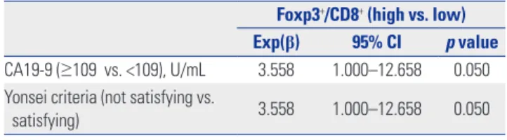
retrieved LNs) were significantly higher in the high Foxp3+/CD8+ group (p=0.020). Pathologic tumor stage, nodal stage, tumor differentiation, lymphovascular invasion, and perineural inva-sion were not associated with CD8+ levels or Foxp3+/CD8+ ratio (Table 3). In multivariate analysis, high levels of CA19-9 and not satisfying Yonsei criteria were significantly associated with a high Foxp3+/CD8+ ratio [Exp(β)=3.558; 95% CI: 1.000–12.658; p=0.050] (Table 4). When the two factors, Yonsei criteria and CA19-9, were combined for analysis of factors affecting the CD8+ counts and Foxp3+/CD8+ ratio, high levels of CA19-9 and not satisfying Yonsei criteria were significantly associated with a high Foxp3+/CD8+ ratio, compared to other conditions (p=0.024) (Table 5).
DISCUSSION
In this study, we investigated the effects of TILs on the prog-Table 2. Survival Analysis According to Tumor Infiltrating Lymphocyte Counts
DFS OS
Univariate Multivariate Univariate Multivariate
Months p value Exp(β) 95% CI p value Months p value Exp(β) 95% CI p value
Absolute count (stained cell number)
CD3+ 0.918 0.724 Low (≤265, n=24) 12.5 35.8 High (>265, n=26) 11.1 27.9 CD4+ 0.594 0.903 Low (≤143, n=25) 11.1 29.7 High (>143, n=25) 13.7 33.2 CD8+ 0.002 0.016 Low (≤121, n=25) 8.4 3.436 1.701–6.944 0.001 18.9 2.140 1.035–4.426 0.040 High (>121, n=25) 18.7 43.7 Granzyme B+ 0.033 0.136 Low (≤24, n=24) 10.2 29.9 High (>24, n=26) 18.0 37.2 Foxp3+ 0.435 0.368 Low (≤25, n=25) 13.5 39.3 High (>25, n=25) 10.3 27.9 Relative ratio Foxp3+/CD3+ 0.151 0.296 Low (≤0.11, n=26) 15.1 39.3 High (>0.10, n=24) 10.2 23.9 Foxp3+/CD4+ 0.041 0.034 Low (≤0.17, n=25) 15.1 48.3 High (>0.17, n=25) 10.2 21.3 Foxp3+/CD8+ 0.005 0.012 Low (≤0.23, n=26) 18.7 48.3 High (>0.23, n=24) 9.4 2.505 1.262–4.974 0.009 21.2 2.235 1.074–4.650 0.032 Foxp3+/Granzyme B+ 0.014 0.040 Low (≤0.17, n=25) 18.7 39.3 High (>0.17, n=25) 9.4 20.2 DFS, disease-free survival; OS, overall survival; CI, confidence interval.
nosis of left-sided PDAC and examined whether the Yonsei criteria were associated with differences in TIL distributions.
In addition to clinicopathologic factors, the host immune re-sponse against tumors plays a critical role in survival outcomes for numerous types of cancers.3-5,8,10,11 In terms of TIL subsets, a ratio of low Treg density to high T-cells (CD4+, CD8+, and gran-zyme B+ T-cell) has been reported to be a promising indepen-dent favorable factor in various tumors.4,5,9 In our previous study,16 we showed that patients with a low Foxp3+/granzyme B+ ratio had significantly improved DFS (25 months vs. 8 months; p=0.008) and OS (47 months vs. 17 months; p=0.003). Multi-variate analysis of the data accumulated since our previous study supports that a high level of CD8+ cells and a low Foxp3+/ CD8+ ratio are independent predictors for favorable DFS (p=0.001 and p= 0.009, respectively) and OS (p=0.040 and
p=0.032, respectively) (Table 2, Figs. 2 and 3).
In our previous study, we analyzed clinicopathologic factors associated with differences in the distribution of TIL subsets: only low levels of CA19-9 were significantly associated with a low Foxp3+/granzyme B+ ratio. Even though several studies have demonstrated that distributions of TILs are associated with survival outcomes in patients with PDAC,20-22 only a few stud-ies have described clinicopathologic factors associated with differences in TILs distribution. Fukunaga, et al.20 reported that positivity for both tumor infiltrating CD4+ T-cells and CD8+ T-cells was significantly associated with a lower grade of depth of invasion and tumor stage, compared with both negative group. Hiraoka, et al.23 demonstrated that a high prevalence of Treg in CD4+ T-cells was significantly correlated with distant metastasis, advanced tumor stage, and high tumor grade.
1.0 0.8 0.6 0.4 0.2 0.0 1.0 0.8 0.6 0.4 0.2 0.0 0 25 50 75 100 125 0 25 50 75 100 125 Months after distal pancreatectomy Months after distal pancreatectomy
p=0.001 p=0.040 CD8+>121 CD8+>121 CD8+≤121 CD8+≤121 DFS rate OS rate A B
Fig. 2. Kaplan-Meier analysis of DFS (A) and OS (B) according to CD8+ levels (low vs. high; number of cells stained with immunohistochemical
stain-ing). The group with high levels of CD8+ showed favorable survival outcomes in regards to DFS (p=0.001) and OS (p=0.040). DFS, disease-free survival;
OS, overall survival.
Fig. 3. Kaplan-Meier analysis of DFS (A) and OS (B) according to Foxp3+/CD8+ ratio (low vs. high). Low Foxp3+/CD8+ ratio showed favorable survival
outcomes in regards to DFS (p=0.009) and OS (p=0.032). Foxp3, forkhead/winged helix transcription factor. DFS, disease-free survival; OS, overall sur-vival. 1.0 0.8 0.6 0.4 0.2 0.0 1.0 0.8 0.6 0.4 0.2 0.0 0 25 50 75 100 125 0 25 50 75 100 125 Months after distal pancreatectomy Months after distal pancreatectomy
p=0.009 p=0.032 Foxp3+/CD8+≤0.23 Foxp3+/CD8+≤0.23 Foxp3+/CD8+>0.23 Foxp3 +/CD8+>0.23 DFS rate OS rate A B
These studies have investigated pathologic findings that could only be analyzed postoperatively. To our knowledge, there are no other studies that have analyzed preoperative clinical
fac-tors associated with TILs to predict patient survival outcomes. In the present study, among the preoperatively detected clini-cal factors, high levels of CA19-9 (p=0.022) and not satisfying Table 3. Association between Clinicopathologic Factors and CD8+ Counts and Foxp3+/CD8+ Ratio
CD8+
p value
Foxp3+/CD8+
p value
Low group
(≤121, n=25) (>121, n=25)High group (≤0.23, n=26)Low group (>0.23, n=24)High group
Preoperative factors Age, yr 1.000 >0.999 <63 13 (52) 12 (48) 13 (50) 12 (50) ≥63 12 (48) 13 (52) 13 (50) 12 (50) Gender 0.387 0.729 Male 13 (52) 17 (68) 15 (57.7) 15 (62.5) Female 12 (48) 8 (32) 11 (42.3) 9 (37.5) Tumor size, cm 0.256 0.586 <3.5 11 (44) 16 (64) 15 (57.7) 12 (50) ≥3.5 14 (56) 9 (36) 11 (42.3) 12 (50) CA19-9, U/mL 0.778 0.022 <109 11 (45.8) 13 (52) 17 (65.4) 7 (30.4) ≥109 13 (54.2) 12 (48) 9 (34.6) 16 (69.6) Yonsei criteria 0.777 0.022 Satisfying 11 (44) 12 (48) 16 (61.5) 7 (29.2) Not satisfying 14 (56) 13 (52) 10 (38.50 17 (70.8) NCCN resectability 0.667 >0.999 Resectable 21 (84) 23 (92) 23 (88.5) 21 (87.5) Borderline 4 (16) 2 (8) 3 (11.5) 3 (12.5) Pathologic factors pT stage 0.187 0.164 T1 0� 1 (4) 1 (3.8) 0� T2 4 (16) 1 (4) 4 (15.4) 1 (4.2) T3 21 (84) 21 (84) 19 (73.1) 23 (95.8) T4 0� 2 (8) 2 (7.7) 0� pN stage 0.571 0.093 N0 12 (48) 15 (60) 17 (65.4) 10 (41.7) N1 13 (52) 10 (40) 9 (34.6) 14 (58.3) Number of metastatic LNs 0.020 0.049 <2 15 (62.5) 21 (91.3) 22 (88) 14 (63.6) ≥2 9 (37.5) 2 (8.7) 3 (12) 8 (36.4) LN ratio 0.09±0.13 0.04±0.08 0.149 0.03±0.05 0.1±0.14 0.020 Differentiation 0.421 0.798 Well 4 (16) 7 (28) 6 (23.1) 5 (20.8) Moderate 17 (68) 17 (68) 17 (65.4) 17 (70.8) Poor 3 (12) 1 (4) 2 (7.7) 2 (8.3) Undifferentiation 1 (4) 0� 1 (3.8) 0� Lymphovascular invasion 0.752 0.374 No 18 (75) 16 (69.6) 18 (78.3) 16 (66.7) Yes 6 (25) 7 (30.4) 5 (21.7) 8 (33.3) Perineural invasion 0.387 0.882 No 14 (58.3) 10 (43.5) 12 (52.2) 12 (50) Yes 10 (41.7) 13 (56.5) 11 (47.8) 12 (50) NCCN, National Comprehensive Cancer Network; LNs, lymph nodes.
Yonsei criteria (p=0.022) were significantly associated with a high Foxp3+/CD8+ ratio. Among the postoperative pathologic factors, having more than two of metastatic LNs was also relat-ed to low CD8+ levels (p=0.020) and high Foxp3+/CD8+ ratio (p=0.049) (Table 3). In multivariate analysis, high levels of CA19-9 and not satisfying Yonsei criteria were associated with a high Foxp3+/CD8+ ratio [Exp(β)=3.558; 95% CI: 1.000–12.658; p=0.050] (Table 4). These two factors are preoperatively detect-able clinical parameters. When we analyzed the survival out-comes between the patients with low and high CD8+ T cells and low and high FOXP3+/CD8+ T cells ratios in each subgroup of patients according to metastatic LN (<2 and ≥2) and CA19-9 (≤10CA19-9 and >10CA19-9), as well as Yonsei criteria (satisfying versus not satisfying), even the number of patients and the number of events in each group were too small to find meaningful re-sults in all subgroups with Kaplan-Meier method, high CD8+ T cell and low FOXP3+/CD8+ T cell ratio were significantly as-sociated with better survival in the subgroup with CA19-9 >109 (p=0.022 and p=0.022, respectively). Also, high CD8+ T cell levels and a low FOXP3+/CD8+ T cell ratio had marginal significance in the metastatic LN <2 subgroup (p=0.055 and p=0.069, respectively) (Supplementary Figs. 1, 2, and 3, only online).
Several clinicopathologic factors that can predict the prog-nosis of PDAC have been analyzed in various studies.1,24 Mul-tivariate analysis in this study showed that more than two met-astatic LNs [Exp(β)=3.354; 95% CI: 1.582–7.110; p=0.002] and intraoperative transfusion [Exp(β)=2.615; 95% CI: 1.248–5.482; p=0.011] were independent risk factors for DFS. Not satisfying Yonsei criteria [Exp(β)=2.590; 95% CI: 1.151–5.829; p=0.021] and more than two metastatic LNs [Exp(β)=2.536; 95% CI: 1.083– 5.936; p=0.032] were also independent risk factors for OS (Ta-ble 1).
Slidell, et al.24 investigated the impact of nodal stage and
LNRs affecting survival outcomes in a large population-based (with Surveillance, Epidemiology, and End Results, SEER data) study of patients with PDAC. In their study, N1 disease was associated with a worse 5-year survival rate, compared with N0 disease (4.3% vs. 11.3%, respectively, p<0.001). For N1 patients, LNR was one of the most powerful factors associated with survival (LNR >0–0.2, 15 months; LNR >0.2–0.4, 12 months; LNR >0.4, 10 months) (p<0.001). We did not find differences in survival outcomes according to pathologic nodal stage or LNR in N1 stage in this study; however, having more than two metastatic LNs was an independent risk factor for DFS and OS (Table 1).
Intraoperative transfusion has been shown to have an ad-verse oncologic impact on various kinds of tumors, including PDAC.25-28 Although the actual mechanism by which intraop-erative transfusion negatively affects survival outcomes is un-known, it is thought to be an important factor in patient im-mune system suppression.29
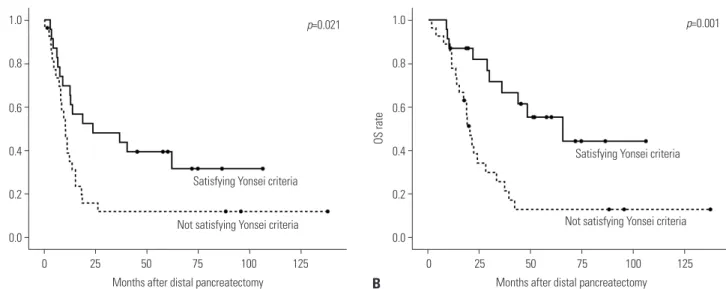
Recently, although the number of patients diagnosed with early staged PDAC has increased due to routine medical check-up, it is still difficult to diagnose PDAC early, which is associat-ed with a good prognosis. Furthermore, it is not easy to clearly define what is truly an early staged PDAC. The preoperative resectability definition of the National Comprehensive Cancer Network (NCCN) guidelines,30 which are based on imaging studies, are the most widely used method for determining surgical or non-surgical treatment strategies for PDAC. How-ever, even in the NCCN guideline for ‘resectable’ status, it is difficult to distinguish early stage PDAC. In the present study, we were unable to discern any differences in survival outcomes according to NCCN guidelines. In our previous studies,17-19 we found that the Yonsei criteria for left-sided PDAC, 1) tumor con-fined to the pancreas, 2) intact fascia layer between the distal pancreas and the left adrenal gland and kidney, and 3) tumor located more than 1–2 cm from the celiac axis, were valuable selection criteria for surgeons seeking to apply minimally in-vasive distal pancreatectomy. In the present study, patients that satisfied the Yonsei criteria showed favorable survival out-comes in regards to DFS (p=0.021) and OS (p=0.001) (Table 1, Fig. 4) and were significantly associated with favorable TIL subset distributions. Although we merely noted an association between preoperatively detectable parameters and host im-mune status against tumor, there are some reports20,31,32 that Table 4. Multivariate Analysis of Clinicopathologic Factors Affecting
Foxp3+/CD8+ Ratio
Foxp3+/CD8+ (high vs. low)
Exp(β) 95% CI p value
CA19-9 (≥109 vs. <109), U/mL 3.558 1.000–12.658 0.050 Yonsei criteria (not satisfying vs.
satisfying) 3.558 1.000–12.658 0.050 CI, confidence interval.
Table 5. Association between Combined Factors and CD8+ Counts and Foxp3+/CD8+ Ratio
CD8+ Foxp3+/CD8+
Low group
(≤121, n=24) (>121, n=25)High group p value (≤0.23, n=26)Low group (>0.23, n=23)High group p value
Combined factors 0.102 0.024
Satisfying Yonsei criteria+Ca19-9<109 8 (33.3) 5 (20) 11 (42.3) 2 (8.7) Satisfying Yonsei criteria+CA19-9≥109 3 (12.5) 7 (28) 5 (19.2) 5 (21.7) Not satisfying Yonsei criteria+CA19-9<109 3 (12.5) 8 (32) 6 (23.1) 5 (21.7) Not satisfying Yonsei criteria+CA19-9≥109 10 (41.7) 5 (20) 4 (15.4) 11 (47.8)
suggest that patients with a high density of TILs (CD3+, CD4+, and CD8+ T cells) in pancreatic cancer have a better prognosis than those who have a low density of TILs. Accordingly, fur-ther well-designed translational research is warrant to better determine the biologic mechanisms for this phenomenon.
In conclusion, the host immune response against tumors plays a critical role in survival outcomes. High levels of tumor infiltrating CD8+ T cells and low Foxp3+/CD8+ ratios were sig-nificantly associated with long DFS and OS in this study. The Yonsei criteria can play a role as clinically detectable biologic marker with which to predict tumor-immunologic status and survival outcomes of patients with resected left-sided PDAC.
ACKNOWLEDGEMENTS
This study was supported by a faculty research grant from Yonsei University College of Medicine for 2015 (6-2015-0162).
AUTHOR CONTRIBUTIONS
Conceptualization: Ho Kyoung Hwang, Sung Hwan Lee, Hyoung-Il
Kim, and Chang Moo Kang. Data curation: Ho Kyoung Hwang, Sung Hwan Lee, Hyoung-Il Kim, and Chang Moo Kang. Formal analysis: Ho Kyoung Hwang, Sung Hwan Lee, Hyoung-Il Kim, Se Hoon Kim, Junjeong Choi, and Chang Moo Kang. Funding acquisition: Ho Kyoung Hwang and Chang Moo Kang. Investigation: all authors. Methodolo-gy: all authors. Project administration: Ho Kyoung Hwang and Chang Moo Kang. Resources: Ho Kyoung Hwang and Chang Moo Kang.
Software: Ho Kyoung Hwang, Sung Hwan Lee, Hyoung-Il Kim, and
Chang Moo Kang. Supervision: Chang Moo Kang and Woo Jung Lee.
Validation: Se Hoon Kim and Junjeong Choi. Visualization: Ho
Kyo-ung Hwang, HyoKyo-ung-Il Kim, Se Hoon Kim, and Junjeong Choi.
Writ-ing—original draft: Ho Kyoung Hwang, Sung Hwan Lee, Hyoung-Il
Kim, and Chang Moo Kang. Writing—review & editing: Se Hoon Kim, Junjeong Choi, Chang Moo Kang, and Woo Jung Lee. Approval of final
manuscript: all authors.
ORCID iDs
Ho Kyoung Hwang https://orcid.org/0000-0003-4064-7776 Sung Hwan Lee https://orcid.org/0000-0003-3365-0096 Hyoung-Il Kim https://orcid.org/0000-0002-6134-4523 Se Hoon Kim https://orcid.org/0000-0001-7516-7372 Junjeong Choi https://orcid.org/0000-0003-1339-593X Chang Moo Kang https://orcid.org/0000-0002-5382-4658 Woo Jung Lee https://orcid.org/0000-0001-9273-261X
REFERENCES
1. Hidalgo M. Pancreatic cancer. N Engl J Med 2010;362:1605-17. 2. Jung YS, Park JH, Park DI, Sohn CI, Lee JM, Kim TI. Physical
inac-tivity and unhealthy metabolic status are associated with de-creased natural killer cell activity. Yonsei Med J 2018;59:554-62. 3. Bates GJ, Fox SB, Han C, Leek RD, Garcia JF, Harris AL, et al.
Quan-tification of regulatory T cells enables the idenQuan-tification of high-risk breast cancer patients and those at high-risk of late relapse. J Clin Oncol 2006;24:5373-80.
4. Fu J, Xu D, Liu Z, Shi M, Zhao P, Fu B, et al. Increased regulatory T cells correlate with CD8 T-cell impairment and poor survival in he-patocellular carcinoma patients. Gastroenterology 2007;132:2328-39.
5. Kim HI, Kim H, Cho HW, Kim SY, Song KJ, Hyung WJ, et al. The ratio of intra-tumoral regulatory T cells (Foxp3+)/helper T cells (CD4+) is a prognostic factor and associated with recurrence pat-tern in gastric cardia cancer. J Surg Oncol 2011;104:728-33. 6. Zhang L, Conejo-Garcia JR, Katsaros D, Gimotty PA, Massobrio
M, Regnani G, et al. Intratumoral T cells, recurrence, and survival in epithelial ovarian cancer. N Engl J Med 2003;348:203-13. 7. Balch CM, Riley LB, Bae YJ, Salmeron MA, Platsoucas CD, von
Eschenbach A, et al. Patterns of human tumor-infiltrating lym-phocytes in 120 human cancers. Arch Surg 1990;125:200-5. 8. Clemente CG, Mihm MC Jr, Bufalino R, Zurrida S, Collini P,
Casci-nelli N. Prognostic value of tumor infiltrating lymphocytes in the vertical growth phase of primary cutaneous melanoma. Cancer 1996;77:1303-10.
9. Gao Q, Qiu SJ, Fan J, Zhou J, Wang XY, Xiao YS, et al. Intratumoral balance of regulatory and cytotoxic T cells is associated with
prog-Fig. 4. Kaplan-Meier analysis of DFS (A) and OS (B) according to the Yonsei criteria. Satisfaction of the Yonsei criteria indicated favorable survival out-comes in regards to DFS (p=0.021) and OS (p=0.001). DFS, disease-free survival; OS, overall survival.
1.0 0.8 0.6 0.4 0.2 0.0 1.0 0.8 0.6 0.4 0.2 0.0 0 25 50 75 100 125 0 25 50 75 100 125 Months after distal pancreatectomy Months after distal pancreatectomy
p=0.021 p=0.001
Satisfying Yonsei criteria
Satisfying Yonsei criteria
Not satisfying Yonsei criteria Not satisfying Yonsei criteria
DFS rate OS rate
nosis of hepatocellular carcinoma after resection. J Clin Oncol 2007;25:2586-93.
10. Petersen RP, Campa MJ, Sperlazza J, Conlon D, Joshi MB, Harpole DH Jr, et al. Tumor infiltrating Foxp3+ regulatory T-cells are asso-ciated with recurrence in pathologic stage I NSCLC patients. Can-cer 2006;107:2866-72.
11. Cho Y, Miyamoto M, Kato K, Fukunaga A, Shichinohe T, Kawara-da Y, et al. CD4+ and CD8+ T cells cooperate to improve progno-sis of patients with esophageal squamous cell carcinoma. Cancer Res 2003;63:1555-9.
12. Duffour MT, Chaux P, Lurquin C, Cornelis G, Boon T, van der Bruggen P. A MAGE-A4 peptide presented by HLA-A2 is recog-nized by cytolytic T lymphocytes. Eur J Immunol 1999;29:3329-37. 13. Bleackley RC. A molecular view of cytotoxic T lymphocyte
in-duced killing. Biochem Cell Biol 2005;83:747-51.
14. Sakaguchi S. Naturally arising CD4+ regulatory t cells for immu-nologic self-tolerance and negative control of immune responses. Annu Rev Immunol 2004;22:531-62.
15. Hori S, Nomura T, Sakaguchi S. Control of regulatory T cell devel-opment by the transcription factor Foxp3. Science 2003;299:1057-61.
16. Hwang HK, Kim HI, Kim SH, Choi J, Kang CM, Kim KS, et al. Prog-nostic impact of the tumor-infiltrating regulatory T-cell (Foxp3+)/ activated cytotoxic T lymphocyte (granzyme B+) ratio on resected left-sided pancreatic cancer. Oncol Lett 2016;12:4477-84.
17. Choi SH, Kang CM, Lee WJ, Chi HS. Multimedia article. Laparo-scopic modified anterior RAMPS in well-selected left-sided pan-creatic cancer: technical feasibility and interim results. Surg En-dosc 2011;25:2360-1.
18. Choi SH, Kang CM, Hwang HK, Lee WJ, Chi HS. Robotic anterior RAMPS in well-selected left-sided pancreatic cancer. J Gastroin-test Surg 2012;16:868-9.
19. Lee SH, Kang CM, Hwang HK, Choi SH, Lee WJ, Chi HS. Mini-mally invasive RAMPS in well-selected left-sided pancreatic can-cer within Yonsei criteria: long-term (>median 3 years) oncologic outcomes. Surg Endosc 2014;28:2848-55.
20. Fukunaga A, Miyamoto M, Cho Y, Murakami S, Kawarada Y, Os-hikiri T, et al. CD8+ tumor-infiltrating lymphocytes together with CD4+ tumor-infiltrating lymphocytes and dendritic cells improve the prognosis of patients with pancreatic adenocarcinoma. Pan-creas 2004;28:e26-31.
21. Ikemoto T, Yamaguchi T, Morine Y, Imura S, Soejima Y, Fujii M, et al. Clinical roles of increased populations of Foxp3+CD4+ T cells in peripheral blood from advanced pancreatic cancer patients.
Pancreas 2006;33:386-90.
22. Wachsmann MB, Pop LM, Vitetta ES. Pancreatic ductal adenocar-cinoma: a review of immunologic aspects. J Investig Med 2012;60: 643-63.
23. Hiraoka N, Onozato K, Kosuge T, Hirohashi S. Prevalence of FOXP3+ regulatory T cells increases during the progression of pancreatic ductal adenocarcinoma and its premalignant lesions. Clin Cancer Res 2006;12:5423-34.
24. Slidell MB, Chang DC, Cameron JL, Wolfgang C, Herman JM, Schulick RD, et al. Impact of total lymph node count and lymph node ratio on staging and survival after pancreatectomy for pan-creatic adenocarcinoma: a large, population-based analysis. Ann Surg Oncol 2008;15:165-74.
25. Hwang HK, Jung MJ, Lee SH, Kang CM, Lee WJ. Adverse oncolog-ic effects of intraoperative transfusion during pancreatectomy for left-sided pancreatic cancer: the need for strict transfusion policy. J Hepatobiliary Pancreat Sci 2016;23:497-507.
26. Burrows L, Tartter P. Effect of blood transfusions on colonic ma-lignancy recurrent rate. Lancet 1982;2:662.
27. Katz SC, Shia J, Liau KH, Gonen M, Ruo L, Jarnagin WR, et al. Oper-ative blood loss independently predicts recurrence and survival after resection of hepatocellular carcinoma. Ann Surg 2009;249: 617-23.
28. Nagai S, Fujii T, Kodera Y, Kanda M, Sahin TT, Kanzaki A, et al. Impact of operative blood loss on survival in invasive ductal ade-nocarcinoma of the pancreas. Pancreas 2011;40:3-9.
29. Blajchman MA, Bardossy L, Carmen R, Sastry A, Singal DP. Alloge-neic blood transfusion-induced enhancement of tumor growth: two animal models showing amelioration by leukodepletion and passive transfer using spleen cells. Blood 1993;81:1880-2. 30. Tempero MA, Malafa MP, Behrman SW, Benson AB 3rd, Casper ES,
Chiorean EG, et al. Pancreatic adenocarcinoma, version 2.2014: featured updates to the NCCN guidelines. J Natl Compr Canc Netw 2014;12:1083-93.
31. Yoshida S, Ito Z, Suka M, Bito T, Kan S, Akasu T, et al. Clinical sig-nificance of tumor-infiltrating T cells and programed death li-gand-1 in patients with pancreatic cancer. Cancer Invest 2019;37: 463-77.
32. Sideras K, Biermann K, Yap K, Mancham S, Boor PPC, Hansen BE, et al. Tumor cell expression of immune inhibitory molecules and tumor-infiltrating lymphocyte count predict cancer-specific survival in pancreatic and ampullary cancer. Int J Cancer 2017; 141:572-82.