저작자표시-비영리-변경금지 2.0 대한민국 이용자는 아래의 조건을 따르는 경우에 한하여 자유롭게 l 이 저작물을 복제, 배포, 전송, 전시, 공연 및 방송할 수 있습니다. 다음과 같은 조건을 따라야 합니다: l 귀하는, 이 저작물의 재이용이나 배포의 경우, 이 저작물에 적용된 이용허락조건 을 명확하게 나타내어야 합니다. l 저작권자로부터 별도의 허가를 받으면 이러한 조건들은 적용되지 않습니다. 저작권법에 따른 이용자의 권리는 위의 내용에 의하여 영향을 받지 않습니다. 이것은 이용허락규약(Legal Code)을 이해하기 쉽게 요약한 것입니다. Disclaimer 저작자표시. 귀하는 원저작자를 표시하여야 합니다. 비영리. 귀하는 이 저작물을 영리 목적으로 이용할 수 없습니다. 변경금지. 귀하는 이 저작물을 개작, 변형 또는 가공할 수 없습니다.
A THESIS FOR THE DEGREE OF MASTER OF SCIENCE
Conversion of primed porcine embryonic stem cells to
a naïve state through the
overexpression of reprogramming factors
돼지 배아 줄기세포의 리프로그래밍 팩터
과발현을 통한 만능성 상태 전환
February, 2018
By
TAE YEONG PARK
Department of Agricultural Biotechnology
Graduate School
농학석사학위논문
Conversion of primed porcine embryonic stem cells to
a naïve state through the
overexpression of reprogramming factors
돼지 배아 줄기세포의 리프로그래밍 팩터
과발현을 통한 만능성 상태 전환
2018년 2월
서울대학교 대학원
농생명공학부
박 태 영
3
*This thesis will be published in elsewhere as a partial fulfilment of Tae-Yeong Park’s M.S. program
ABSTRACT
Conversion of primed porcine embryonic stem cells
to a naïve state through the overexpression of
reprogramming factors
TAE YEONG PARK
Department of Agricultural Biotechnology
Graduate School
Seoul National University
Establishing pig embryonic stem cells (pESCs) remains a challenge due to differences in the genetic backgrounds of mouse, human, and pig. Therefore, pig-specific pluripotency markers and their cellular signaling must be identified. In this study, doxycycline (DOX)-inducible vectors carrying Oct4, sex determining region Y-box 2 (Sox2), Nanog, Kruppel-like family 4 (Klf4), and Myc, which are known reprogramming factors, were transduced into pESCs to analyze the pluripotent gene network. Pig ESCs were stably maintained in basic fibroblast growth factor (bFGF)-supplemented media, when cultured without DOX. However, when treated with DOX,
4
the cells lost their alkaline phosphatase activity and differentiated within two weeks. Subsequently, we investigated the expression of genes related to pluripotency in DOX-treated pESCs using quantitative reverse transcription PCR. Expression levels of Oct4,
E-cadherin, and Fut4 were significantly increased by Oct4 overexpression, and Fut4
were upregulated in the Sox2 overexpressed group. When a combination of two reprogramming factors, Oct4 or Sox2 was introduced, weak alkaline phosphatase activity remained. In addition, Oct4 and Nanog, Oct4 and Klf4, or Sox2 and Nanog combinations transduction groups could be maintained after subculturing with transgene activation. Although a long-term culture failed, pESCs transduced with Oct4 and Nanog, Oct4 and Klf4, or Sox2 and Nanog combinations could be subcultured even under transgene activation conditions. Analysis of the cause of long-term culture failure by quantitative real-time PCR (qPCR) confirmed that the expression of intermediate reprogramming markers such as Lin28 and Sall4 was not maintained. Given these results, additional strategies are needed to support the completion of each reprogramming phase to succeed in the conversion of the pluripotent state of pESCs. The present study improve our understanding of pluripotent networks and could be used to aid in the establishment of bona fide pig pluripotent stem cells.
5
CONTENTS
ABSTRACT ... 3 CONTENTS ... 5 LIST OF TABLES ... 6 LIST OF FIGURES ... 7 LIST OF ABREVIATIONS ... 8 LITERATURE REVIEW ... 101. Overview of Embryonic stem cells (ESCs) ... 11
2. Naïve and primed pluripotent states ... 14
3. Pluripotency state conversion ... 18
4. Phase of reprogramming ... 22
5. Studies for pESCs ... 24
INTRODUCTION ... 27
MATERIALS AND METHODS ... 31
RESULTS ... 43
CONCLUSION ... 65
6
LIST OF TABLES
Table 1. Naïve and primed pluripotent cell properties ... 17
Table 2. Primer sets used to detect differentiation marker. ... 37
Table 3. Primer sets used for the detection of transgene insertion in
cDNA. ... 39
7
LIST OF FIGURES
Figure 1. Characterization of pESCs. ... 47
Figure 2. Reprogramming factor-transduced pESCs morphology
and alkaline phosphatase activity. ... 50
Figure 3. Analysis of pluripotent-related gene expression using
quantitative reverse transcription (qRT)-PCR. ... 52
Figure 4. Morphology and alkaline phosphatase activity of
two-reprogramming-factor-transduced pESCs. ... 56
Figure 5. Quantitative reverse transcription PCR analysis of
8
LIST OF ABREVIATIONS
2i Two inhibitors; GSK and ERK pathway inhibitors
ANOVA One-way analyses of variance
AP Alkaline phosphatase
bFGF Basic fibroblast growth factor
BMP cDNA
Bone morphogenetic protein Complementary DNA
ChIP Chromatin immunoprecipitation
Ct Threshold cycle
DMEM DNA
Dulbeco’s modified Eagle’s medium Deoxyribonucleic acid
DOX Doxycycline
DPBS Dulbecco’s phosphate-buffered saline Dppa2 Developmental pluripotency associated 2
EBs Embryoid bodies
EpiSCs Epiblast stem cells
ERK Extra –cellular signal-regulated kinase
ESCs Embryonic stem cells
Esrrb Oestrogen-related receptor beta
FBS Fetal bovine serum
FCS Fetal calf serum
9
hESCs Human embryonic stem cells
ICM Inner cell mass
Id Inhibitor of differentiation
iPSCs Klf4
Induced pluripotent stem cells Kruppel-like family 4
LIF Leukemia inhibitory factor
MAPK Mitogen-activated protein kinase
MEFs Mouse embryonic fibroblasts
mEGCs Mouse embryonic germ cells
mESCs Mouse Embryonic stem cells
MET Mesenchymal-to-epithelial transition
NBT/BCIP Nitro blue tetrazolium chloride/5-bromo-4-chloro-3-indoly phosphate toluidine salt
pESCs Pig embryonic stem cells
PESM pESCs medium
PI-3K Phosphatidylinositol-3-kinase
qPCR Quantitative reverse transcription PCR
RNA Ribo nucleic acid
Sox2 Sex determining region Y-box 2
TE Trophectoderm
TGF Transforming growth factor
10
11
1. Overview of Embryonic stem cells (ESCs)
All creatures start from fertilized eggs. Embryos have totipotency, which is the ability to differentiate into tissues and organs to form an organism. Embryos form blastocysts as development progresses. The outside of the blastocysts consists of trophoblast cells that differentiate into the placenta, and the inside consists of an inner cell mass (ICM) that becomes the fetus. The cells of the ICM are no longer totipotent, but have pluripotency which is the ability to differentiate into all layered cells. ESCs derived from the ICM also have pluripotency and self-renewal capacity. One of the earliest and most well established stem cell models for pluripotency is mouse embryonic stem cells (mESCs), which were derived from the ICM of the mouse blastocyst nearly three decades ago(Evans and Kaufman 1981; Martin 1981). mESCs have met the criteria that define all pluripotency, such as pluripotency marker expression, single cell clonogenecity, and chimeric mice formation as well as successful tetraploid complementation assay. Based on these results, mESCs were used as a useful model for molecular studies of pluripotent states. Early studies have led to the discovery of the core pluripotency factors, namely Oct4, Sox2 and Nanog in mouse embryos and mESCs (Chambers and Tomlinson 2009). Disruption of Oct4 caused aberrant differentiation of the ICM to the trophectoderm (TE) lineage instead of the embryo
12
(Nichols et al. 1998), Nanog null ICM failed to develop into epiblast (Silva et al. 2009), while Sox2 Knockout in mice conferred early embryonic lethality (Avilion et al. 2003). The fact that the three genes play an important role in pluripotency formation was found by the Oct4, Sox2 and Nanog blocking experiments in early embryos.
Genome-wide chromatin immunoprecipitation (ChIP) studies revealed that Oct4,
Sox2 and Nanog co-bind to the promoters of many genes in both mESCs and human
ESCs (hESCs) (Boyer et al. 2005; Loh et al. 2006). Notably categories of co-bound target genes include signaling intermediaries, micro-RNAs, chromatin-remodeling and histone modifying proteins, hence establishing a direct link between the core transcriptional network and other key aspects of pluripotency regulation. These studies also indicate that the core transcriptional factors maintain ESCs by activating other pluripotency factors, such as Esrrb and Zic3, coincident with repressing developmental genes such as Cdx2 to suppress differentiation. Oct4, Sox2 and Nanog activate themselves and also each other, constituting a core transcriptional regulatory network with features of auto-activation and feed-forward loops (Boyer et al. 2005; Loh et al. 2006; Chew et al. 2005; Kuroda et al. 2005; Okumura-Nakanishi et al. 2005; Rodda et al. 2005). In addition to transcription factors, external stimuli, such as culture conditions, also contribute to the maintenance of stemness by engaging in the signaling pathways of the ESCs. Initially, mESCs were cultured on mouse embryonic fibroblasts (MEFs)
13
by using a medium supplemented with fetal calf serum (FCS). Later, FCS could be replaced with BMP4 (Ying et al. 2003) and MEFs could be replaced with cytokine, Leukemia inhibitory factor (LIF) (Niwa et al. 1998). The chemicals required for the cultivation of mESCs affect a variety of transcription factors and signaling pathways. BMP4 induces transcription factor, Smad1 and inhibitor of differentiation (Id) proteins, which inhibit neural-specific differentiation (Ying et al. 2003). On the other hand, LIF mediates its action by multiple signaling pathway for cellular proliferation, including the LIF-STAT3, mitogen-activated protein kinase (MAPK) and phosphatidylinositol-3-kinase (PI-3K) pathways. The use of MEK inhibitors in the culture of mESCs results in a much better self-renewal status, since the MEK inhibitor blocks the MAPK pathway (Burdon et al. 1999). When the MAPK pathway was inhibited, PI-3K was inactivated and helped to maintain undifferentiated state (Paling et al. 2004). In the absence of LIF, BMP4 redirects cells to non-neural differentiation, and the basis of this differentiation propensity is due to activation of the fibroblast growth factor (FGF)-extra –cellular signal-regulated kinase (ERK) pathway (Kunath et al. 2007). In order to maintain the characteristics of ESCs, various factors inside and outside of the cell must be harmonized. If any of these factors are not working properly, ESCs lose pluripotency and become differentiated. Therefore, it is important to understand functions of the factors that make up the pluripotency network and to study the mutual relationship. A
14
deep understanding of pluripotency can be useful in a variety of ways, including cell therapy, developmental models, and disease research.
2. Naïve and primed pluripotent states
It was two decades after the discovery of mESCs that hESCs were established in culture (Thomson et al. 1998). Although hESCs are derived from the ICM, many striking differences from mESCs were observed such as morphology, marker expression, transcription factor binding activities and culture requirements. Intriguingly, it was later found that hESCs resemble more closely to mouse epiblast stem cells (EpiSCs), which were derived from post-implantation late epiblasts of the mouse (Brons et al. 2007; Tesar et al. 2007). Like embryonic stem cells, EpiSCs had the ability to differentiate into three germ layers and has self-renewal ability, but there were slight differences in the characteristics of cells and molecules. Morphology also grew into flat single layer form completely was different from the compact form of the mESCs colonies, whereas mESCs were capable of single cell cloning, but EpiSCs were vulnerable to single cell cloning.
Apart from morphological and molecular differences, EpiSCs differ from mESCs on the level of pluripotency that can be attained by various standard assays (Kuijk et al. 2011). EpiSCs can form teratomas in vivo, but they neither form chimeras nor contribute to the germline like mESCs. (Brons et al. 2007; Tesar et al. 2007) Recently, a number
15
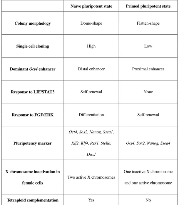
of studies have reported that pluripotent stem cells can be categorized as naïve or primed according to the level of pluripotency (Nichols and Smith 2009; Hanna et al. 2010). mESCs represent naïve pluripotent stem cells, whereas human and large animal ESCs and EpiSCs are considered to be in the primed state. A vigorous comparison and analysis of naïve and primed states revealed features of the naïve and primed pluripotent states as shown below (Table. 1).
Naïve pluripotent stem cells include mESCs and mouse embryonic germ cells (mEGCs). Naïve pluripotent cells form dome-shaped colonies, which are self-renewing via LIF signaling. In addition, ESCs derived from female cells have two active X chromosomes. On the other hand, primed pluripotent stem cells with a flat colonies and undergo self-renewal via FGF signaling. Primed pluripotent stem cells exhibit a greater differentiated status than naïve pluripotent stem cells in terms of developmental capacity, gene expression, and epigenetic signature. In addition, naïve pluripotent stem cells have a higher transgenic efficiency than primed pluripotent stem cells. Naïve pluripotent stem cells can produce chimeric embryos via injected into recipient blastocysts. Furthermore, the differentiation ability of naïve pluripotent stem cells can be confirmed in vitro, as well as to form teratomas via subcutaneous injection. However, primed pluripotent stem cells have only been verified for their ability to differentiate in
vitro, and have failed in vivo differentiation assays such as the production of chimeric
fetus and teratoma assays. Taken together, these characteristics indicate that naïve pluripotent stem cells have a higher degree of pluripotency than primed pluripotent stem cells.Thus, many studies have been attempted to the conversion of primed pluripotent
16
17
Table 1
. Naïve and primed pluripotent cell properties.
Naïve pluripotent state Primed pluripotent state
Colony morphology Dome-shape Flatten-shape
Single cell cloning High Low
Dominant Oct4 enhancer Distal enhancer Proximal enhancer
Response to LIF/STAT3 Self-renewal None
Response to FGF/ERK Differentiation Self-renewal
Pluripotency marker
Oct4, Sox2, Nanog, Ssea1, Klf2, Klf4, Rex1, Stella,
Dax1
Oct4, Sox2, Nanog, Ssea4
X chromosome inactivation in female cells
Two active X chromosomes
One inactive X chromosome and one active chromosome
Tetraploid complementation Yes No
18
3. Pluripotency state conversion
Interest in pluripotency was heightened after the report that it was classified as naïve state and primed state according to the level of pluripotency. In addition, many researchers have tried to overcome the limitations of primed pluripotent stem cells by various artificial methods. Stem cells in the naive state include mESCs, and representative stem cells in the primed state included EpiSCs and hESCs. mESCs were pluripotent stem cell models with the most ideal pluripotency and EpiSCs had similar characteristics of hESCs and were being studied extensively as hESCs models. So studying mESCs and EpiSCs will be very helpful in converting hESCs to naïve pluripotent stem cells.
In terms of similarity to induced pluripotent stem cells (iPSCs) reprogramming, transcription factor mediated approaches has been a prominent choice. As EpiSCs do not express Klf4 or Klf2 as opposed to mESCs, it may be intuitive to reintroduce these key pluripotency transcription factors in an attempt to recapitulate mESC expression levels. Transfection with Klf4 under GSK and ERK pathway inhibitor (2i)/LIF conditions generated converted cells, termed as Epi-iPSC, which exhibited mESC morphology and marker profile, and could contribute to germline-competent chimeras (Guo et al. 2009). Given the high functional redundancy among members of the Kruppel-like family, Klf2 can substitute for Klf4 in the generation of Epi- iPSCs under GSK and ERK pathway inhibitor (2i)/LIF conditions (Hall, Guo, et al. 2009). In
19
addition, Klf2 was introduced into EpiSCs instead of Klf4 and cultured with 2i and LIF to support the conversion into a naïve-like state. Klf2 and Klf4 target several pluripotency genes in common with Nanog, which regulate Nanog expression (Hall, Guo, et al. 2009). Therefore it is likely that Nanog could potentially mediate the same reprogramming process. Indeed, transfection of Nanog induced a similar reversion of EpiSCs to a mESCs like phenotype under 2i/LIF conditions at a 10-fold higher efficiency than Klf4 transfection (Silva et al. 2009). Unlike Klf4, however, Nanog-mediated EpiSC reprogramming can occur under LIF/BMP4 conditions without the need for 2i. These Nanog-induced Epi-iPSCs exhibit an mESC-like expression profile, including upregulation of Klf4. Transient transfection of Nanog, however, did not yield any stable Epi-iPSCs and chimera formation, which was only observed upon co-transfection with Klf4 (Yang et al. 2010). Therefore, Klf4 and Nanog may function synergistically in the conversion of EpiSC to a mESC-like state. Similar success has been achieved by other chemical-assisted reprogramming approaches that do not involve the exogenous introduction of specific transcription factors. mESC-like cells can also be generated from EpiSCs under purely 2i/LIF conditions without the need for transcription factors, and these cells have displayed chimera formation as well as germline transmission (Greber et al. 2010). One possible explanation is that inhibition of the FGF/ERK pathway induces Klf2 expression in EpiSCs (Greber et al. 2010). A more diverse inhibitor cocktail has also been established for the conversion of EpiSC to a mESC-like state. A combination of parnate and transforming growth factor (TGF)-ß inhibitor, can reprogrammed EpiSCs to cells that can contribute to germline (Zhou et
20
al. 2010). Parnate can induce a global increase in the activating H3K4 methylation and is postulated to activate previously silenced pluripotency genes (Lee et al. 2006), highlighting the importance of epigenetic modification in facilitating cellular conversions. Unlike other studies that require 2i conditions, Surani and co-workers (Bao et al. 2009) reported that EpiSCs could be reprogrammed into mESC-like cells in the presence of LIF/FCS and MEF feeder cells. These reprogrammed epiblast ESC-like cells show gradually increasing similarity to mESCs in their gene expression profile with more passages, including an upregulation of E-cadherin. E-cadherin, a transmembrane molecule involved in intercellular adhesion, was previously shown to be stimulated by LIF/BMP4 and correlated with differentiation capability (Chou et al. 2008). Furthermore, knockout of E-cadherin traps cells in a partially reprogrammed state (Lee et al. 2006). Taken together, these studies indicate a potential requirement for
E-cadherin in attainment of pluripotency. LIF is an essential component of traditional
mESC culture medium, and is frequently added to 2i cultures. Therefore, it would be informative to determine the effect of LIF, especially LIF-STAT3 pathway, in reprogramming. It was shown that STAT3, while insufficient on its own, can increase the efficiency of EpiSCs reprogramming by several fold when used in combination with
Klf4 or Nanog transfection, indicating that the enhancing effects of STAT3 extends
beyond direct activation of these pluripotency factors (Lee et al. 2006). A transient activation of STAT3, followed by transfer to 2i/LIF culture can recapitulate the effects of continuous STAT3 activation (Lee et al. 2006), suggesting that a transient activation of LIF-STAT3 is sufficient to prime EpiSCs for reprogramming. A recent study
21
demonstrated that hESC can be converted to a naïve, or mESC-like state by the constitutive expression of Oct4 and Klf4 or Klf4 and Klf2 under 2i/LIF conditions (Hanna et al. 2010). These naïve hESCs gained a mESC-like colony morphology, displayed pre-X-inactivation status and survived single-cell passaging as well as inhibition of Activin/Nodal signaling. However, the naïve hESCs rapidly differentiated when the exogenous transcription factors were removed, unless Forskolin was added, which prolonged the maintenance of the cells (Hanna et al. 2010). Forskolin acts partly by inducing Klf4 and Klf2, emphasizing the importance of KLF factors in maintenance of the naïve state. There have been other efforts towards achieving certain characteristics of mESCs. In the presence of LIF, the combination of MEK and p38 inhibitors can convert hESCs to mESC-like cell type (Xu et al. 2010). Cell passaging, a property that was correlated to an increased expression of E-cadherin and reduced dependent on integrin signaling. Although further characterization of the converted hESCs is required, this study suggests that a switch in cell adhesion systems may be required for hESCs to acquire certain mESC-like properties.
Besides cell adhesion molecules, atmospheric oxygen levels can also possibly affect maintenance of pluripotency. According to Lenger et al., when the hESCs were incubated at 5% oxygen concentration, the XIST promoter region was methylated and the inactive X chromosome becomes active. According to Lenger et al., when the hESCs were incubated at 5% oxygen concentration, the XIST promoter region was methylated and the inactive X chromosome became active. In addition, many researchers were trying to establish naïve pluripotent stem cells in large animals.
22
Roberts et al., 2011, transduced Oct4 and Klf4 to ICM of porcine blastocysts and derived pESCs. These pESCs were self-renewed by LIF and succeeded to differentiate into three germ layers in the teratoma formation assay (Telugu et al. 2011). Many studies had been conducted to induce pluripotent state conversion through various methods such as introducing of exogenous factors, addition of chemicals to culture media, and oxygen concentration control.
4. Phase of reprogramming
In 2006, Takshi and Yamanaka published their milestone strategy to reprogramme somatic mammalian cells to iPSCs by overexpression of only four transcription factors (Takahashi et al. 2007; Takahashi and Yamanaka 2006). Since then, a number of scientists conducted research to uncover the reprogramming process. The most obvious method of analyzing the complex molecular mechanism of reprogramming was to examine how the cell population changes over time after the introduction of reprogramming factors. According to this study, reprogramming can be divided into two stages. The first step was the stochastic phase, which takes a relatively long time and applies only to a few of the many candidate cells(Buganim, Faddah, and Jaenisch 2013). Cells that have undergone the stochastic step go into the second step and undergo a process of rapid reprograming. This second step was called deterministic phase.
23
When Oct4, Sox2, Klf4 and Myc were introduced into fibroblasts, some cells may have various fates when stochastic gene expression begins (apoptosis, aging, transformation, transdifferentiation and reprogramming). Among them, the cells that become iPSCs were largely reprogrammed through the early, middle and the late stages. When reprogramming factors are introduced into somatic cells, Ccnd1, Ccnd2 and genes involved in DNA replication, which are related in cell proliferation, are expressed at an early stage. The ratio of S phase in the cell cycle increases. The genes expressed in somatic cells (Snai1, Snai2, Thy1) are less expressed by histone modification. At the early stage, the reprogrammed cells are transferred to the intermediate stage and become round and dense in shape. Interior of the cells, TGF-ß signaling is suppressed inside the cells and pro-mesenchymal to epithelial transition (MET) micro RNAs are activated. As a result, bone morphogenetic protein (BMP) signaling is enhanced and MET begun (Mikkelsen et al. 2008; Samavarchi-Tehrani et al. 2010; Koche et al. 2011).
Cells at this stage exhibit stochastic activation of markers related with pluripotency (Buganim et al. 2012), and developmental regulators (Polo et al. 2012) and glycolysis (Hansson et al. 2012) are activated. After this step, genes like undifferentiated embryonic cell transcription factor1 (Utf1), oestrogen-related receptor beta (Esrrb), developmental pluripotency associated 2 (Dppa2), Sall4 and Lin28 are highly expressed and these genes activate Sox2. When Sox2 is activated, a deterministic event occurs either directly or indirectly. Cells that reached the late stage are stably pluripotent and could remain pluripotent despite inactivating the transgene. In later phase, the cytoskeleton redesigns the cell morphology to an ESC-like shape. And
24
repressive chromatin characters such as H3k27 or H3k9 disappear from the pluripotency core transcription factor and activate the pluripotency circuitry. When the pluripotency circuitry is activated, Oct4, Nanog, Rex1 and Esrrb are highly expressed, and pluripotent state is maintained even when ectopically introduced reprogramming factors are removed (Golipour et al. 2012; Polo et al. 2012; Hansson et al. 2012; Buganim et al. 2012).
5. Studies for pESCs
Pigs are physiologically similar to humans, and pESCs are of interest as models of human disease research (Brevini et al. 2007; Hall 2008; Houdebine 2009). Many research groups have long tried to derive pluripotent cells from porcine early embryos. In 1990, several reports on the first pESC derivation were published (Notarianni et al. 1990; Evans et al. 1990; Strojek et al. 1990; Piedrahita, Anderson, and Bondurant 1990). The first pESCs were identified based on morphological criteria, and there were no reports of reliable markers. Alkaline phosphatase (AP) (Talbot et al. 1993) and stage-specific embryonic antigen-1 (Wianny, Perreau, and Hochereau de Reviers 1997) were then identified in pESCs that have been demonstrated to be undifferentiated. However, these studies were based on undifferentiated mESCs markers and lacked studies on pig specific markers. pESCs are primed pluripotent stem cells that can differentiate into all three germ layers in vitro (Piedrahita et al. 1999; Piedrahita, Anderson, and Bondurant
25
1990; Wianny, Perreau, and Hochereau de Reviers 1997), but do not exhibit in vivo differentiation ability because the pluripotency level of pESCs are not as high as the pluripotent level of naïve pluripotent cells (Mueller et al. 1999; Piedrahita et al. 1998; Shim et al. 1997). It is fair to say that research on the specific characteristics of pESCs are lacking.
If overcome these barriers, studies are underway to convert pig embryonic stem cells into naïve state. Most studies on pluripotent state conversion to porcine embryonic stem cells have proceeded in a way that applies research that had succeeded in converting primed pluripotent stem cells such as hESCs or EpiSCs into a naïve pluripotent state. However, this approach did not apply to pESCs. Because of the different molecular mechanisms in embryonic development, the reprogramming conditions of mouse and humans could not be easily applied to pigs. Pigs require a longer period of time to implant embryos than mouse and humans (Alberio and Perez 2012). Thus, the signal regulating the pluripotency of pig ICM is different from that of mouse embryos. Unlike mouse, pigs show Oct4 and Cdx2 in ICM and TE. Conversely, Sox2 is expressed from the beginning of embryo cavitation, and when it is divided into TE/ICM, it appears only specifically in the embryo and progressively disappeared as the differentiation progresses. ICM cells and epiblasts of pig blastocysts do not have LIF receptors, but FGF receptors are highly expressed. Thus, FGF signaling plays an important role in maintaining the pluripotency of pigs over LIF signaling (Hall, Christensen, et al. 2009). In addition, strong expression of BMPr and SMAD in porcine ICM cells and EpiSCs was detected by in vitro immunostaining and BMP signaling inhibitor experiments.
26
Treatment of porcine ICM cells and EpiSCs cells with BMP inhibitors in vitro reduced cell viability (Hall and Hyttel 2014). BMP signaling also plays an important role in maintaining pig pluripotency. (Zhang et al. 2015) recently succeeded in generating ‘intermediate’ porcine pluripotent stem cells with naïve and primed characteristics even when the transgene was removed using three growth factors (LIF, FGF2, and BMP4) and the inhibitor 2i. These results show that the pluripotent stem cells of pigs maintain pluripotency by unique cell signaling that differs from those of mouse and human.
27
28
Pigs are physiologically similar to humans, and pESCs have attracted attention as a human disease research model (Brevini et al. 2007; Hall 2008; Houdebine 2009). Many research groups have long tried to isolate pluripotent cells from porcine early embryos. In 1990, several reports of the first pESC isolation were published (Evans et al. 1990; Notarianni et al. 1990; Piedrahita, Anderson, and Bondurant 1990; Strojek et al. 1990) The first pESCs were identified based on morphological criteria, and there were no reports on the reliable markers. Then, AP (Talbot et al. 1993) and stage-specific embryonic antigen-1(Wianny, Perreau, and Hochereau de Reviers 1997) were identified in pESCs, which were proven to be undifferentiated. However, these studies markers were based on markers of undifferentiated mESCs, and studies on pig-specific markers were lacking. pESCs are primed pluripotent stem cells that can differentiate into all three germ layers in vitro, (Piedrahita et al. 1999; Piedrahita, Anderson, and Bondurant 1990; Wianny, Perreau, and Hochereau de Reviers 1997), but fail to show any in vivo differentiation ability because the pluripotency level of pESCs is not as high as that of naïve pluripotent cells (Mueller et al. 1999; Piedrahita et al. 1998; Shim et al. 1997). It is fair to say that there a lack of research on the specific characteristics of pESCs. Recently, a number of studies have reported that pluripotent stem cells can be categorized as naïve or primed according to the level of pluripotency (Nichols and Smith 2009; Hanna et al. 2010). mESCs represent naïve pluripotent stem cells, whereas human and large animal ESCs and EpiSCs are considered to be in the primed state. Naïve pluripotent cells form dome-shaped colonies and are self-renewing via LIF signaling. In addition, ESCs obtained from female cells have two active X
29
chromosomes. In contrast, primed pluripotent stem cells form flat colonies and undergo self-renewal via FGF signaling. Primed pluripotent stem cells show a greater differentiated status than naïve pluripotent stem cells in terms of developmental capacity, gene expression, and epigenetic signature. Meanwhile, naïve pluripotent stem cells have a higher transgenic efficiency than primed pluripotent stem cells. Naïve pluripotent stem cells can be injected into recipient blastocysts to produce chimeric embryos. Furthermore, the differentiation ability of naïve pluripotent stem cells can be confirmed in vitro, and can form teratomas via subcutaneous injection. However, primed pluripotent stem cells have only been verified for their ability to differentiate in
vitro, and have failed in vivo differentiation assays such as the chimeric fetus and
teratoma assays. Together, these characteristics indicate that naïve pluripotent stem cells have a higher degree of pluripotency than primed pluripotent stem cells. Thus, many studies have been conducted on the conversion of primed pluripotent cells into naïve pluripotent cells (Bao et al. 2009; Guo et al. 2009; Yang et al. 2010).
Many recent studies have compared the conversion of primed pluripotent stem cells into naïve pluripotent stem cells and the reprogramming of somatic cells into iPSCs. The reprogramming of somatic cells into iPSCs requires sequential steps. When reprogramming factors are introduced into somatic cells, Ccnd1, Ccnd2, and genes involved in DNA replication, which are involved in cell proliferation, are expressed at an early stage. This increases the ratio of the S phase in the cell cycle. The genes expressed in somatic cells (Snai1, Snai2, and Thy1) are downregulated via histone modification. Then, the reprogrammed cells in the early stage are transferred to the
30
intermediate stage, and their shape becomes rounder and denser (Mikkelsen et al. 2008; Samavarchi-Tehrani et al. 2010; Koche et al. 2011). Inside the cells, TGF-ß signaling is suppressed and pro-MET microRNAs are activated. As a result, BMP signaling is enhanced and MET occurs. Cells at this stage exhibit stochastic activation of markers associated with pluripotency (Buganim et al. 2012), and developmental regulators (Polo et al. 2012) and glycolysis are activated (Hansson et al. 2012). After this step, genes such as Utf1, Esrrb, Dppa2, Sall4, and Lin28 are highly expressed, which activate Sox2. The activation of Sox2 causes deterministic events either directly or indirectly. In the late phase, the cytoskeleton redesigns the cell morphology into an ESC-like shape. Repressive chromatin characteristics, such as H3k27 or H3k9, disappear from the pluripotency core transcription factors, activating pluripotency circuitry. When pluripotency circuitry is activated, Oct4, Nanog, Rex1, and Esrrb are highly expressed, and the pluripotent state is maintained even when the reprogramming factors that were introduced ectopically are removed (Buganim et al. 2012; Golipour et al. 2012; Hansson et al. 2012; Polo et al. 2012).
In this study, we overexpressed reprogramming factors in primed pESCs and analyzed the changes that occurred in endogenous genes related to pluripotency. This study improves our understanding of pluripotent networks and can be used to aid in the establishment of bona fide pig pluripotent stem cells.
31
32
1. Animal care
The care and experimental use of pigs and mice was approved by the Institutional Animal Care and Use Committee at Seoul National University [Approval No.: ILAR-17-06-192 for MEF isolation]. The ovaries used in the present study were donated from local slaughterhouses (Dodram, Korea; Samsung, Korea) for research purposes. Pregnant ICR mice were purchased from Samtaco Bio Inc., Korea. The mice were cared for according to the standard protocol of the Institute of Laboratory Animal Resources and sacrificed by cervical dislocation after anesthesia.
2. Lentiviral transduction of reprogramming factors
Production and transduction of lentiviral vectors with inducible systems containing human Oct4, Sox2, Klf4, Myc, and pig Nanog were performed following the protocols described by (Choi et al. 2016).
3. Culture and growth of pESCs
pESCs were derived from blastocysts produced in vitro. The produced and hatched blastocysts were seeded onto feeder cells composed of mitotically inactivated MEFs according to previous studies (Son et al. 2009; Park et al. 2013). After 5–7 days, primary colonies of ESCs were observed and cultured for approximately 7–10 days. Fully
33
expanded colonies, mechanically dissociated via trituration with pulled-glass pipettes, were transferred onto new feeder cells for subculturing.
pESCs were cultured in pESC medium (PESM) consisting Dulbecco’s modified Eagle’s medium (DMEM; low glucose), Ham’s F10 medium containing 15% fetal bovine serum (FBS; collected and processed in the USA), 2 mM GlutaMAX, 0.1 mM ß-mercaptoethanol, and 1× antibiotic/antimycotic (Gibco, USA). To support pluripotency and self-renewal, the ESCs were cultured in PESM with 20 ng/mL human recombinant bFGF (hrbFGF; R&D Systems), and 100 ng/mL heparin sodium salt (Sigma-Aldrich, USA). For the analysis of overexpressed genes, the transfected cells were cultured in medium supplemented with 2 ng/mL DOX and 1,000 unit/mL LIF (Millipore, MA, USA). The medium was changed every 24 h and all cells were cultured in humidified conditions with 5% CO2 at 37°C. pESCs were subcultured every 5–7 days
using pulled-glass pipettes. Expanded colonies were detached from the feeder cells and dissociated into small clumps. They were transferred onto new feeder cells containing mitomycin-C-treated (Roche, Germany) MEFs.
4. AP staining
AP staining was performed using the nitro blue tetrazolium chloride/5-bromo-4-chloro-3-indolyl phosphate toluidine salt (NBT/BCIP; Roche) method. Before staining, all cells were preincubated for 10 min at 4°C and fixed with 4% paraformaldehyde for 20 min. After washing, fixed cells were incubated for 30 min at room temperature in
34
the dark with NBT/BCIP stock solution diluted in buffer solution (0.1 M Tris-HCl, 0.1 M NaCl, pH 9.5). The cells were examined under an inverted microscope.
5. Embryoid body formation and in vitro differentiation of pESCs
To evaluate the capacity for in vitro differentiation, embryoid bodies (EBs) were generated from pESCs. Cultured ESC colonies were detached from the feeder cells, and colonies were mechanically dissociated into small clumps. These clumps were cultured in Petri dishes without cytokines for 5 days. After suspending the culture, the dissociated cells aggregated and formed EBs. The cultured EBs were seeded onto 0.1% gelatin-coated plates and further cultured for 2 weeks with DMEM containing 15% FBS. After 2 weeks, differentiated cells were analyzed using reverse transcription (RT)-PCR, qPCR, and immunostaining with differentiation-specific antibodies.
6. Immunocytochemistry staining
Immunocytochemistry analyses were performed to evaluate the expression of genes related to pluripotency and differentiation. Before staining, all cells were preincubated for 10 min at 4°C and fixed with 4% paraformaldehyde for 30 min. After washing twice with Dulbecco’s phosphate-buffered saline (DPBS; Welgene), samples were treated for
35
1 h with 10% goat serum in DPBS to prevent nonspecific binding. Then, the cells were incubated overnight at 4°C with the primary antibodies as follows: anti-Oct4 (Santa Cruz Biotechnology, CA, USA; 1:200), anti-Sox2 (Millipore; 1:200), anti-Nanog (Santa Cruz Biotechnology; 1:200), anti-Neurofilament (Millipore, 1:100), anti-Vimentin (Millipore; 1:100), and anti-Cytokeratin17 (Millipore; 1:100). When using antibodies for intracellular proteins such as Oct4, Sox2, and Nanog, fixed cells were treated for 5 min with 0.2% Triton X-100 (Sigma-Aldrich. After incubating with the primary antibody, the cells were treated for 3 h at room temperature with Alexa Fluor-conjugated secondary antibodies. Nuclei were stained with Hoechst 33342 (Molecular Probes, USA). Images of the stained cells were captured using a TE2000-U inverted microscope (Nikon, Japan).
36
7. RT-PCR
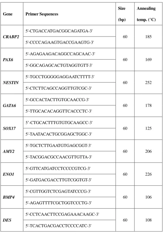
Total RNA was extracted from the cells using TRIzol Reagent (Invitrogen, USA) according to the manufacturer’s instructions. Complementary DNA (cDNA) was synthesized using the High Capacity RNA-to-cDNA Kit. cDNA was amplified with 2× PCR master mix solution (iNtRON, Korea) and 2 pmol of primers, as shown in Tables 2 and 3. PCR reactions were performed in a thermocycler under the following conditions: 94°C for 5 min, 35 cycles of denaturation at 95°C for 30 s, annealing for 30 s (where the annealing temperatures depended on each primer set), extension at 72°C for 30 s, and a final extension at 72°C for 7 min. Amplified PCR products were visualized using electrophoresis on 1% agarose gel stained with ethidium bromide.
37
Table 2. Primer sets used to detect differentiation markers.
Gene Primer Sequences
Size (bp) Annealing temp. (°C) CRABP2 5'-CTGACCATGACGGCAGATGA-3' 60 185 5'-CCCCAGAAGTGACCGAAGTG-3' PAX6 5'-AGAGAAGACAGGCCAGCAAC-3' 60 169 5'-GGCAGAGCACTGTAGGTGTT-3' NESTIN 5'-TGCCTGGGGGAGGAATCTTTT-3' 60 252 5'-CTCTTCAGCCAGGTTGTCGC-3' GATA6 5'-GCCACTACTTGTGCAACCG-3' 60 178 5'-TTGCACACAGGTTCACCCTC-3' SOX17 5’-CTGCACTTTGTGTGCAAGCC-3' 60 125 5'-TAATACACTGCGGAGCTGGC-3' AMY2 5'-TGCTCTTGAATGTGAGCGGT-3' 60 206 5'-TACGGACGCCAACGTTGTTA-3' ENO1 5'-GTTCATGATCCTCCCCGTCG-3' 60 226 5'-GATGACGACCTTGTCGGTGT-3' BMP4 5'-CGTTGGTCTCGAGTATCCCG-3' 60 106 5'-AGAGTTTTCGCTGGTCCCTG-3' DES 5'-CCTCAACTTCCGAGAAACAAGC-3' 60 108 5'-TCACTGACGACCTCCCCATC-3'
38
ACTB
5'-GTGGACATCAGGAAGGACCTCTA-3'
60 137
39
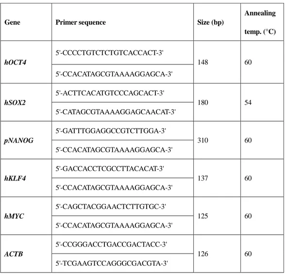
Table 3. Primers sets used for the detection of transgene insertion in cDNA.
Gene Primer sequence Size (bp)
Annealing temp. (°C) hOCT4 5'-CCCCTGTCTCTGTCACCACT-3' 148 60 5'-CCACATAGCGTAAAAGGAGCA-3' hSOX2 5'-ACTTCACATGTCCCAGCACT-3' 180 54 5'-CATAGCGTAAAAGGAGCAACAT-3' pNANOG 5'-GATTTGGAGGCCGTCTTGGA-3' 310 60 5'-CCACATAGCGTAAAAGGAGCA-3' hKLF4 5'-GACCACCTCGCCTTACACAT-3' 137 60 5'-CCACATAGCGTAAAAGGAGCA-3' hMYC 5'-CAGCTACGGAACTCTTGTGC-3' 125 60 5'-CCACATAGCGTAAAAGGAGCA-3' ACTB 5'-CCGGGACCTGACCGACTACC-3' 126 60 5'-TCGAAGTCCAGGGCGACGTA-3'
40
8. Quantitative real-time polymerase chain reaction (qPCR)
To verify the gene expression levels in pESCs, I performed qPCR. Total RNA from individual samples was extracted using TRIzol Reagent (Invitrogen) according to the manufacturer’s instructions. cDNA was synthesized using the High-capacity RNA-to-cDNA Kit (Applied Biosystems, CA, USA) according to the manufacturer’s instructions, and a final volume of 20 µL of cDNA was produced. Extracted cDNA samples were amplified with the DyNAmo HS SYBR Green qPCR Kit (Thermo Scientific, MA, USA) containing 1 pmol of each primer set listed in Table 4 in a 10-µL reaction volume. Amplification and detection were conducted using the ABI 7300 Real-Time PCR System (Applied Biosystems) under the following conditions: one cycle of denaturation at 50°C for 2 min and 95°C for 10 min, 40 cycles of denaturation at 95°C for 15 s, and annealing/extension for 1 min (where the annealing/extension temperatures depended on each primer set). The relative expression level was calculated by normalizing the threshold cycle (Ct) values of each gene to that of ACTB using the Δ–Ct method (Livak and Schmittgen 2001).
41
Table 4. Primers sets used for qPCR.
Gene Primer sequence
Size (bp) Annealing temp. (°C) OCT4a 5'-CTTGGAGAGCCCTGGTTTTACT-3' 159 64 5'-GCCAGGTCCGAGGATCAAC-3' REX1 5'-TCTGAACCCCTCGTGGAAGA-3' 100 60 5'-AGCTTGCTGTAAGCACCTGT-3' FUT4 5'-AGACCGGGCCAATTATGAGC-3' 113 60 5'-CTGGGTTTCGGTCGAGGAAT-3' E-cadherin 5'-ATTCTGGGAGGCATCCTTGC-3' 117 64 5'-GTTGTCCCGGGTGTCATCTT-3' DAX1 5'-GGTACCAGGCCAGATTTGCT-3' 183 60 5'-CAGCTCCTGTACTTGGGTGG-3' SALL4 5'-TACCAGAGCCGAAGTCCAGA-3' 143 60 5'-ATCTCAGTGCGGCTGTTCTC-3' LIN28 5'-AAACGCAGATCCAAAGGAGA-3' 242 60 5'-GCTCAATTCTGAGCCTCTGG-3' KLF2 5'-TTCGGTATCTTTGACGACCCG-3' 122 64 5'-GGCTTGGCCTCTAGTAGCTC-3' ACTB 5'-GTGGACATCAGGAAGGACCTCTA-3' 131 64 5'-ATGATCTTGATCTTCATGGTGCT-3'
42
9. Statical anlaysis
The gene expression data from the qPCR analysis data were statistically analyzed using GraphPad Prism 6 statistical software (GraphPad Software, CA, USA). All Statistical differences in the datasets (except those presented in Fig. 2E) were determined using one-way analyses of variance (ANOVAs) followed by Fisher’s least significant difference test. The statistical differences in the data presented in Figure 2E were investigated using ANOVA followed by the Bonferroni post-hoc test. Differences were considered significant when p < 0.05.
43
44
Characterization of pESCs
The pESC cell line used in this study was generated from porcine preimplantation blastocysts using in vitro fertilization in a manner reported previously (Lee et al. 2007). The established pESC cell line was flat, similar to previous reports of hESCs and mouse EpiSCs (Fig. 1A) (Park et al. 2013). In addition, they possessed AP activity (Fig. 1B), were stably maintained in their pluripotent state over > 40 passages in 1 year. The cells were analyzed for pluripotent marker expression and their capacity for in vitro differentiation according to standards reported previously (Park et al. 2013). Expression of transcription factors related to pluripotency such as Oct4, Sox2, and Nanog was examining using immunocytochemistry (Fig. 1C). The cells were detached from feeder cells and cultured in suspension to verify their capacity for differentiation. The pESCs cultured in suspension formed EBs (Fig. 1D, left panel). The generated EBs spontaneously differentiated into the three germ layers upon placement on gelatin-coated plates (Fig. 1D, right panel). In the differentiated cells, the expression of differentiation marker genes in the three germ layers was detected by RT-PCR and immunostaining (ectoderm: PAX6, NESTIN, CRABP2, Neurofilament; mesoderm:
ENO1, BMP4, DES, Vimentin; endoderm: SOX17, GATA4, AMY2, Cytokeratin17).
Thus, we confirmed that the established cell line was pluripotent with the differentiation potential to generate the three germ layers (Fig. 1E-F).
We performed an experiment to observe the changes that occurred when reprogramming factors were overexpressed in the pESCs. First, DOX-inducible vectors carrying Oct4, Sox2, Nanog, Klf4, or Myc, known as reprogramming factors, were
45
transduced into pESCs. The cells were stabilized, and then cultured in medium supplemented with LIF and DOX or bFGF and DOX. Some of the single-gene overexpression groups were used for gene expression analysis and others were maintained for morphological observations. Based on the results of the gene expression analysis, combinations of two reprogramming factors were introduced into the pESCs and the culture medium was replaced with LIF- and DOX-supplemented medium. The two-gene transduced pESCs were cultured in exchanged medium for 2, 4, or 6 days, and then sampled for analysis of endogenous gene expression patterns and pESC morphology (Fig. 1G).
47
Figure 1. Characterization of pESCs.
(A) Colony morphology of pESCs. (B) pESCs stained for alkaline phosphatase activity. (C) Oct4, Sox2, and Nanog expressed via immunostaining. (D) Embryoid bodies (EBs) formed by pESCs cultured in suspension. The EBs differentiated into three germ layers on gelatin-coated plates (left panel: EBs, right panel: differentiated EBs). (E) Reverse transcription PCR analysis for marker genes of the three germ layer expressed in the differentiated EBs (M: size marker; ectoderm markers: Pax6, Nestin,
Crabp2; mesoderm markers: Eno1, Bmp4, Des; endoderm markers: Sox17, Gata4, Amy2). Immunostaining of Cytokeratin17 (endoderm), Vimentin (mesoderm), and Neurofilament (ectoderm) expressed in differentiated EBs. (G) Schematic overview of
48
Observation of pESCs with single-factor overexpression
A single reprogramming factor was introduced into pESCs using the tetracycline -inducible system (Fig. 2D). In the Tet-Off state, the pESCs with single-factor overexpression formed flat colonies and exhibited AP activity, similar to typical pESCs. However, when DOX was added to the medium to activate the transgene, the pESCs lost their typical characteristics. The cell mass of the pESCs decreased gradually, the boundary of the colony became obscured, and AP activity was lost (Fig. 2B). As the subculture progressed, the transgene-activated experimental groups became differentiated (Fig. 2C). To analyze these results based on endogenous gene expression, we performed qPCR (Fig. 3A–H). Prior to this experiment, we found that lentiviral infection had no effect on gene expression (Fig. 2E). The pESCs with single-factor overexpression were cultured for 3 days on LIF- and DOX-supplemented medium, because long-term culture was not possible. We selected gene candidates for qPCR based on a reprogramming process (early phase: E-cadherin; intermediate phase: Lin28 and Sall4; late phase: Rex1, Fut4, Klf2 and Dax1) and a representative pluripotent marker (Oct4) (Buganim et al. 2012; Golipour et al. 2012; Hansson et al. 2012; Polo et al. 2012). Analysis of the qPCR results revealed that most ectopic factors did not downregulate endogenous genes in the pESCs. However, the experimental group with
Oct4 and Sox2 exhibited significant upregulation of several endogenous genes in
pESCs. The Oct4 overexpression group induced upregulation in endo-Oct4, Fut4, and
50
Figure 2. Reprogramming factor-transduced pESCs morphology and
alkaline phosphatase activity.
(A) Without doxycycline (DOX), reprogramming factor-transduced pESCs showed colony morphologies and alkaline phosphatase activity. (B) When cultured with medium containing leukemia inhibitory factor (LIF) + DOX or bFGF + DOX, colony morphologies and alkaline phosphatase activity of reprogramming factor -transduced pESCs. (C) After subculturing treated with LIF + DOX or bFGF + DOX, colony morphologies and alkaline phosphatase activity of reprogramming factor -transduced pESCs. (D) The integration of reprogramming factors into the genome was confirmed. (hOCT4-pESCs: hOct4 transduced pESCs; hSOX2-pESCs: hSox2 transduced pESCs; pNANOG-pESCs: pNanog transduced pESCs; hKLF4-pESCs: hKlf4 transduced pESCs; hMYC-pESCs: hMyc transduced pESCs) (E) To test whether lentiviral infection affects gene expression, we performed quantitative PCR analysis of M2rtTA infected pig embryonic stem cells. (scale bar = 200 µ m).
52
Figure 3. Analysis of pluripotent-related gene expression using quantitative
reverse transcription (qRT)-PCR.
Three days after treatment with LIF and DOX, the expression of endogenous pluripotent-related genes, including (A) Oct4, (B) Rex1, (C) Fut4, (D) Klf2, (E) Dax1, (F) E-cadherin, (G) Lin28, and (H) Sall4, was analyzed by qRT-PCR in transduced pESCs.
53
Characteristics of pESCs with two overexpressed factors
Based on the analysis of overexpressed pESCs, we transduced two factors into the pESCs. DOX-inducible lentiviral vectors with a combination of Oct4 and Sox2, Oct4 and Nanog, Oct4 and Klf4, or Sox2 and Nanog were introduced into pESCs. Typical colony morphology and AP activity of pESCs were observed in the Tet -Off state (Fig. 4A). However, when the transgene was activated using LIF- and DOX-supplemented media, the pESCs with two overexpressed factors showed a different appearance from the control group, including decreased cell mass and AP activity (Fig. 4B). Interestingly, when one reprogramming factor was introduced, AP activity was lost completely, but when two reprogramming factors were introduced, weak AP activity remained. In addition, several of the groups transduced with two reprogramming factors could be maintained after subculturing, with transgene activation. Although long-term culture failed, pESCs with Oct4 and Nanog, Oct4 and Klf4, or Sox2 and Nanog could be subcultured even under transgene activation conditions. We were able to observe the colonies for about two weeks. However, the remaining groups were differentiated after subcultivation in transgene-activated states (Fig. 4C). Based on these results, we observed changes in endogenous genes related to pluripotency when two reprogramming factors were overexpressed in pESCs. Transgenes were activated and qPCR was performed in samples collected after culturing for 2, 4, and 6 d ays. In total, four groups, including the three experimental groups that could be subcultivated in the transgene activation state and a group with Oct4 and Sox2, were observed. The analysis of the qPCR results showed that E-cadherin, an early-phase reprogramming marker, was upregulated on day 2, and then decreased slightly on day 4 and maintained
54
a similar level on day 6. Conversely, the intermediate-phase reprogramming markers Lin28 and Sall4 increased more than ten-fold on day 2, and 40-fold in the Sox2 and Nanog transduction group. However, on day 4, they tended to decrease sharply and showed little expression on day 6. The late-phase reprogramming markers Fut4, Rex1, and Klf2 were not expressed throughout the experiment. Meanwhile, Dax1 was upregulated on day 2, but showed little expression on days 4 and 6 (Fig. 5A –H).
56
Figure 4. Morphology and alkaline phosphatase activity of
two-reprogramming-factor-transduced pESCs.
(A) Without doxycycline, two-reprogramming-factor-transduced pESCs showed typical colony morphologies. (B) When cultured with medium containing LIF and DOX, colony morphologies and alkaline phosphatase activity of transduced pESCs. (C) Colony morphologies of two-reprogramming-factor-transduced pESCs over time after subculturing. (scale bar = 200 µ m).
58
Figure 5. Quantitative reverse transcription PCR analysis of
pluripotent-related genes in two-factor-transduced pESCs.
To verify the effects of ectopic gene expression on endogenous genes, expression of endogenous genes representing pluripotency and the reprogramming phase [pluripotency markers: Oct4 (A); early phase marker: E-cadherin (B); intermediate phase markers: Lin28 (C) and Sall4 (D); late phase markers: Fut4 (E), Rex1 (F), Klf2 (G), and Dax1 (H)] were determined by quantitative PCR. Each group was harvested at days 2, 4, and 6 after treatment with DOX- and LIF-supplemented medium. All data were normalized to the control groups.
59
60
The experimental groups overexpressing one reprogramming factor in pESCs may have differentiated because of an imbalance in their pluripotency network. Pluripotency networks are balanced by a number of genes with complex mechanisms (e.g., antagonism, synergistic action, and additive interaction). (Niwa, Miyazaki, and Smith 2000) reported that Oct-3/4 expression should be maintained at an appropriate level for mESCs to remain undifferentiated. In this study, a two-fold increase in the expression of Oct-3/4 in mESCs led to differentiation into primitive endoderm and mesoderm. Conversely, after (Niwa, Miyazaki, and Smith 2000) lowered the expression Oct-3/4 level, the cells differentiated into TE. In another study, Nanog was overexpressed in EpiSCs and hESCs. The results of this study suggested that hESCs with overexpressed Nanog failed to differentiate into ectoderm and definitive endoderm. Based on the results of this experiment and previous studies (Niwa, Miyazaki, and Smith 2000), if the precise expression levels of reprogramming factors in pESCs are not maintained, the pluripotent balance can be broken. Meanwhile, EpiSCs transduced with Klf4 resulted in the successful formation of germline-competent chimeras and naïve marker expression (Guo et al. 2009). In addition, Klf2 was introduced into EpiSCs instead of Klf4 and cultured with 2i and LIF to support the conversion into a naïve-like state (Hall, Guo, et al. 2009). Together, these studies show that even in the same primed pluripotent state, cells differ in their pluripotent networks depending on species and strains.
Although pESCs with one overexpressed reprogramming factor were not maintained in this experiment, the results showed that Oct4 and Sox2 acted as triggers for the transient high expression of endogenous Oct4, Fut4, and E-cadherin in pESCs. In many
61
studies, Oct4 has an important role in the maintenance of pluripotency and reprogramming of differentiated cells (Vallier et al. 2009; Zafarana et al. 2009; Li et al. 2010). The results from Oct4-centered protein interaction analysis and genome-wide identification of the Oct4 target gene indicate that Oct4 co-binds with pluripotency regulators. These results demonstrate that Oct4 has an important role in maintaining the undifferentiated state of ESC (Pardo et al. 2010; van den Berg et al. 2010). Sox2 is another essential element in maintaining the pluripotency of ESCs, as verified by Sox2 null ESCs that differentiated into TE-like cells because Sox2 is related in the maintenance of orphan nuclear receptors such as Nr5α2. Nr5α2 is associated with the activity of Oct4 combined with the Oct4 proximal promoter (Masui et al. 2007). In addition, endogenous Sox2 expression is an event necessary to reprogram somatic cells into iPSCs. Endogenous Sox2 expression induces the reprogramming of the next event to occur and the expression of genes related to pluripotency such as Lin28, Esrrb, and Sall4 (Buganim et al. 2012). These results and previous reports suggest that Oct4 and Sox2 are key transcription factors in pluripotency networks and that they interact with a variety of genes related to pluripotency.
Based on the experimental results, all of the two-factor transduction pESC treatment groups failed long-term culturing. The qPCR analysis offered several reasons for this failure. Self-renewal by LIF signaling is a characteristic of cells with naïve pluripotency. Therefore, to cultivate primed pESCs with primed pluripotency in LIF -supplemented media, reprogramming is required. However, in the pESCs transduced with two reprogramming factors, the expression of genes expressed at the intermediate phase of reprogramming was not maintained. E-cadherin, which was expressed early
62
in reprogramming, showed an mRNA expression level increased by six-fold or more on day 2, which decreased slightly on day 4 and was maintained until day 6. However,
Lin28 and Sall4, which are expressed in the intermediate phase of reprogramming,
were expressed only on day 2, and then decreased rapidly and were not expressed on day 6. Therefore, the naïve marker genes Rex1, Fut4, and Klf2 were not expressed after day 2. These results demonstrate that reprogramming factor transduction in pESCs failed to induce reprogramming.
Like pESCs, the pluripotency states of hESCs and EpiSCs with primed pluripotency have been successfully converted by introducing two reprogramming factors (Silva et al. 2009; Hanna et al. 2010; Yang et al. 2010). However, the reprogramming conditions of mouse and humans cells do not apply to pigs, because the molecular mechanisms during embryo development differ. Pigs require a longer period of time before embryos are implanted than mouse and human (Alberio and Perez 2012). Therefore, the signaling that regulates the pluripotency of pig ICM differs from that of mouse embryo. Unlike mouse, pigs express Oct4 and Cdx2 in ICM and TE. Conversely, Sox2 is expressed from the beginning of embryo cavitation, and when it is divided into TE/ICM, it appears specifically only in the embryonic portion and gradually disappears with the progression of differentiation. ICM cells and epiblasts of pig blastocysts do not have LIF receptors, whereas FGF receptors are highly expressed. Therefore, FGF signaling has a critical role in maintaining pluripotency in pigs than LIF signaling (Hall, Christensen, et al. 2009). In addition, strong expression of BMPr and SMAD in porcine ICM cells and EpiSCs was detected by immunostaining and BMP signaling inhibitor experiments in vitro. When pig ICM cells and EpiSCs cells were treated with BMP
63
inhibitors in vitro, the survival rate of the cells decreased (Hall and Hyttel 2014). BMP signaling also has an important role in maintaining pig pluripotency. Recently, (Zhang et al. 2015) succeeded in generating ‘intermediate’ pig pluripotent stem cells with both naïve and primed characteristics even when the transgene was removed using three growth factors (LIF, FGF2, and BMP4) and the inhibitor 2i. These results show that pig pluripotent stem cells maintain pluripotency by unique cell signaling that differs from those of mouse and human.
There are cases of the successful establishment of pig naïve -like pluripotent stem cells by introducing two reprogramming factors. Roberts et al. (2011) transduced Oct4 and Klf4 into the ICM of porcine blastocysts and isolated pESCs. These pESCs were self-renewed by LIF, and their ability to differentiate into the three germ layers was verified using the teratoma formation assay (Telugu et al. 2011). This suggests that transducing reprogramming factors to establish porcine naïve-like pluripotency cells via reprogramming factor transduction methods can be effectively used to gain a pluripotent cell source with low epigenetic memory. Overall, these results indicate that pig-specific culture systems or reprogramming methods are required to obtain pig naïve pluripotent stem cells.
These results indicate that pig-specific strategies are required to obtain pig naïve pluripotent stem cells. Present experiment was the first attempt to convert pluripotency state into naïve state by transducing ectopic factor into established pESCs. In this experiment, we were also able to easily understand how the introduction of exogenous factors, commonly used reprogram in other primed pluripotent stem cells, affects the
64
regulation of endogenous pluripotent related genes in pESCs. In the experiment, human factor was used to distinguish between endogenous gens and ectopic factors. I wonder that results would have been obtained if pig originated factors were introduced into pESCs. Later, I would like to study the gene network analysis of pESCs by introducing pig derived factors. Especially in a recent report, Sox2 has been reported as a true porcine pluripotency marker (Liu et al. 2015), and it is expected that some changes will occur when pig originated Sox2 is introduced into pESCs. In addition to the expressed gens, there were notable differences in naïve and primed pluripotent cells (Table 1). It was also possible to attempt to perform a pluripotency state conversion using these characteristics. In particular, the naïve and primed pluripotent states differ in the intracellular signaling, which maintains pluripotency. Using these characteristics, we could try switching the pluripotency state by screening the signaling mechanisms that make up pESCs and mESCs, artificially using chemical to induce pESCs signaling similar to mESCs. By accessing the pESCs pluripotent network in these various ways, we could better understand the functions and systems of each factor of the network. Ultimately, we could establish bona fide pluripotent pESCs.
65
66
In this study, all experimental groups in which one or two reprogramming factors were transduced into pESCs failed to show long-term cultivation in LIF-supplemented medium. Based on qPCR analysis performed to determine the reasons for this failure, the expression of both Lin28 and Sall4 genes was not maintained. Since these two genes are markers expressed at the intermediate stage of reprogramming, it can be presumed that the expression of both genes was not maintained because the intermediate stage of reprogramming was not completed. Based on these results, additional strategies are required to complete each reprogramming phase to succeed in converting pESCs into a pluripotent state. Overall, to support the conversion of pESCs into a pluripotent state, a greater understanding of the process is required to identify the unique pluripotency network in pig and to determine whether bona fide pig pluripotent stem cells can be established.
67