의학
의학
의학
의학 석사학위
석사학위
석사학위
석사학위 논문
논문
논문
논문
Expression Profiles of Molecules Involved
in Antigen Presentation
in Gangliosides-stimulated Primary
Astrocytes from Rat brain
아
아
아
아 주
주
주
주 대
대
대
대 학
학
학
학 교
교 대
교
교
대
대
대 학
학
학
학 원
원
원
원
의
의
의
의 학
학
학 과
학
과
과
과
윤
윤
윤
윤 희
희
희 정
희
정
정
정
Expression Profiles of Molecules Involved
in Antigen Presentation
in Gangliosides-stimulated Primary Astrocytes
from Rat brain
by
HeeJung Yoon
A Dissertation Submitted to The Graduate School of Ajou University
in Partial Fulfillment of the Requirements for the Degree of
MASTER OF MEDICAL SCIENCES
Supervised by
Ilo Jou, M.D., Ph.D.
Department of Medical Sciences
The Graduate School, Ajou University
윤희정의
윤희정의
윤희정의
윤희정의 의학
의학
의학
의학 석사학위
석사학위
석사학위
석사학위 논문을
논문을
논문을
논문을 인준함
인준함
인준함....
인준함
심사위원장
심사위원장
심사위원장
심사위원장
주
주
주
주
일
일
일
일
로
로
로
로
인
인
인
인
심
심
심
심 사
사
사
사 위
위
위 원
위
원
원
원
조
조
조
조
은
은
은
은
혜
혜
혜
혜
인
인
인
인
심
심
심
심 사
사
사 위
사
위
위 원
위
원
원
원
박
박
박
박
선
선
선
선
인
인
인
인
아
아
아
아 주
주
주
주 대
대
대
대 학
학
학
학 교
교 대
교
교
대
대
대 학
학
학
학 원
원
원
원
2004년
년
년
년 12월
월
월
월 22일
일
일
일
- ABSTRACT -
Expression Profiles of Molecules Involved in Antigen Presentation
in Gangliosides-stimulated Primary Astrocytes from Rat brain
Astrocytes can function as antigen-presenting cells (APC) by expressing class II major histocompatibility complex (MHC) antigens and co-stimulatory molecules upon activation. In vitro, astrocytes are activated by lipopolysaccharides (LPS), and interferon-γ (IFN-γ). We previously reported that gangliosides, membrane components of brain cells, especially neurons, can activate brain glial cells in vitro, thus could play roles in brain physiology and pathology. In this study, I tested whether gangliosides could induce the expression of immune molecules related to antigen presentation, using cultured rat brain glial cells, astrocytes and microglia. I treated cells with 50 µg/ml gangliosides mixture (Gmix) for 6 h to 3 d, and assayed the mRNA expression of class II transactivator (CIITA) and class II major histocompatibility (MHCII) molecules, using RT-PCR and real-time PCR. Gangliosides induce CIITA mRNA expression, followed by MHC II in astrocytes, but not in microglia. Critical for the development of mature APC capability is surface expression of class II MHC molecules and co-stimulatory molecules expression. Cell surface molecules of central importance to costimulation are CD40, ICAM-1, CD80
were constitutive and that of CD86 was further induced by treatment of Gmix. Those expression patterns are the same in IFN-γ-treated cells. Next, I tested the surface expression of MHC II by FACS and immunocytochemistry analysis. Gmix treatment failed to induce it in both FACS and immunocytochemistry analysis, in contrast to IFN-γ treatment. This imply that Gmix could induce the mRNA expressions of CIITA and MHCII, but they could not induce surface expression of MHCII. To clarify the mechanisms, I assayed the molecules involved in it, including invariant
chain (Ii) and thenon-classical class II molecule RT1-DMs. Treatment of Gmix could
not induce the mRNA expression of Ii, while IFN-γ could. Still further studies are needed, I presumably suggest that Gmix are defective in fully presenting antigens due to a lack of expressing Ii molecules.
Key words: Ganglioside, Astrocyte, antigen-presentig cell, MHCII, CIITA, co-stimulatory molecules.
TABLE OF CONTENTS
ABSTRACT ---ⅰⅰⅰⅰ TABLE OF CONTENTS ---ⅲⅲⅲⅲ LIST OF FIGURES ---ⅴⅴⅴ ⅴ LIST OF TABLE --- ⅵⅵⅵⅵ LIST OF ABBREVIATION --- ⅶⅶⅶⅶ Ⅰ ⅠⅠ Ⅰ. INTRODUCTION --- 1 Ⅱ Ⅱ Ⅱ Ⅱ. MATERIAL AND METHOD --- 5A. Reagents --- 5
B.Cell cultures --- 5
C.Flow cytometry (FACS) --- 6
D. RNA isolation and Reverse Transcription Polymerase Chain Reaction (RT-PCR) --- 6
E. SYBR Green Real-time PCR--- 8
F. Immunocytochemistry--- 9
Ⅲ ⅢⅢ Ⅲ. RESULTS--- 11
A. Gmix induces the mRNA of CIITA and MHCII in primary rat astrocytes but not in primary rat microglia. --- 11
B. Gmix failed to induce MHCII expression at cell surface. --- 16 C. B7-2 transcript was induced, while those of B7-1, CD40
induced by Gmix. --- 19
D. Individual components of Gmix have the same effect on the transcript and surface expression of
antigen-presenting molecules. --- 22 E. Gmix failed to induce Ii transcript in astrocytes. --- 25
Ⅳ ⅣⅣ Ⅳ. DISCUSSION --- 28 Ⅴ ⅤⅤ Ⅴ. CONCLUSION --- 33 BIBLIOGRAPHY--- 34 국문요약 국문요약국문요약 국문요약 --- 41
LIST OF FIGURES
Fig. 1. mRNA expression of CIITA and MHCII in rat glial cells.--- 12
Fig. 2. Expression of MHCII on astrocytes surface. ---16
Fig. 3. Expression of adhesion and co-stimulatory molecules in
Gmix-treated astrocytes. --- 21
Fig. 4. mRNA expression of adhesion and co-stimulatory molecules
in gangliosides components treated astrocytes.--- 22
LIST OF TABLE
LIST OF ABBREVIATION
APC, antigen presenting cell
MHCII, class II major histocompatibility complex LPS, lipopolysaccharides
IFN-γ, interferon-gamma Gmix, ganglioside mixture CIITA, class II transactivator FACS, flow cytometry
Ii, invariant chain
GAPDH, glyceraldehydes-3-phosphate dehydrogenase ICAM-1, intercellular adhesion molecule-1
Ⅰ
Ⅰ
Ⅰ
Ⅰ. INTRODUCTION
Following injury in the CNS, two main glial populations, astrocytes and microglia, show an immediate response in the form of proliferation cytokine production and expression of immunocompetent molecules (Mucke and Eddleston,
1993; Kreutzberg, 1996). One marks of the immunocompetence of astrocytes and
microglia is their capacity to modulate class II major histocompatibility (MHCII) molecules expression under disease conditions in vivo as observed in multiple sclerosis (Lee and Moore, 1990), experimental allergic encephalitis, Toxoplasma encephalitis (Deckert-Schlüter and Schlüter, 1994), viral CNS infection (Sedgwick and Schwender, 1991) or after intrathecal application of interferon gamma (IFN-γ).
Processing of antigen by APC for presentation to CD4+ T cells occursthrough
the endocytic pathway, which requires the coordinate regulationof molecules that
participate in class II biosynthesis and maturation (Wolf and Ploegh, 1995). The class
II transactivator (CIITA) is the key intermediateresponsible for constitutive and
IFN-γ-inducible expression ofclass II (Mach and Steimle, 1996). CIITA also activates other genes involved in antigen presentation, such as the invariant chain (Ii) and HLA-DM (HLA-DM in the human, RT1-DM in the rat, H-2M in the mice) genes. MHCII binds Ii in the endoplasmic reticulum (ER). MHCII-Ii complex move out of the ER, through the Golgi apparatus. And these complexes go into lysosome-like antigen processing compartments (Demotz and Grey, 1990). Ii is degraded by
proteases and dissociates from MHCII (Hiltbold and Roche, 2002). CLIP, derived from Ii, remains associated with the peptide-binding groove of the MHCII (Blum and Cresswell, 1988). HLA-DM catalyzes the exchange of the MHCII-associated CLIP fragment with high affinity antigenic peptides that are generated by proteolysis of internalizes foreign antigen. The antigenic peptide binds MHCII and express on cell surface (Jensen and Weber, 1999; Busch and Doebele, 2000).
MHCII, expressed on the surface of APC, binds TCR on T cells. TCR-MHCII interactions trigger multiple signaling pathways. The TCR-MHCII complex enhances the expression of CD40 on the APC. CD40 binds CD40 ligand (CD40L) on T cell (Grakoui and Bromley, 1999; Monks and Freiberg, 1998). The interaction of CD40 and CD40L induced expression B7 family (B7-1, B7-2) on the cell surface. The B7 family interact CD28 on the surface of the T cell. B7-CD28 complex makes signal to finish the antigen presentation process (Lanzavecchia, 1997; Lenschow and Walunas, 1996).
Gangliosides are sialic acid-containing glycosphingolipids that are constituents
of mammalian cell membranes. Gangliosidesare particularly abundant in neuronal
cell membranes and participate in various cellular events of the nervous system
(Rodden and Wiegandt, 1991).The major types of gangliosides in the brain are GM1,
GD1a, GD1b,GT1b, and GQ1b, which differ in their profiles of sialic acidresidues
and carbohydrate moieties. Several lines of evidencepoint to the importance of the
brain-derived gangliosides in immune responses and the pathogenesis of brain
damaged neuronal cells into the extracellular space, which may lead to pathophysiological conditions (Blennow and Davidsson,, 1991; Gisslen and Hagberg,
1997; Michikawa and Gong, 2001). Despite the evidence of a role for gangliosidesin
brain pathology, there appears to be little known about howgangliosidesact.
On the other hand, gangliosides are potent inhibitor of cell function (Ladisch and Becker, 1992; McKallip and Ladisch, 1999). Gangliosides profoundly and
specifically suppress IFN-γ-inducible expression of both MHC class I and II on
astrocytes (Massa, 1993). Also, gangliosides have an effect on the expression of CD80 by mature APC (Caldwell and Heitger, 2003). In antitumor immunity, a reduction or absence of CD80 in activated-APC has been associated with increased tumorigenicity. Reduced expression of CD80 by gangliosides treated mature APC may be linked to the reduced expression of CD40. The reduced expression CD40 and CD80 caused by gangliosides treatment may cause inhibition of T cell proliferation.
Despite the immune suppressive effects of gangliosides, I tested whether gangliosides could induce the expression of immune molecules related to antigen presentation, using cultured rat brain glial cells, astrocytes and microglia. I show that gangliosides induce class II expression in the astrocytes but not in microglia. I tested the surface expression of MHC II by FACS analysis and immunocytochemistry. Gmix treatment failed to induce it in contrast to IFN-γ treatment. These imply that Gmix could induce the mRNA expressions of CIITA and MHCII, but they could not induce surface expression of MHCII. I have examined the mRNA expression patterns
gangliosides-treated astrocytes are the same in IFN-γ-treated. But Gmix could not induce the mRNA expression of Ii, while IFN-γ could. These results suggest that Gmix are defective in fully presenting antigens due to a lack of expressing Ii molecules.
Ⅱ
Ⅱ
Ⅱ
Ⅱ. MATERIAL AND METHOD
A. Reagents
Lipopolysaccharide (LPS), poly-D-lysine, DABCO were purchased from Sigma
(St. Louis, MO), and IFN-γ was from Calbiochem (Cambridge, MA). Minimal
essential media (MEM) was from Life Technologies (Grand Island, NY). Bovine brain gangliosides mixture (Gmix), GD1a, GT1b and GM1 were purchased from Matreya (Pleasant Gap, PA). RT-PCR primers were from Bioneer (Seoul, Korea).
RNAzolTMB and reverse transcriptase from Avian Myeloblastosis Virus were
purchased from TEL-TEST Inc. (Friendswood, TX) and Promega (Japan). Fluorescein isothiocyanate (FITC) conjugated MHCII was purchased from Ebioscience. Inc. (San Diego, CA)
B. Cell cultures
Primary microglia was cultured from the cerebral cortices of 1-3 day Sprague-Dawley rats as previously described (Pyo, 1998). Briefly, the cortices were triturated into single cells in MEM containing 10% fetal bovine serum (Hyclone,
Logna, UT) and plated into 75cm2 T-flasks (0.5 hemisphere/flask) for 2 weeks. Then,
microglia were detached from the flasks by mild shaking and applied to a nylon
mesh to remove astrocytes and cell clumps. Cells were plated in 6-well plates (5 x10⁵
One hour later, the cells were washed to remove unattached cells before being used in experiments. Primary astrocytes were prepared using trypsin after microglia were
removed. Detached astrocytes were seeded in 6-well plates or 60mm dishes.
C. Flow cytometry (FACS)
Cells were washed in the plates with ice-cold phosphate buffered saline (PBS)/0.1% fetal bovine serum (FBS), then gently removed from the wells using cell scrapers to avoid trypsinization. Cells were centrifuged at 200 ×g for 5 min and the resulting pellet was resuspended in 100 µl staining buffer. Direct staining for B7-1, B7-2 and MHCII was performed with FITC- and PE- conjugated antibodies (PharMingen, Inc., Ebioscience. Inc.) or PBS/0.1%FBS (negative control) for 1 h on ice in the dark. Stained cells were centrifuged at 200 ×g for 5 min and the resulting pellet was resuspended in 500 µl PBS. Analysis was performed using a FACScalibur flow cytometer (Becton Dickinson, San Jose, CA, USA). The analysis was completed using CellQuest software (Becton Dickinson, San Jose, USA). In each run at least 10,000 (PI-excluded) gated events were analyzed.
D. RNA isolation and Reverse Transcription Polymerase Chain Reaction (RT-PCR)
Total RNA was isolated using RNAzolTMB and cDNA was prepared using
the manufacturer’s instructions. PCR was performed with 30 cycles of sequential reactions: 94 ℃ for 30 sec, 55 ℃ for 30 sec, and 72 ℃ for 30 sec. Oligonucleotide primers were purchased from Bioneer (Seoul, Korea). The sequences of PCR primers were shown on table 1. PCR products were separated by
electrophoresis in a 1.5% agarose gel and detected under UV light.Primers used for
RT PCR are different from those used in real-time RT PCR. TABLE 1. Primer sequences for RT-PCR
NAME SEQUENCES
GAPDH F-TCC CTC AAG ATT GTC AGC AA
R-AGA TCC ACA ACG GAT ACA TT
MHCII F-TCT GCC CCA ATT CAT CGT
R-TCA ATC AGG GTC TCG GAA TC
CIITA F-CGA ACC CAG AGG CTG AGA AAC
R-GTA CAA GCT CAG CCT TAG GAG
B7-1 F-GCT CGT AGT ATT TTG GCA GGA CCG
R-GCT GGA GGA TAA TTG ATC CCG TGG
B7-2 F-CTC CAC GGT CAG GTG TTT CAC
R-ACT CCT CGG GTT TCC ACG TCT CAG
ICAM-1 F-CTG GAG AGC ACA AAC AGC AGA G
R-AAG GCC GCA GAG CAA AAG AAG C
CD40 F-GTG TGT TAC GTG CAG TGA CAA
R-ATC CTC ACA GCT TGT CCA
Ii F-TGA AGA ACG TTA CCA AGT ACG G
R-CTC CAC CTA AAG TAC TGA CCA A
RT1-DMa F-AAC ATA GGG CTC TCC GAG
R-ATG AAA CAG ACC AGC GTG
RT1-DMb F-GTC CAA GTA GCC CAA ACC
E. SYBR Green Real-time PCR
The PCR reactions were carried out in 0.2ml MicroAMP® optical tubes
(Applied Biosystems, Foster City, CA) in a 25 ul reaction volume with SYBR Green Master Mix (Applied Biosystems, Foster City, CA) with optimized concentrations of specific primers. An ABI Prism 7000 Sequence Detector (Applied Biosystems, Foster City, CA) was programmed for an initial step of 2 min at 50 °C and 10 min at 95 °C, followed by 40 cycles of 15 s at 95 °C and 1 min at 60 °C. Every assay included duplicate, 4-fold or 10-fold serial dilutions of the calibrator cDNA, test cDNA samples, and controls. Specificity of PCR amplification of each primer pair was confirmed by analyzing PCR products by agarose gel electrophoresis and by melting curve analysis (Ririe and Rasmussen, 1997).
Primer concentrations for each primer set were optimized by checkerboard titration. Briefly, 900, 300 and 50 nM concentrations of each -5′ primer were tested with 900, 300 and 50 nM concentrations of the corresponding -3′ primer, with and without template (18 total reactions for each primer set). This optimization step identified primer concentrations that provided the highest sensitivity and specificity for each target sequence.
Standard curves were generated from calibrator cDNAs made from decreasing amounts of total RNA (10-fold dilutions) to monitor the efficiency of real-time RT-PCR for each assay. Prior to acceptance of data for quantitative work, we required at least four of the five standard curve dilutions in an assay to yield specific product
(based on dissociation curve analysis) and that no product was seen in the no-template control. The quality of standard curves can be judged from their slopes and
correlation coefficients (r). The PCR efficiency (Ex) was determined using the
equation: Ex = (10−1/slope) −1) × 100%. The threshold cycle (Ct) value was defined as
the number of PCR cycles required for the fluorescence signal to exceed the
detection threshold value(Livak, 1997). Fold differences for gene expression were
calculated by the comparative Ct method. This method compares test samples to a
calibrator sample and uses results obtained with a uniformly expressed control gene to correct for differences in the amount of RNA present in the two samples being
compared to generate a ∆Ct value.
The sequences for PCR primers were as followings. (F) 5’-CTA CAA TGA GCT GCG TGT GG-3’ and (R) 5’-CGG TGA GGA TGT TCA TGA GG-3’ for β-Actin; (F) 5’-GTC TGC AGA CA AAC TAC GAG G-3’ and (R) 5’-TAA GCT GTG TGG ACA CGA CC -3’ for MHCII.
F. Immunocytochemistry
Cells were plated on 12-mm round coverslips (Fisher Scientific, Pittsburgh, PA, USA) that were coated with poly-D-lysine. Cells were fixed with ice-cold 100% methanol (Merck & Co., Whitehouse station, NJ, USA). For immunostaining, the fixed cells were washed twice with PBS and then incubated for 30 min with 1% bovine serum albumin (Sigma). The cells were then washed three times with PBS
containing 0.01% triton X-100 (PBST) and then incubated overnight at 4ºC with fluorescein isothiocyanate (FITC)-conjugated MHC antibodies. Each coverslip was rinsed three times with PBST. After washed once with PBS, the cell mounted on slides with mounting solution (DABCO in 70% glycerol), and observed with a confocal laser scanning microscope FV1000 (Olympus, Tokyo).
Ⅲ
Ⅲ
Ⅲ
Ⅲ. RESULTS
A. Gmix induces the mRNA of CIITA and MHCII in primary rat astrocytes but not in primary rat microglia.
Previous studies have shown that MHCII, the major histocompatibility complex class II molecule, are key players in antigen presentation and allogeneic immune responses. MHCII molecules are displayed at surface of APC where they
present peptides to the TCR of CD4+ T cell. And the importance of CIITA in
regulating the transcription of MHCII-associated molecules has been previously shown (Steimle and Otten, 1993). Cell surface expression of MHCII is congruent with expression of CIITA, whereas inactivated APCs lack CIITA expression. Therefore, I tested mRNA expression of CIITA and MHCII were expressed in rat primary glial cells (Fig. 1).
Gangliosides can activate brain glial cells in vitro. But gangliosides have a
character as immunosuppressive factor. IFN-γ-inducible expression of MHC class I
and II molecules was suppressed by Gmix treatment of astrocytes (Caldwell and Heitger, 2003). I confirmed whether expression of MHCII, CIITA, and Ii were
affected by Gmix in IFN-γ treated astrocytes. Rat primary astrocytes were co-treated
or pretreated with 50 μg/ml Gmix for 3 h and stimulated with 10 U/ml IFN-γ for 12
h to 3 d (Fig 1A). In Gmix and IFN-γ co-treated astrocytes, mRNA expression of
whether mRNA expression of CIITA and MHCII was induced in Gmix treated primary rat glial cells. When astrocytes were stimulated with 10 U/ml IFN-γ and 50
μg/ml Gmix for 6 h to 3 d, mRNA expression of CIITA and MHCII increased (Fig.
1B). Primary microglia cells were stimulated with 10 U/ml IFN-γ and 50 μg/ml Gmix for 30 min to 12 h, mRNA expression of CIITA and MHCII increased by
IFN-γ, but not by Gmix (Fig. 1C). To determine the optimal concentration of Gmix
demonstrated the induction of CIITA and MHCII mRNA, I investigated the effect of Gmix to astrocytes in the range of 25 to 75 μg/ml (Fig. 1D) and the expressions of
them are most prominent in 50 µg/ml-treated astrocytes. I confirmed RT-PCR data by
SYBR Green Real time PCR (Fig. 1E). These results show that Gmix induced CIITA mRNA expression followed by MHC II in astrocytes, but not in microglia.
GAPDH
0 6 12 24 48 72 0 6 12 24 48 72 (h)
Gmix 50 μμμμg/ml IFN-γ 10 U/ml
CIITA MHCII
B.
GAPDH MHC II CIITA Gmix 50 μμμμg/ml IFN-γγγγ 10 U/mlC.
0 1 3 6 12 0 1 3 6 12 (h)A.
GAPDH MHCII Ii CIITA Gmix 50 µµµµg/ml-
+ + ++
-
-
+ ++
+
-IFN-γγγγ 10 unit/ml
-
+ + ++ +
-
+ + + + + 0 12 24 48 72 72 0 12 24 48 72 72 (h) Gmix 3hr pre-treatGmix & IFN-γγγγ
co -treat Time Time Time Time 0.5 0.5
E.
M H C II m R N A L e v e l Gmix 50 µµµµg/ml 6 12 24 72Gmix 25μμμμg/ml Gmix 50μμμμg/ml Gmix 75μμμμg/ml
D.
0 48 GAPDH MHC II CIITA (h) 0 0.5 1 1.5 2 2.5 3 3.5 4 0 6 12 24 48 72 6 12 24 72 0 48 6 12 24 72 0 48Fig. 1. mRNA expression of CIITA and MHCII in rat glial cells. Rat primary
astrocyte were co-treated or pretreated with 50μg/ml Gmix for 3h and stimulated with 10unit/ml IFN-γ for 12 h to 3 d (A). Astrocytes (B) and microglia cells (C) were serum-starved for 48 h or 5 h respectively and then stimulated with 50 μ g/ml Gmix and 10 U/ml IFN-γ for the indicated times. Gmix demonstrated the induction of CIITA and MHCII mRNA in the range of 25, 50 and 75 μg/ml (D). Products shown were electrophoresised on a 1.5% agarose gel. The transcription of glyceraldehyde-3-phosphate dehydrogenase (GAPDH) was measured for normalization (E). Confirmation of RT-PCR data by SYBR Green Real-time PCR. The result from Real-time PCR was normalized to the expression of the β-actin. Results representations of three separate experiments are shown.
B. Gmix failed to induce MHCII expression at cell surface.
Mature APC express MHCII on surface. Expression of MHCII on surface can binds TCR on the T cell. TCR-MHCII interactions dictate the antigen specificity of the response and play an essential role in initiating T cell activation. I tested whether synthesized MHCII was expressed on Gmix-treated astrocytes surface. To detect
astrocytes surfaces expression, IFN-γ, LPS, Gmix-stimulated astrocytes were
analyzed by FACS and immunocytochemistry analysis. Astrocytes were stimulated with 50 μg/ml Gmix, 10 U/ml IFN-γ and 100 ng/ml LPS for 3 d (Fig 2A). To
measure expression of MHCII, I used the mAbs I-Ek. Through stimulation with
IFN-γ and LPS, an elevation in MHCII expression could be observed (Fig 2A upper right
panel, middle left panel). But Gmix failed to induce MHCII expression on astrocytes (Fig 2A. middle right panel). I also confirmed these results by using immunocytochemistry (B). In Gmix-treated astrocytes, MHCII was detectable in
intracellular localization (Fig 2B upper right panel). In IFN-γ and LPS-treated
astrocytes, MHCII was detectable at cell surface (Fig 2.B lower panels). These
results suggest that Gmix could not expression of MHCII on surface, unlikely IFN-γ
B.
100 101 102 103 104 FL1-H 0 1 0 0 E v e n ts 100 101 102 103 104 FL1-H 0 1 00 E v e n ts M1 M2 Control Gmix 50 μμμμg/ml IFN-γ10 U/ml LPS 100 ng/ml 100 101 102 103 104 FL1-H 0 1 0 0 E v en ts M1 M2 100 101 102 103 104 FL1-H 0 1 00 E v e n ts M1 M2 100 101 102 103 104 FL1-H 0 1 00 E v e n ts M1 M2Control IFN-γγγγ 10 U/ml LPS 100 ng/ml
Gmix 50 µµµµg/ml overall
A.
Control IFN-γγγγ LPS GmixFig. 2. Expression of MHCII on astrocytes surface. MHCII expression on
astrocytes is determined by FACS (A). Astrocytes were stimulated with 50 μ g/ml Gmix, 10 U/ml IFN-γ and 100 ng/ml LPS for 3d. Black line; control, Yellow line; Gmix treatment, Blue line; LPS treatment, Green line; IFN-γ
treatment. Confirmation of MHCII expression on surface by
immunocytochemistry (B). Rat primary astrocyte cells were stimulated with 50
µg/ml Gmix, 10 U/ml IFN-γ and 100 ng/ml LPS for 1 d. Arrow; expression
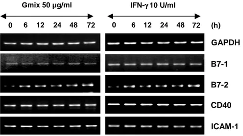
C. B7-2 transcript was induced, while those of B7-1, CD40 and ICAM-1 were constitutively expressed, not being further induced by Gmix.
APC express adhesion and co-stimulatory molecules that bind to receptors on T cells. Adhesion molecules are critical for normal functioning of the immune system in that they regulate the recruitment of immunocompetent cells such as lymphocytes, monocytes and granulocytes, and they mediate antigen presentation (Springer, 1990). Intercellular adhesion molecule-1 (ICAM-1) is such a cell surface glycoprotein belonging to the Immunoglobulin supergene family. ICAM-1 can abrogate leukocyte, monocyte recruitment and aggregation, antigen presentation, T-cell activation and aggregation, and cytotoxic function of T cells and NK cells (Makgoba and Sanders, 1988). The stimulation of a TCR is necessary, but not sufficient, to induce complete T cell activation. Complete T cell activation requires a second signal, the so-called
co-stimulatory signals. Gangliosidesprevented the induction and maintenance of the
expression of CD80 andCD40 on the stimulated APC cell surface and reduced the
production of cytokines (IL-6, IL-12, and TNF-α) interfered with the nuclear binding
of NF-κB, a key regulator of the co-stimulatory regulatory cycle.These results may
diminish antitumorimmune responses (Massa, 1993). Therefore, I have questioned
whether Gmix only affect expression of adhesion and co-stimulatory molecules or not. I treated astrocytes for 6 h to 3 d with 50 µg/ml Gmix and 10 U/ml IFN-γ that induce expression of adhesion/co-stimulatory molecules such as ICAM-1, CD40, CD80 (B7-1) and CD86 (B7-2). Expression of these molecules was evaluated by
astrocytes examined in vitro have also conflicted (Williams and Ulvestad, 1994; Wittwer and Herrmann, 1997; Tan and Gordon, 1998). Some investigators have
reported that IFN-γ-stimulated astrocytes express both B7-1 and B7-2 molecules
(Satoh and Lee, 1995). In contrast, otherinvestigators have observed that astrocytes
do not express B7-1or B7-2 co-stimulatory molecules (Nikcevich and Gordon, 1997).
In my experimental system, IFN- γ induces mRNA expression of B7-2, but no effect on B7-1, CD40 and ICAM-1. Gmix also up-regulates B7-2 expression, whereas Gmix-treated astrocytes show enhanced not mRNA expression of B7-1, CD40,
ICAM-1 molecules. B7-2 was induced by stimultaneous treatment with IFN-γ and
Gmix in astrocytes. Other molecules constitutively expressed. These results show effect of Gmix on the expression of adhesion/co-stimulatory molecules are the same of IFN- γ.
GAPDH
CD40 B7-2 B7-1
Gmix 50 µg/ml IFN-γ γ γ γ 10 U/ml
ICAM-1
0 6 12 24 48 72 0 6 12 24 48 72 (h)
Fig. 3. Expression of adhesion and co-stimulatory molecules in Gmix-treated astrocytes. Rat primary astrocyte cells were stimulated with 50 μg/ml
Gmix and 10 U/ml IFN-γ for 6 h to 72 h. B7-1, B7-2 and CD40; co-stimulatory molecules, ICAM-1; adhesion molecule.
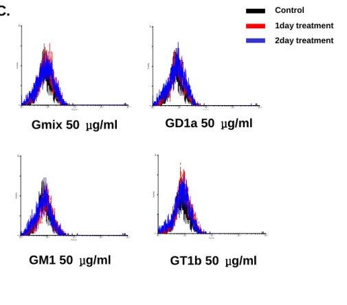
D. Individual components of Gmix have the same effect on the transcript and surface expression of antigen-presenting molecules.
The major types of gangliosides in the brain are GM1, GD1a, GD1b,GT1b,
and GQ1b, which differ in their profiles of sialic acidresidues and carbohydrate
moieties. To investigate possible mechanisms responsible in astrocytes, the effectsof
gangliosides mixture on the expression of adhesion/co-stimulatory molecules
including MHCII, B7-1, B7-2, CD40 and ICAM-1 were examined.. When astrocytes
from rat brain were stimulated with 50 μg/ml GD1a, 50 μg/ml GM1, 50 μg/ml GT1b, for 6 h to 3 d, mRNA expression of CIITA and MHCII increased (Fig. 4A). However, these expression in Gangliosides components treated astrocytes were induced weakly than in Gmix treated. Next, I tested whether gangliosides components could induce the expression of adhesion/co-stimulatory molecules (Fig. 4B). I confirmed that expression of MHCII were not detectable on gangliosides components treated astrocytes surface (Fig. 4C). Astrocytes were stimulated with 50
μg/ml Gmix, 50 μg/ml GD1a, 50 μg/ml GM1, 50 μg/ml GT1b for 1 d or 2 d. GD1a,
GM1, GT1b failed to induce MHCII expression on astrocytes. Those expression patterns are the same in Gmix-treated cells.
A.
B.
06 12 24 48 72 (h) 0 6 12 24 48 72 GD1a 50 µµµµg/ml GM1 50 µµµµg/ml GT1b 50 µµµµg/ml GAPDH MHC II CIITA 0 6 12 24 48 72 GD1a 50 µµµµg/ml GM1 50 µµµµg/ml GT1b 50 µµµµg/ml GAPDH CD40 B7-2 B7-1 ICAM-1 0 6 12 24 48 72 0 6 12 24 48 72 0 6 12 24 48 72 (h)
C.
100 101 102 103 104 FL1-H 0 4 0 E v en ts 100 101 102 103 104 FL1-H 0 4 0 E ve n ts 100 101 102 103 104 FL1-H 0 4 0 E v en ts 100 101 102 103 104 FL1-H 0 4 0 E ve ntsFig. 4.mRNA expression of adhesion and co-stimulatory molecules in gangliosides components treated astrocytes. Astrocytes were stimulated with
50 μg/ml GD1a, 50 μg/ml GM1, 50 μg/ml GT1b for 6 h to 3d. Total RNA was isolated and analyzed for levels of CIITA, MHCII (A), B7-1, B7-2, CD40 and ICAM-1 (B) using an RT-PCR. The transcription of GAPDH was measured for normalization. MHCII expression on astrocytes is determined by FACS. (C) Astrocytes were stimulated with 50 μg/ml Gmix, 50 μg/ml GD1a, 50 μg/ml GM1, 50 μg/ml GT1b for 1 d or 2 d. Black line; control, Red line;
1day treatment, Blue line; 2day treatment.
Gmix 50 μμμμg/ml GD1a 50 μμμμg/ml
GM1 50 μμμμg/ml GT1b 50 μμμμg/ml
Control 1day treatment 2day treatment
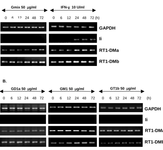
E. Gmix failed to induce Ii transcript in astrocytes.
My data have shown that Gmix-treated astrocytes were induced mRNA expression of MHCII, but could not expression on cell surface (Fig. 1, 2). These findings indicate that the failure to expression MHCII on Gmix-treated astrocytes surface was due to deficient MHCII processing and presentation pathway. Before MHCII molecules can bind antigen-derived peptides for presentation to T cells at the cell surface, the MHCII αβ heterodimers must first be released from their interaction with Ii. This is achieved through stepwise removal of Ii fragments generated by proteolytic cleavage by the cysteine protease cathepsin S (Riese and Wolf, 1996), which leaves a peptide associated with the MHCII molecule known as CLIP (Denzin and Cresswell, 1995). The final removal of CLIP from the class II αβ complex is
facilitated by HLA-DM (RT1.DM in the rat)(Kelly and Monaco, 1991), a molecule
encoded within the class II region of the MHC (Williams and Ulvestad, 1994). This mature MHCII molecule is loaded with peptide and ready for export to the cell surface for presentation to T cells in conjunction with appropriate costimulatory molecules. I tested whether Gmix induced mRNA expression of Ii, RT1-DMa and RT1-DMb in primary rat astrocytes using RT-PCR (Fig. 5A). Astrosytes were stimulated with IFN-γ for 6 h to 3 d, mRNA expression of Ii, DMa and DMb increased. Gmix-treated astrocytes were induced mRNA expression of RT1-DMa and RT1-DMb but not expression of Ii. Next, I tested whether mRNA expression of Ii, RT1-DMa and RT1-DMb were induced by gangliosides components (Fig. 5B). In gangliosides components treated astrocytes, expression pattern of Ii and
RT1-DMs mRNA are the same in Gmix treated astrocytes. These findings demonstrate that Gmix could not sufficiently activate the MHCII processing and presentation pathway mechanism.
Fig. 5. mRNA expression of Ii and RT1-DMs in astrocytes. Astrocytes were
stimulated with 50 μg/ml Gmix and 10 U/ml IFN-γ for 6 h to 3 d. mRNA expression of Ii, RT1-DMa and RT1-DMb was induced by Gmix in astrocytes using RT-PCR (Fig. 5A). Astrocytes were stimulated with 50 μg/ml GD1a, GM1, GT1b for 6 h to 3 d (Fig. 5B). Products shown were electrophoresised on a 1.5% agarode gel. The transcription of GAPDH was measured for normalization.
GAPDH
RT1-DMb RT1-DMa Ii
Gmix 50 μμμμg/ml IFN-γγγγ 10 U/ml 0 6 12 24 48 72 0 6 12 24 48 72 (h) GAPDH Ii RT1-DMa RT1-DMb B. GD1a 50 μμμμg/ml GT1b 50 μμμμg/ml 0 6 12 24 48 72 0 6 12 24 48 72 0 6 12 24 48 72 (h) GM1 50 μμμμg/ml A.
Ⅳ
Ⅳ
Ⅳ
Ⅳ. DISCUSSION
The main findings of this study are as follows; (ⅰ) mRNA expression of CIITA and MHCII were induced in Gmix-treated astrocytes, but not in microglia; (ⅱ) Gmix could not induce astrocytes cell surface expression of MHCII; (ⅲ) Gmix induced the mRNA expression of RT1-DMs but not that of Ii. These results demonstrate that the Gmix-treated astrocytes up-regulated mRNA CIITA, MHCII, RT1-DMs but could not mature astrocytes to act as a full APC. MHCII play a critical role in induction of immune responses through presentation of processed antigens to
CD4+ T-helper cells. Although MHCII are normally expressed on professional APC,
such as B cells, macrophages, dendritic cells and thymic epithelium, expression on other cell types, including astrocytes, can be induced and/or regulated by cytokines,
neurotransmitters, and neuropeptides. In this regard, IFN-γ is the most potent inducer
of MHCII expression on astrocytes. Although regulated MHCII expression is required for normal immune responses by APCs, inappropriate MHCII expression has been implicated in several autoimmune diseases, including rheumatoid arthritis, inflammatory bowel disease, and multiple sclerosis (Grusby and Glimcher, 1995). Conversely, the lack of MHCII expression in the hereditary disease bare lymphocyte syndrome leads to patient death from recurring viral and bacterial infections (Mach, 1996).
Gangliosides consist of an oligosaccharide core with an attachedsialic acids
and a ceramide and are found primarily in theouter leaflet of the cell membrane.
Several lines of evidencepoint to the importance of the brain-derived gangliosides in
immuneresponses and the pathogenesis of brain disease. There are reportsthat brain
injury can cause release of gangliosides from damaged neuronal cells into the
extracellular space, which may lead topathophysiological conditions (Massa, 1993;
Caldwell and Heitger, 2003).
In contrast, previously reported that gangliosides shed by tumor cells exert
potent inhibitory effects on cellular immune responses. Many tumors, such as
neuroblastoma, medulloblastoma, and renal cell carcinoma, shed membrane
gangliosides into the microenvironment. These biologically active molecules are
efficiently bound to target cells. In vivo, coinjection of gangliosides with poorly
tumorigenic cells increases their tumorigenicity (McKallip and Ladisch, 1999). I had a question whether Gmix only treated primary rat glial cells function as APC. I investigated whether expression of MHCII, CIITA and adhesion/co-stimulatory molecules in Gmix-treated primary rat glial cells. I observed mRNA expression of MHCII and CIITA were induced in Gmix-treated astrocytes but not in microglia cells. But Gmix-treated astrocytes could not express MHCII on astrocytes surface. These results suggested Gmix induce mRNA expression of MHCII but could not surface expression of MHCII. Also, these results led us to search for MHCII processing and presentation pathway.
molecule ICAM-1. Astrocytic expression of the costimulatory molecules B7 and CD40 is also controversial. Human astrocytes do not express B7-1 or B7-2, either
constitutively or after IFN-γ exposure. In the murine system, conflicting reports have
been obtained regarding B7 expression in astrocytes. Primary murine astrocytes have been shown to express B7-2 constitutively, and expression of B7-1 and B7-2 is
induced and upregulated, respectively, by the cytokine IFN-γ. Some investigators
(Soos and Ashley, 1999)demonstrated in primary murine astrocytes and transformed
murine astrocytic cell lines that B7-2, but not B7-1, was induced upon IFN-γ
stimulation. In contrast, other investigators (Aloisi and Ria, 1998) documented
induction of MHCII expression in primary murine astrocytes upon IFN-γ treatment,
but not that of B7-1 or B7-2. In vivo studies are also conflicting. Some investigators (Issazadeh and Navikas, 1998) documented B7-2 expression on APCs (macrophages, microglia, and astrocytes) that correlated with the clinical signs of EAE, while other investigators (Cross and Ku, 2000) did not detect expression of either B7-1 or B7-2 by astrocytes during acute, remitting, relapsing, or chronic EAE. In vivo studies on MS, CNS tissue indicates that B7-1 and B7-2 are observed on microglia and macrophages, but not on astrocytes. In my experimental system, B7-2 is induces on
rat astrocytes by stimultaneous treatment with IFN-γ and Gmix. And
activated-astrocytes constitutively expressed B7-1, ICAM-1, and CD40. In addition, the effects of each gangliosides components on the expression of co-stimulatory molecules including MHCII, B7-1, B7-2, CD40 and ICAM-1 were examined. When astrocytes were stimulated with 50 µg/ml GD1a, 50 µg/ml GM1, 50 µg/ml GT1b for 6 h to 3 d,
mRNA expression of CIITA and MHCII increased. Next, I tested whether gangliosides components could induce the expression of adhesion/co-stimulatory molecules. Those expression patterns are the same in Gmix-treated cells. These findings indicated that effect of Gmix and each gangliosides components on the astrocytes are the same effect of IFN-γ. MHCII expression and function is dependent on the action of two main chaperones, Ii and DM, whose expression is co-regulated
with MHCII. Newly synthesizedclass II α and β chains form a nonameric complex
with three Iimolecules in the ER where Ii blocksthe peptide-binding groove and
guides class II dimers tothe endocytic pathway where Ii is proteolytically cleaved
leavingthe CLIP fragment bound to class II. Peptide loading andediting mainly
occurs in late endocytic compartments designatedMHCII-containing compartments.
HLA-DM, an endocytic chaperone,protects empty class II dimers until they bind a
peptide capableto confer enough stability to the complex. To further examine the
capacity of astrocytes to serve as potential APCs, I investigated the effect of the
Gmix and IFN-γ treatment on the expressionof two additional molecules required for
antigen processing and presentation, Ii and RT1-DMs. I tested whether mRNA
expression of Ii, RT1-DMa and RT1-DMb were induced by Gmix in primary rat astrocytes using RT-PCR. Astrocytes were stimulated with IFN-γ for 6 h to 3 d, mRNA expression of Ii, RT1-DMs increased. Gmix-treated astrocytes induced mRNA expression of RT1-DMs but not expression of Ii.
On the basis of these data, I have formulated a pathway by which Gmixinhibit
expression of MHCII, RT1-DMs. But Gmix could not induce the mRNA level of Ii and surface expression of MHCII. Thus, Gmix-treated astrocytes did not function as mature APC. Additional studies are needed to define the roles of this pathway in Gmix-inhibited APC function.
Ⅴ
Ⅴ
Ⅴ
Ⅴ. CONCLUSION
Microglia and astrocytes play an important role in CNS inflammation. By using RT-PCR and Real-time PCR, I confirmed that Gmix induced CIITA, leading to
increased expression of MHCII, RT1-DMs. But Gmix could not induce the mRNA
level of Ii and surface expression of MHCII. In this report, I suggest that Gmix are defective in fully presenting antigens due to a lack of expressing Ii molecules.
REFERENCES
1. Aloisi F, Ria F, Penna G, Adorini L : Microglia are more efficient than astrocytes in antigen processing and in Th1 but not Th2 cell activation. J
Immunol. 160: 4671-80, 1998
2. Blennow K, Davidsson P, Walin A, Fredman P, Gottfries CG, Karlsson I, Mansson JE, Svennerholm L : Gangliosides in cerebrospinal fluid in 'probable Alzheimer's disease'. Arch. Neurol. 48:1032-1035, 1991
3. Blum JS, P Cresswell : Role for intracellular proteases in the processing and transport of class II HLA antigens. Proc Natl Acad Sci USA. 85:3975-9, 1988
4. Busch R, RC Doebele, NS Patil, A Pashine, ED Mellins : Accessory molecules for MHC class II peptide loading. Curr. Opin. Immunol. 12:99-106, 2000
5. Caldwell S, Heitger A, Shen W, Liu Y, Taylor B, Ladisch S : Mechanisms of ganglioside inhibition of APC Function. J Immunol. 171:1676-83, 2003
6. Cross AH, Ku G : Astrocytes and central nervous system endothelial cells do not express B7-1 (CD80) or B7-2 (CD86) immunoreactivity during experimental autoimmune encephalomyelitis. J Neuroimmunol 110: 76-82, 2000
7. Deckert-Schlüter M, Schlüter D, Hof H, Wiestler OD, Lassmann H : Differential expression of ICAM-1, VCAM, and their ligands LFA-1, Mac-1, CD43, VLA-4, and MHC class II antigens in murine Toxoplasma encephalitis: a light microscopic and ultrastructural immunohistochemical study. J Neuropathol Exp
Neurol. 53:457-468, 1994
8. Denzin LK, Cresswell P : HLA-DM induces CLIP dissociation from MHC class II alpha beta dimers and facilitates peptide loading. Cell. 82:155-165, 1995.
9. Demotz S, HM Grey, A Sette : The minimal number of class II MHC-antigen complexes needed for T cell activation. Science. 249:1028-30, 1990
10. Gisslen M, Hagberg L, Norkransn G, Lekman A, Fredman P : Increased cerebrospinal fluid ganglioside GM1 concentrations indicating neuronal involvement in all stages of HIV-1 infection. J Neurovirol. 3:148-152, 1997
11. Grakoui A, Bromley SK, Sumen C, Davis MM, Shaw AS, Allen PM, DustinML : The immunological synapse: a molecular machine controlling T cell activation. Science. 285: 221-227, 1999
12. Grusby MJ, Glimcher LH : Immune Responses in MHC Class II-Deficient MICE., Annual Review of Immunology, 13: 417-435, 1995
13. Hiltbold EM, PA Roche : Trafficking of MHC class II molecules in the late secretory pathway. Curr. Opin. Immunol. 14:30-5, 2002
14. Issazadeh S, Navikas V, Schaub M, Sayegh M, Khoury S : Kinetics of expression of costimulatory molecules and their ligands in murine relapsing experimental autoimmune encephalomyelitis in vivo. J Immunol. 161:1104-1112, 1998.
15. Jensen PE, DA Weber, WP Thayer, LE Westerman, CT Dao : Peptide exchange in MHC molecules. Immunol. Rev. 172:229-38, 1999
16. Kelly AP, Monaco JJ, Cho SG, Trowsdale J : A new human HLA class II-related locus, DM. Nature. 353: 571-3, 1991
17. Kreutzberg GW : Microglia: a sensor for pathological events in the CNS.
Trends Neurosci. 19: 312-8, 1996
18. Ladisch S, H Becker, L Ulsh : Immunosuppression by human gangliosides. I. Relationship of carbohydrate structure to the inhibition of T cell responses.
Biochim Biophys Acta. 1125:180-8, 1992
19. Lanzavecchia A : Understanding the mechanisms of sustained signaling and T cell activation. J Exp Med. 185: 1717-9, 1997
20. Lee SC, Moore GRW, Golenwsky G, Raine CS : Multiple sclerosis: a role for astroglia in active demyelination suggested by class II MHC expression and ultrastructural study. J Neuropathol Exp Neurol. 49:122-36, 1990
21. Lenschow DJ, Walunas TL, Bluestone JA : CD28/B7 system of T cell costimulation. Annu Rev Immunol 14: 233-58, 1996
22. Livak K : ABI Prism 7700 Sequence Detection System, User Bulletin 2. PE Applied Biosystem, 1997
23. Nikcevich KM., Gordon KB, Tan L, Hurst SD, Kroepfl JF, Gardiner M, Barrett
TA, Miller SD : IFN-γ activated primary murine astrocytes express B7
co-stimulatory molecules and prime naive antigen-specific T cells. J. Immunol. 158:614-21, 1997
24. Mach B, V Steimle, E Martinez-Soria, W Reith : Regulation of MHC class II genes: lessons from a disease. Annu. Rev. Immunol. 14:30-311, 1996.
25. Makgoba MW, Sanders ME, Lece GEG, Dustin ML, Springer TA, Clark EA, Mannoni P, Shaw S : ICAM-1, a ligand for LFA-1-dependent adhesion of B, T and myeloid cells, Nature. 331: 86-8, 1988
26. Massa PT : Specific suppression of major histocompatibility complex class I and class II genes in astrocytes by brain-enriched gangliosides, J Exp Med. 178:1357-63, 1993
27. McKallip R, R Li, S Ladisch : Tumor gangliosides inhibit the tumor-specific immune response. J. Immunol. 163:3718-26, 1999
28. Michikawa M, Gong J, Fan Q, Sawamura N, Yanagisawa KA : Novel Action of Alzheimer's Amyloid -Protein (A): Oligomeric A Promotes Lipid Release.
J. Neurosci. 21:7226-35, 2001
29. Monks CRF, Freiberg BA, Kupfer H, Sclaky N, Kupfer A : Three-dimensional segregation of supramolecular activation clusters in T cells. Nature. 395:82-6, 1998
30. Mucke L, Eddleston M : Astrocytes in infectious and immune-mediated diseases of the central nervous system. FASEB J. 7:1226-32, 1993
31. Pyo H, Jou I, Jung S, Hong S, Joe EH : Mitogen-activated protein kinases activated by lipopolysaccharide and beta-amyloid in cultured rat microglia.
Neuroreport., 9:871-4, 1998
32. Riese, RJ, Wolf PR, Bromme D, Natkin LR, Villadangos JA, Ploegh HL, Chapman HA : Essential role for cathepsin S in MHC class II-associated invariant chainprocessing and peptide loading. Immunity. 4:357-66, 1996
33. Ririe KM, Rasmussen RP, Wittwer CT : Product differentiation by analysis of DNA melting curves during the polymerase chain reaction. Anal Biochem. 245:154-60, 1997
34. Rodden FA, Wiegandt H, Bauer BL : Gangliosides: the relevance of current research to neurosurgery. J Neurosurg . 74:606-19, 1991
35. Satoh J, Lee YB, Kim SU : T-cell co-stimulatory molecules B7-1 (CD80) and B7-2 (CD86) are expressed in human microglia but not in astrocytes in culture. Brain Res. 704:92-6, 1995
36. Sedgwick JD, Schwender S, Imrich H, Dörries R, Butcher GW, ter Meulen V : Isolation and direct characterization of resident microglial cells from the normal and inflamed central nervous system. Proc Natl Acad Sci U S A. 88: 7438-42, 1991
37. Soos JM, Ashley TA, Morrow J, Patarroyo JC, Szente BE, Zamvil SS : Differential expression of B7 co-stimulatory molecules by astrocytes correlates with T cell activation and cytokine production. Int Immunol. 11: 1169-79, 1999
38. Springer TA : Adhesion receptors of the immune system. Nature. 346:425-34. 1990
39. Steimle V, Otten LA, Zufferey M, Mach B : Complementation cloning of an MHC class II transactivator mutated in hereditary MHC class II deficiency (or bare lymphocyte syndrome). Cell. 75:135-146, 1993
40. Tan L, Gordon KB, Mueller JP, Matis LA, Miller SD : Presentation of proteolipid protein epitopes and B7-dependent activation of encephalitogenic
41. Vass K, Lassmann H : Intrathecal application of interferon-gamma: progressive appearance of MHC antigens within the rat nervous system. Am J Pathol. 137:789-800, 1990
42. Williams K, Ulvestad E, Antel JP : B7/BB-1 antigen expression on adult human microglia studied in vitro and in situ. Eur J Immunol. 24:3031-7, 1994
43. Wittwer CT, Herrmann MG, Moss AA, Rasmussen RP : Continuous fluorescence monitoring of rapid cycle DNA amplification. Biotechniques. 22:130-8, 1997
44. Wolf PR, HL Ploegh : How MHC class II molecules acquire peptide cargo: biosynthesis and trafficking through the endocytic pathway. Annu Rev
- 국문요약국문요약국문요약 -국문요약 --
Ganglioside
Ganglioside
Ganglioside
Ganglioside 자극
자극
자극
자극 신경
신경
신경
신경 성상세포
성상세포
성상세포
성상세포 활성에서
활성에서
활성에서
활성에서
항원
항원
항원
항원 제시
제시
제시 물질
제시
물질
물질
물질 발현에
발현에
발현에 관한
발현에
관한
관한
관한 연구
연구
연구
연구
아주대학교 아주대학교아주대학교 아주대학교 대학원의학과대학원의학과대학원의학과대학원의학과 윤 윤윤 윤 희희희희 정정정정 ((((지도교수지도교수지도교수지도교수: : : : 주주주 일주 일일일 로로로로)))) 신경교세포는 lipopolysaccharides(LPS)와 interferon-γ(IFN-γ) 자극에 의한 class II major histocompatibility complex (MHCII)와 동시 자극 물질 (co-stimulatory molecules)의 활성화에 의해 항원 제시 세포(APC)로 작용할 수 있다. 최근 보고에 의하면 뇌세포의 막성분인 gangliosides(Gmix)에 신경교세포를 자극하여 활성화 시키나 이미 다른 자극제에 의해 활성화된 MHCII와 동시 자극 물질의 발현을 저해한다. 그러나 Gmix가 쥐 뇌의 신경교세포로부터 면역 관련 물질들을 발현시키는데 단독으로는 어떤 작용을 하는지에 대해서는 보고가 되어 있지 않다.본 연구에서 Primary microglia와 astrocytes를 Gmix로 자극하면 CIITA와 MHCII는 astrocytes에서는 발현되나 microglia에서는 발현되지 않음을 RT-PCR로 확인하였으며, astrocytse에서 MHCII의 발현도는 Real-time PCR로 관찰하였다. 그러나 astrocytes에서 발현된 MHCII는 IFN-γ와 LPS에 의해
발현된 MHCII와 달리 세포 표면으로 발현되지 않는 것을 FACS와 immunohistochemistry 실험에서 확인하였다. IFN-γ와 Gmix에 의한 동시 자극 물질 발현은 같은 경향을 보이는 것을 확인하였다. 그러나 MHCII가 세포 표면으로 발현되는 경로에 관여하는 invariant chain(Ii)과 RT1-DMs을 RT-PCR로 관찰하였을 때, RT1-DMs의 발현은 IFN-γ와 Gmix 모두 같은 경향을 보였으나, Ii는 Gmix에 의해서 발현되지 않았다. 지금까지 결과를 모두 종합하여, Gmix는 이전에 보고된 바와 같이 신경교세포를 자극하여 활성화시키나 신경교세포가 APC로 작용하기 위한 MHCII의 발현에 있어, mRNA은 증가시키나 항원 제시 경로에서 Ii의 발현이 일어나지 않아 APC로 작용하지 못하게 된다는 결론을 얻을 수 있었다.