V
␥4
ⴙ
␥␦ T Cells Regulate Airway Hyperreactivity to
Methacholine in Ovalbumin-Sensitized and Challenged Mice
1
Youn-Soo Hahn,*
‡Christian Taube,
†Niyun Jin,* Katsuyuki Takeda,
†Jung-Won Park,
†J. M. Wands,* M. Kemal Aydintug,* Christina L. Roark,* Michael Lahn,*
Rebecca L. O’Brien,* Erwin W. Gelfand,
†and Willi K. Born
2*
The V␥4ⴙpulmonary subset of␥␦ T cells regulates innate airway responsiveness in the absence of ␣ T cells. We now have examined the same subset in a model of allergic airway disease, OVA-sensitized and challenged mice that exhibit Th2 responses, pulmonary inflammation, and airway hyperreactivity (AHR). In sensitized mice, V␥4ⴙcells preferentially increased in number following airway challenge. Depletion of V␥4ⴙcells before the challenge substantially increased AHR in these mice, but had no effect on airway responsiveness in normal, nonchallenged mice. Depletion of V␥1ⴙcells had no effect on AHR, and depletion of all TCR-␦ⴙ cells was no more effective than depletion of V␥4ⴙ cells alone. Adoptively transferred pulmonary lymphocytes containing V␥4ⴙcells inhibited AHR, but lost this ability when V␥4ⴙcells were depleted, indicating that these cells actively suppress AHR. Eosinophilic infiltration of the lung and airways, or goblet cell hyperplasia, was not affected by depletion of V␥4ⴙ cells, although cytokine-producing␣ T cells in the lung increased. These findings establish V␥4ⴙ␥␦ T cells as negative regulators of AHR and show that their regulatory effect bypasses much of the allergic inflammatory response coincident with AHR. The
Journal of Immunology, 2003, 171: 3170 –3178.
A
hallmark of asthma is airway hyperresponsiveness (AHR)3to the cholinergic agonist and bronchoconstric-tor, methacholine (MCh). Although the mechanisms leading to AHR are not yet fully understood, it is typically asso-ciated with an inflammatory response involving several cell types, initiated by allergen-specific␣ T cells (1). The role of the aller-gen-specific cells has been studied in animal models, including a murine model in which mice are sensitized i.p. to OVA and later challenged via the airways with nebulized OVA (2). In these mice, the challenge elicits a response of OVA-specific␣ T cells that are recruited to the lung, in particular the Th2 type (3, 4). Th2 cells produce the cytokines IL-4, IL-5, and IL-13 (5, 6); promote the development of OVA-specific IgE Abs; and evoke eosinophilic airway inflammation, goblet cell differentiation, and mucin pro-duction (1).
In contrast to mechanisms leading to airway inflammation and AHR, mechanisms that prevent inflammation and protect airway function have received less attention. Lung surfactant and alveolar macrophages appear to create an immune-suppressive microenvi-ronment in the lung (7–9). CD4⫹CD25⫹␣ T cells have been
described that counteract Th2 responses in the airways (10), yet cells of this phenotype were also found to promote Th2 cell-me-diated allergic airway inflammation (11). Several recent studies have indicated that␥␦ T cells can be protective as well. Responses of ␥␦ T cells during lung inflammation have been recorded in various infections (12–15) and after noninfectious lung injury (16), and it has been suggested that they help in resolving the inflam-mation and limiting collateral tissue damage (13, 16, 17). A re-sponse of␥␦ T cells has also been found following repeated airway challenges with OVA. Here,␥␦ T cells arose that were capable of suppressing primary IgE responses to OVA upon transfer to naive recipients, in parallel with a loss of responsiveness to this Ag in the challenged cell donors (18, 19). In OVA-sensitized and challenged mice that develop AHR, a deficiency in␥␦ T cells was associated with exacerbated AHR, suggesting a regulatory role for these cells (20). The regulatory effect of the ␥␦ T cells was dependent on TNF-␣ (21) and signaling through TNFR p75 (22), an observation consistent with the finding that␥␦ T cells preferentially respond to TNF-␣ signals mediated by this receptor (23). A deficiency in ␥␦ T cells was also associated with some increase in airway respon-siveness in mice that had only been challenged, but not sensitized (20). For this latter condition of AHR,␣ T cells were not re-quired, revealing the existence of an innate mechanism of hyper-reactivity that triggers regulatory␥␦ T cells in the lung. The reg-ulatory cells were found to be part of the V␥4⫹ population of pulmonary lymphocytes (24), one of three major pulmonary␥␦ T cell subsets (24, 25). Furthermore, the regulatory effect of V␥4⫹ cells in the lungs of nonsensitized mice was found to require IFN-␥ (24), consistent with our earlier study implicating V␥4⫹cells in Th1 like reactivity in a model of virus-induced myocarditis (26). The V␥4⫹cells in the lung also required normal MHC class I expression for their regulatory function (24).
However,␥␦ T cells have conversely been proposed to promote allergic AHR, based on observations of decreased inflammation and Th2 cytokine production in␥␦ T cell-deficient mice (27, 28).
*Department of Immunology,†Division of Cell Biology, Department of Pediatrics at
National Jewish Medical and Research Center, Denver, CO 80206; and‡Department
of Pediatrics, Chungbuk National University and College of Medicine, Cheongju, Korea
Received for publication March 17, 2003. Accepted for publication July 9, 2003. The costs of publication of this article were defrayed in part by the payment of page charges. This article must therefore be hereby marked advertisement in accordance with 18 U.S.C. Section 1734 solely to indicate this fact.
1This work was supported by National Institutes of Health Grants RO1HL65410 and
AI40611 (to W.K.B.), HL36557 and HL61005 (to E.W.G.), and AI-44920 (to R.L.O.).
2Address correspondence and reprint requests to Dr. Willi K. Born, Department of
Immunology, National Jewish Medical and Research Center, 1400 Jackson Street, Denver, CO 80206. E-mail address: [email protected]
3Abbreviations used in this paper: AHR, airway hyperreactivity; BAL,
bronchoal-veolar lavage; Cdyn, dynamic compliance; MBP, major basic protein; MCh,
metha-choline; NAD, nylon wool nonadherent; PAS, periodic acid-Schiff; Penh, enhanced pause; RL, lung resistance.
This seeming discrepancy may be explained by functional differ-ences between␥␦ T cell subsets (29).
Because V␥4⫹cells appeared to regulate airway responsiveness in the lungs of nonsensitized mice (24), we considered that these cells might also regulate AHR in OVA-sensitized and challenged mice. However, AHR in these mice is strictly dependent on Ag-specific␣ T cells, whereas the challenge response in nonsensi-tized mice was not (20, 24). Furthermore, the sensinonsensi-tized and chal-lenged mice exhibit airway inflammation and several other characteristic features of asthma (6, 30), whereas the nonsensitized mice that are only challenged do not, further emphasizing the dif-ference between the two models. Nevertheless, pulmonary V␥4⫹ cells accumulated in the lungs of the sensitized and challenged mice, and our data suggest that they actively suppressed AHR.
Materials and Methods
Animals
Female C57BL/6, B6.TCR-⫺/⫺, and BALB/c mice were obtained from The Jackson Laboratory (Bar Harbor, ME). TCR-V␥4/6⫺/⫺mice were a gift from Dr. K. Ikuta (Kyoto, Japan). They were backcrossed to the C57BL/6 genetic background and used after 11 backcross generations. All mice were maintained on OVA-free diets. All experimental animals used in this study were under a protocol approved by the institutional animal care and use committee of the National Jewish Medical and Research Center. All mice were 8 –12 wk old at the time of the experiments.
Sensitization and airway challenge
Groups of mice, 8 –12 wk of age, were sensitized by i.p. injection of 20g of OVA (grade V; Sigma-Aldrich, St. Louis, MO) emulsified in 2.25 mg aluminum hydroxide (AlumImuject; Pierce, Rockford, IL) in a total vol-ume of 100l on days 0 and 14. Mice were challenged via the airways with OVA (1% in saline) for 20 min on days 28, 29, and 30 by ultrasonic nebulization (particle size, 1–5m; De Vilbiss, Somerset, PA). Lung re-sistance (RL) and dynamic compliance (Cdyn) were assessed 48 h after the last allergen challenge, and the mice were sacrificed to obtain tissues and cells for further assay.
Administration of anti-TCR mAbs
Hamster anti-TCR␦ mAbs GL3 (31) and 403.A10 (32), anti-V␥4 mAb UC3 (33), and anti-V␥1 mAb 2.11 (34) were purified from hybridoma culture supernatants using a protein G-Sepharose affinity column (Amer-sham Pharmacia Biotech, Uppsala, Sweden). T cell depletion was achieved after injection of 200g of hamster anti-TCR-␦ mAb (a 1/1 mixture of GL3 and 403.A10), anti-V␥4 or anti-V␥1 mAb into the tail veins of mice 3 days before the first OVA challenge. Depletion was monitored as pre-viously described (20, 35). Sham Ab treatments were performed with non-specific hamster IgG (The Jackson Laboratory). In another set of experi-ments, aerosolized anti-V␥4 mAb was administered to C57BL/6 mice during the last challenge (of three) with OVA (10g of mAb/ml saline) as previously described in detail (24). The treatments with the anti-TCR-␦ and anti-V␥ mAbs did not change significantly ␣ T cell numbers in lung and spleen of nonsensitized or sensitized and challenged mice.
Note, throughout this paper we use the nomenclature and numbering system for murine V␥ genes introduced by Heilig et al. (36).
Determination of airway responsiveness
Airway responsiveness was assessed as the change in airway function after provocation with aerosolized MCh using two different techniques. 1) Air-way responsiveness to MCh in conscious, spontaneously breathing BALB/c mice was measured by barometric plethysmography (Buxco, Troy, NY) as previously described (37). Pressure differences between the main chamber of the plethysmograph containing the animal and a reference chamber were measured (box pressure signal). Mice were challenged with aerosolized saline (for the baseline measurement) or MCh (3–50 mg/ml) for 2 min, and readings were taken and averaged for 3 min after each nebulization. Data are expressed as the fold increase above saline challenge baseline values using the dimensionless parameter enhanced pause (Penh) as previously described (37). B, Anesthetized (pentobarbital sodium, 70 –90 mg/kg i.p.), tracheostomized (stainless steel cannula, 18 gauge) mice were mechanically ventilated, and lung function was assessed using methods described by Takeda and colleagues (38). Mice were placed in a whole-body plethysmograph and ventilated (model 683; Harvard
Appara-tus, South Natick, MA) via the intratracheal tube at 160 breaths/min and a tidal volume of 150l with a positive end-expiratory pressure of 2–4 cm H2O. Transpulmonary pressure, lung volume, and flow were determined. RLwas continuously computed (Labview; National Instruments, Austin, TX) by fitting flow, volume, and pressure to an equation of motion. MCh aerosol was administered for 10 s (60 breaths/min, 500l tidal volume) in increasing concentrations. Maximum values of RLand minimum values of Cdynwere taken and expressed as the percent change from baseline after saline aerosol.
Bronchoalveolar lavage (BAL)
Immediately after assessment of airway responsiveness, lungs were la-vaged via the intratracheal tube with HBSS (1 ml), and total leukocyte numbers were measured with a Coulter counter (Coulter, Hialeah, FL). Differential cell counts were performed by counting at least 200 cells on cytocentrifuged preparations (Cytospin 2; Cytospin, Shandon Ltd., Run-corn, U.K.), stained with Leukostat (Fisher Diagnostics, Fairlawn, NJ), and differentiated by standard hematologic procedures.
Histochemistry
Lungs were fixed by inflation (1 ml) and immersion in 10% formalin. Cells containing eosinophilic major basic protein (MBP) were identified using rabbit anti-mouse MBP (provided by Dr. J. J. Lee, Mayo Clinic, Scottsdale, AZ) by immunohistochemical staining as previously described (39). The slides were examined in a blinded fashion with a Nikon microscope (Ni-kon, Melville, NY) equipped with a fluorescein filter system. The number of eosinophils in the peribronchial tissue was evaluated using IPLab2 soft-ware (Signal Analytics, Vienna, VA) for the Macintosh, counting six to eight different fields per animal. For the detection of mucus-containing cells in formalin-fixed airway tissue, sections were cut and stained with periodic acid-Schiff (PAS) and H&E. For analysis of goblet hyperplasia, PAS-positive goblet cells in the airways were counted, and the length of the basement membrane in each studied section was measured using National Institutes of Health Image software (Bethesda, MD). The results are ex-pressed as the mean number of PAS-positive goblet cells per millimeter of basement membrane.
T cell purification from lung and spleen
Lungs were dissected into small pieces and exposed to an enzymatic di-gestion cocktail containing 0.125% dispase II (Roche, Indianapolis, IN), 0.2% collagenase II Aldrich), and 0.2% collagenase IV (Sigma-Aldrich) for 90 min. After enzymatic digestion, a single-cell suspension was produced by pushing the lung tissue fragments through a 70-m di-ameter (Falcon) mesh. A suspension of splenocytes was prepared by me-chanical dispersion. Suspended cells were treated with Gey’s red cell lysis solution and passed through nylon wool columns to obtain T lymphocyte-enriched cell preparations containing⬎75% T cells as previously de-scribed by others (40, 41). Total cell counts were determined using a Coulter counter.
Adoptive transfer of␥␦ T cell-depleted pulmonary lymphocytes Nylon wool nonadherent (NAD) cells were prepared from the lungs of OVA-sensitized and challenged B6.TCR-⫺/⫺mice. These cells contained no␣ T cells, ⬎50% ␥␦ T cells, and 15–20% V␥4⫹␥␦ T cells. NAD cells (5⫻ 105) in PBS/5% FBS were incubated with biotinylated anti-V␥4 mAb UC3 (15 min, 4oC), washed and incubated with streptavidin-conjugated magnetic beads (streptavidin microbeads; Miltenyi Biotec, Bergisch Glad-bach, Germany) for 10 min at 4oC, and passed twice through magnetic columns to remove V␥4⫹cells. This produced a cell population containing ⬍1% V␥4⫹cells as determined by two-color staining with anti-TCR-␦ and anti-V␥4 mAbs. Pulmonary V␥4⫹ cell-depleted and nondepleted cells were washed in PBS and resuspended to 2⫻ 105cells/ml of PBS, and 2⫻ 104cells/mouse were injected in 100l of PBS via the tail vein of OVA-sensitized B6.TCR-V␥4/6⫺/⫺mice, 1 h before the first airway challenge. Within 30 min to 1 h, i.v. transferred␥␦ T cells reach steady state levels in the lung (N. Jin, unpublished observations).
Flow cytometric analysis
For flow cytometric analyses, mAbs were conjugated with N-hydroxysuc-cinimido-biotin or FITC isomer I on Celite (Sigma-Aldrich). Cells (2⫻ 105/well) in 96-well plates (Falcon; BD Biosciences, Franklin Lakes, NJ) were stained by two-color techniques and analyzed on a FACScan flow cytometer (BD Biosciences) counting a minimum of 25,000 events/gated region. The mAb against murine TCR- (clone H57-597) (42) was purified as previously described (35). PE-conjugated anti-murine IFN-␥, IL-4, IL-5, and IL-10 mAbs and mAbs specific for TCR-V␦4 (43) and TCR-V␦5 (44)
were purchased from BD PharMingen (San Diego, CA) or were provided by Drs. L. Lefrancois (Farmington, CT) and P. Pereira (Institut Pasteur, Paris, France). The anti-TCR-V␦ mAbs were tested for specificity on hy-bridomas expressing the corresponding TCRs. For cytometric analysis of intracytoplasmic cytokines, lung cells were resuspended in RPMI 1640 medium supplemented with 2 mML-glutamine, 20M 2-ME, 100 U of
penicillin/ml, 50g of streptomycin/ml, and 10% FBS and stimulated overnight at 37°C in the presence of 50 ng/ml of PMA, 500 ng/ml of calcium ionomycin, and 10g/ml of brefeldin A. Cells were harvested, washed, and resuspended at 2⫻ 105/well in staining buffer (balanced salt solution containing 1% sodium azide and 2% FCS). Cells were first stained with FITC-conjugated hamster anti-mouse TCR- mAbs for 30 min at 4°C and then fixed in 2% paraformaldehyde for 20 min.
For intracytoplasmic staining, cells were washed and incubated in stain-ing buffer containstain-ing 0.1% saponin for 10 min. Continuously exposed to saponin, cells were then stained with PE-conjugated mouse IgG1 anti-murine IFN-␥, IL-4, IL-5, or IL-10 mAbs for 30 min at 4°C. After washing with staining buffer containing saponin, cells were washed again with staining buffer without saponin to allow membrane closure. Results were analyzed using LYSIS II and CellQuest software, and all subsequent anal-ysis used a light scatter gate designated to include only small lymphocytes. We obtained estimated cell numbers by multiplying relative cell frequen-cies with total numbers of T cell-enriched NAD pulmonary cells, gated for viability and low forward and side scatter. These numbers probably under-represent population sizes, but they indicate absolute (as opposed to rela-tive) changes in cell populations.
Statistical analysis
Data are presented as the mean⫾ SEM. The Mann-Whitney test was used for analysis of the effects of mAb treatment on AHR, and ANOVA was used for analysis of differences in numbers of␥␦ TCR⫹cells and cytokine-producing cells (frequency and number). Pairwise comparisons were per-formed using the Tukey-Kramer honest significant difference test. Statis-tical significant levels were set at a p value of 0.05.
Results
In sensitized mice challenged with OVA, pulmonary V␥4⫹␥␦ T cells increase preferentially
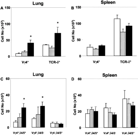
In normal adult C57BL/6 mice, V␥4⫹cells represent⬃20% of the pulmonary␥␦ T cells. After sensitization and challenge, although
not after challenge alone, these cells increased in number by ⬃5-fold, and their relative frequency among total pulmonary␥␦ T cells increased by⬃2.5-fold (Fig. 1A). This increase was large enough to account for the increase in total pulmonary␥␦ T cells in these mice. The spleens of C57BL/6 mice contain a␥␦ T cell population larger than that of the lung, but the relative frequency of V␥4⫹ cells was similar, and numbers of V␥4⫹␥␦ T cells did not increase during sensitization and challenge (Fig. 1B). In both lung and spleen, about half of the V␥4⫹cells expressed either V␦4 or V␦5, and the other half expressed other V␦s (not shown). After sensi-tization and challenge, both the V␥4⫹/V␦4/5⫹ and the V␥4⫹/ V␦4/5⫺population in the lung increased, without a clear prefer-ence for one population over the other (Fig. 1C). In contrast, V␥4⫺/V␦4/5⫹cells did not increase. In the spleen, none of these populations changed significantly (Fig. 1D). Thus, among C57BL/6 pulmonary ␥␦ T cells, V␥4⫹ cells preferentially re-sponded to airway challenge with OVA in OVA-sensitized mice, although a preference for V␦s was not evident.
In adult BALB/c mice, the pulmonary␥␦ T cell population is smaller, and the frequency of these cells relative to all lung lym-phocytes is⬃4 times smaller than that in C57BL/6 mice. How-ever, among pulmonary␥␦ T cells, V␥4⫹cells represent a larger fraction than in C57BL/6 mice (⬃50%), and they also increased preferentially following sensitization and challenge (data not shown). The data with C57BL/6 and BALB/c mice, taken together, indicated that the V␥4⫹subset of␥␦ T cells selectively increases in response to airway challenge with OVA in sensitized mice. Depletion of V␥4⫹␥␦ T cells enhances AHR in sensitized OVA-challenged mice
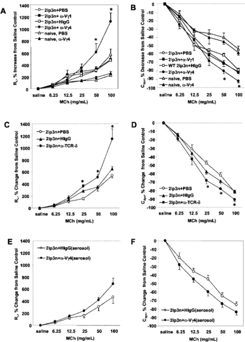
To assess the effects of V␥4⫹T cell depletion on AHR in OVA-sensitized/challenged mice, we administered the anti-TCR V␥4 mAb i.v. before challenge and monitored RL and Cdyn. Fig. 2
shows responses in C57BL/6 mice. The Ab treatment increased RL
FIGURE 1. Increase in V␥4⫹ ␥␦ T cells in the lung of OVA-sensitized and challenged C57BL/6 mice. The numbers of V␥4⫹, TCR-␦⫹, and V␦4/5⫹ (V␦4⫹ or 5⫹) T cells in the lung and spleen of C57BL/6 mice were determined cytofluorimetrically. Mice were untreated (no treatment;䡺); received two i.p. injections of OVA-alum on days 0 and 14, fol-lowed by three consecutive challenges with aerosol-ized OVA on days 27–29 (2ip3n protocol; f); or re-ceived only the challenges without prior sensitization (3n protocol; s). Absolute cell numbers (total cells/ both lungs) were obtained by multiplying the percent-age of positive cells in flow cytometric analysis by the number of NAD gated live cells. Results for each group are expressed as the mean⫾ SEM (n ⫽ 6 in each group).ⴱ, Significant differences (p ⬍ 0.05) be-tween naive and 2ip3n groups.
and decreased Cdyn through much of the MCh dose-response
curve, indicating an enhancement of AHR (Fig. 2, A and B). The changes in RLand Cdynresponses were similar to those in mice
treated with pan-specific anti TCR-␦ mAbs (a 1/1 mixture of mAbs GL3 and 403.A10; Fig. 2, C and D). In contrast to the treatments with anti-V␥4 and anti-TCR-␦ mAbs, treatment with anti-V␥1 mAb 2.11 had no effect on AHR (Fig. 2, A and B). Likewise, treatment of sensitized and challenged mice with nonspecific ham-ster Ig did not significantly alter AHR (Fig. 2, A and B). We also examined responses in nonsensitized, nonchallenged (naive) mice as a further control (Fig. 2, A and B). In these mice, treatment with the anti-V␥4 mAb had no effect on airway responsiveness to MCh. We also used a protocol in which the mice inhaled small quan-tities of this Ab together with OVA during the last challenge. We have previously shown that this treatment selectively targets pul-monary␥␦ T cells and reveals AHR in nonsensitized, OVA-chal-lenged mice (24). The effect of inhaled anti-V␥4 mAb on AHR in the sensitized and challenged mice was small, but reproducible, whereas inhaled hamster Ig was not effective (Fig. 2, E and F).
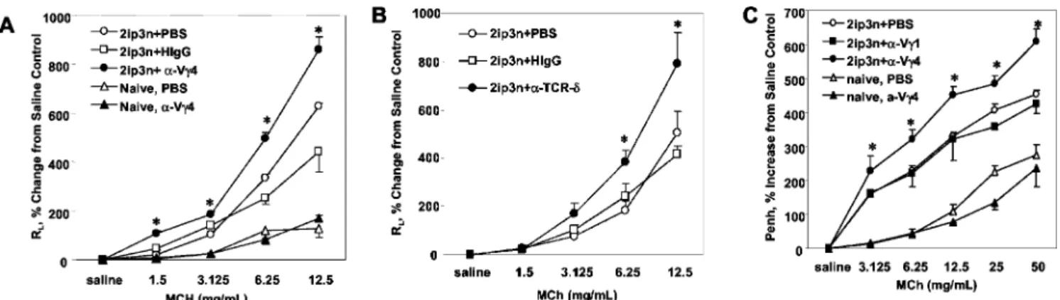
We also tested the effect of the Ab treatments on OVA-sensi-tized and challenged BALB/c mice (Fig. 3). The results closely resembled those with C57BL/6 mice, i.e., anti-V␥4 mAb treatment
increased RLin OVA-sensitized/challenged mice, but had no effect
in naive mice (Fig. 3A). Moreover, the effect of the anti-V␥4 mAb again equaled that of an anti-TCR-␦ mAb (Fig. 3B). When we measured Cdyninstead of RL, the result was essentially the same
(not shown). Finally, we also analyzed the effect of the Ab treat-ments on AHR using noninvasive, barometric, whole-body pleth-ysmography. We measured changes in Penh as a parameter of AHR. Penh values were significantly increased in BALB/c mice treated with the anti-V␥4 mAb (Fig. 3C). In contrast, treatment with anti-V␥1 mAb had no effect (Fig. 3C). Finally, Penh values of naive mice remained essentially unchanged by the treatment with anti-V␥4 mAb (Fig. 3C). Thus, noninvasive plethysmography yielded essentially the same result as invasive plethysmography. Taken together, all these data suggested that in OVA-sensitized and challenged mice, V␥4⫹␥␦ T cells negatively regulate AHR. Adoptively transferred pulmonary lymphocytes containing V␥4⫹ cells reduce AHR in sensitized, OVA-challenged recipients As recipients of adoptive cell transfers, we used mice genetically deficient in both V␥4⫹and V␥6⫹␥␦ T cells, backcrossed to the C57BL/6 genetic background (B6.V␥4/6⫺/⫺). The recipient mice were OVA-sensitized and challenged. By comparison with
FIGURE 2. Increase in AHR in OVA-sensitized and challenged C57BL/6 mice treated with anti-TCR V␥4 mAb. Airway responses to MCh (A, C, and E, RL; B, D, and F, Cdyn) were measured 48 h after the last OVA challenge in C57BL/6 mice and are shown as the percent change from controls that received sa-line. Mice remained either untreated or were sensi-tized and challenged with OVA (2ip3n protocol). 2ip3n-sensitized and challenged mice were further treated with anti-TCR V␥4 mAb, anti-TCR V␥1 mAb, nonspecific hamster IgG (HIgG), or PBS by i.v. injec-tion 3 days before the first OVA challenge (A and B), with anti TCR-␦ mAb and saline by i.v. injection as in A and B (C and D), or with anti-TCR V␥4 mAb or HIgG, by inhalation during the last OVA challenge (E and F). Naive mice were treated with PBS or anti-TCR V␥4 mAb by i.v. injection 5 days before the measurement of airway responses (A and B). Results for each group are expressed as the mean⫾ SEM (n ⫽ 8 in each group). No significant difference from base-line responses to sabase-line were observed in any of these groups.ⴱ, Significant difference (p ⬍ 0.05) between anti-TCR V␥4 mAb-treated and PBS-treated groups.
C57BL/6 mice, B6.V␥4/6⫺/⫺mice contained very small numbers of␥␦ T cells in the lung (Fig. 4A). Their AHR may be slightly increased (not statistically significant; Fig. 4, B and C). As donors of adoptive cell transfers, we used OVA-sensitized and challenged B6.TCR-⫺/⫺mice. These mice are completely deficient in␣ T cells and contain increased numbers of pulmonary␥␦ T cells by comparison with C57BL/6 mice (24). NAD cells prepared from the lungs of B6.TCR-⫺/⫺mice and either nondepleted or depleted of V␥4⫹cells by negative selection on magnetic columns were trans-ferred i.v. into OVA-sensitized B6.TCR-V␥4/6⫺/⫺ mice, shortly before the first airway challenge. Nondepleted NAD cells con-tained 15–20% V␥4⫹cells, whereas depleted NAD cells contained
⬍1% V␥4⫹ cells. Fig. 4, B and C, show the result of adoptive transfers of 2⫻ 104
cells/recipient. The transferred cells signifi-cantly decreased RL and increased Cdyn, but ceased to have this
effect when depleted of V␥4⫹cells, supporting the idea that this particular subset negatively regulates AHR.
Depletion of V␥4⫹␥␦ T cells does not alter pulmonary eosinophilia or goblet cell hyperplasia and enhances OVA challenge-induced shifts in cytokine-producing pulmonary␣ T cells only slightly
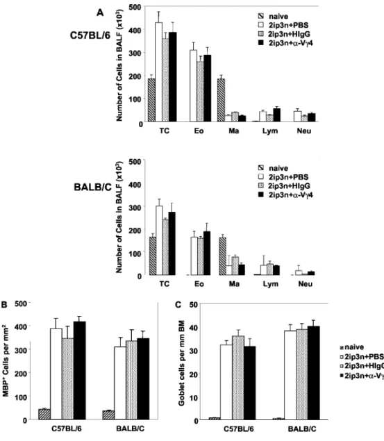
To assess the role of V␥4⫹␥␦ T cells in airway inflammation in OVA-sensitized and challenged C57BL/6 and BALB/c mice,
anti-TCR V␥4 mAb was injected i.v. before the first OVA challenge, following the same protocols as in the airway response studies. No significant changes in eosinophil numbers in BALF were seen in either strain (Fig. 5A). Further, no differences in the numbers of MBP⫹eosinophils in lung parenchyma (Fig. 5B) or in the relative frequencies of goblet cells (Fig. 5C) were found. These data sug-gested that V␥4⫹cells do not alter OVA challenge-induced airway and lung infiltration with eosinophils or goblet cell hyperplasia in previously sensitized mice.
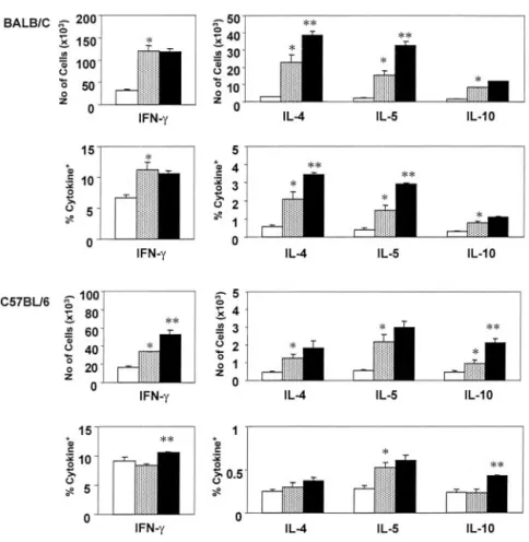
We did not find significant changes in BAL cytokines after the anti-V␥4-treatment in either mouse strain (not shown). To inves-tigate the effects of anti-TCR V␥4 mAb treatment on OVA chal-lenge-induced shifts in Th2 cytokine-committed pulmonary␣ T cells in sensitized mice, we examined NAD, PMA/ionomycin-ac-tivated pulmonary cells from both BALB/c and C57BL/6 mice, costained for TCR- expressed on the cell surface and intracellular cytokines (Fig. 6). Although only a small fraction of cells stained positively for IL-4, IL-5, and IL-10, a shift among pulmonary a T cells toward higher cell numbers and cell frequencies producing 4 and 5 was evident in all sensitized/challenged mice. IL-10-producing cells also increased, albeit to a lesser extent (Fig. 6). The increases in IL-4- and IL-5-producing␣ T cells nearly dou-bled in sensitized/challenged BALB/c mice that had been treated with the anti-V␥4 mAb, but not in identically treated C57BL/6
FIGURE 3. Increase in AHR in OVA-sensitized and challenged BALB/C mice treated with anti-TCR V␥4 mAb. Airway responsiveness (A and B, RL;
C, Penh) in OVA-sensitized and challenged BALB/c mice (2ip3n) was assessed and expressed as the percent change from saline controls in relation to
increasing concentrations of MCh. Groups of mice were treated in addition with anti-TCR-V␥4 mAb, anti-TCR-V␥1 mAb, anti-TCR-␦ mAb, nonspecific hamster IgG (HIgG), or PBS 3 days before the first challenge. Results for each group are expressed as the mean⫾ SEM (n ⫽ 8 in each group). ⴱ, Significant difference (p⬍ 0.05) between anti-TCR mAb-treated and PBS- or HIgG-treated groups.
FIGURE 4. Decrease in AHR in OVA-sensitized and challenged TCR-V␥4/6⫺/⫺mice after adoptive transfer of pulmonary lymphocytes containing V␥4⫹cells. Pulmonary␥␦ T cells in OVA-sensitized and challenged C57BL/6- and B6.TCR-V␥4/6⫺/⫺mice were stained with anti-TCR-␦ mAb GL3 and examined cytofluorimetrically (A). Shown are absolute numbers of TCR-␦⫹cells (total cells/both lungs). Airway responsiveness (RLand Cdyn) was also measured in these mice (B and C). Airway responses are expressed as the percent change from saline controls in relation to increasing concentrations of MCh. In addition to sensitization and challenge, two groups received NAD pulmonary lymphocytes (PL) i.v. before the challenge. The transferred PL were derived from OVA-sensitized and challenged B6.TCR-⫺/⫺mice and remained untreated or were depleted of V␥4⫹cells before the adoptive transfer, as described in Materials and Methods. Results for each group are expressed as the mean⫾ SEM (n ⫽ 4 in each group). ⴱ, Significant difference (p ⬍ 0.05) between adoptive cell transfer recipient that received PL containing V␥4⫹cells and the other two groups.
mice (Fig. 6). In contrast, the Ab treatment increased IFN-␥- and IL-10-producing ␣ T cells in sensitized/challenged C57BL/6 mice, but not or only slightly in the identically treated BALB/c mice (Fig. 6).
Discussion
The present study was prompted by two previous findings: 1)␥␦ T cells negatively regulate AHR in OVA-sensitized and challenged mice (20); and 2) V␥4⫹pulmonary␥␦ T cells negatively regulate AHR evoked by innate mechanisms (in the absence of␣ T cells) in nonsensitized, but airway-challenged, mice (24). In apparent contrast, we and others have shown that␥␦ T cells can enhance allergic airway inflammation, including eosinophilic responses and IgE production (20, 27, 28). In these studies, subsets within␥␦ T cells were not examined, and in some, experimental protocols were different. However, we have found that specific functions within the TCR-V␥-defined ␥␦ T cell subsets vary greatly (26, 29, 45). Since the effects of one subset can negate the effects of another (26), one might predict that experiments based on the deficiency of specific subsets vs the depletion of all␥␦ T cells would have dif-ferent outcomes. In addition, the timing of depletions may be im-portant, as effects of different subsets may be expressed at different stages of the response, even when pan-specific Abs are used. Con-sistently, we report here that anti-TCR-␦ and anti-V␥4 mAbs had essentially the same effect when given just before the airway
chal-lenge of previously sensitized mice, i.e., both treatments resulted in an increase in AHR.
The mechanisms leading to AHR in sensitized and challenged mice vs the development of AHR in mice that are simply chal-lenged with OVA appear to be distinct. AHR in sensitized and challenged mice is dependent on the response of Ag-specific␣ T cells that are recruited to the lung (1, 20), elaborate a characteristic set of cytokines, and initiate airway inflammation (2, 46). In con-trast, AHR in nonsensitized mice does not require␣ T cells, is not associated with airway inflammation, and appears to be driven by an innate response to the airway challenge (20, 24). Neverthe-less, the data presented here suggest that the same␥␦ T cells sim-ilarly regulate AHR under either condition. These regulatory cells probably do not directly target the Ag-specific allergic response, because they appear to be activated and effective regardless of it. In the sensitized and challenged mice, changes in the V␥4⫹ pulmonary (but not splenic) cells were demonstrated, suggesting a response to airway exposure to allergen. In sensitized and chal-lenged mice,␥␦ T cells increased significantly in number, and the increase in V␥4⫹cells accounted for virtually all of this increase. Whether this increase is due to preferential recruitment or local expansion of the V␥4⫹cells is not clear at present. V␥4⫹cells in the spleens of C57BL/6 mice were almost evenly divided into those expressing V␦4 or V␦5 and those expressing other V␦s; others have shown a similar distribution in lymph nodes (44). A
FIGURE 5. Unchanged inflam-matory infiltrate in the airways, tis-sue eosinophilia, and goblet cell hy-perplasia in OVA-sensitized and challenged mice treated with anti-TCR V␥4 mAb. Groups of 2ip3n-treated mice were injected i.v. with anti-TCR V␥4 mAb, nonspecific hamster IgG, or PBS 3 days before the first OVA challenge. For com-parison, untreated naive mice are also included. A, Inflammatory cells in BAL fluid (BALF) were counted 48 h after the last OVA challenge. TC, total cells; Eo, eosinophils; Ma, macrophages; Lym, lymphocytes; Neu, neutrophils. Results for each group are expressed as the mean⫾ SEM (n⫽ 8 in each group). Tissue eosinophilia and goblet cell hyper-plasia were analyzed after anti-MBP staining of eosinophils in lung tissue and PAS staining of airway epithe-lium, respectively. (B) For analysis of tissue eosinophilia, eosinophil numbers per square millimeter in the peribronchial regions were quanti-fied. For analysis of goblet cell hy-perplasia (C), PAS-stained cells were counted and divided by the length (millimeter) of the basement membrane. Results of each group are expressed as the mean⫾ SEM (n ⫽ 8 in each group).
similar partition was seen in the lung. This partition did not change after OVA sensitization and challenge despite the overall increase in V␥4⫹cells. Therefore, it appears that the increase in the V␥4⫹ subset is not driven by a clonal TCR-dependent response to the allergen. Consistently, we have been unable to elicit specific re-sponses of␥␦ T cells to OVA (not shown). In addition to OVA itself, LPS that occurs as contaminant in the OVA preparation (47) could be involved in triggering the polyclonal response of V␥4⫹ cells. LPS is a potent trigger of TNF-␣, and we have already shown that␥␦ T cells are very responsive to this cytokine (23) and depend on its presence in the regulation of AHR (21, 22). How-ever, we have previously observed that in nonsensitized mice chal-lenged with OVA, the relative frequency of CD8⫹cells within the V␥4⫹subset increases in a manner dependent on normal MHC class I expression (24). Others have reported that peripheral selec-tion in naive mice helps to determine the repertoire of V␥4⫹and V␦5⫹␥␦ T cells (48, 49). Thus, even though our data indicate a broad non-Ag-specific response within the V␥4⫹ subset during airway challenge, the possibility remains that the cells involved are subject to some (non-Ag-specific) selection during the challenge, with possible effects on the functional impact of these cells.
Depletion of V␥4⫹cells before the airway challenges resulted in increased airway responsiveness, similar to an earlier study in non-sensitized mice (24). The interpretation that this increase is due to the removal of V␥4⫹cells that act as negative regulators of AHR is now supported by adoptive transfer experiments as well. OVA-sensitized, genetically V␥4/6-deficient mice reconstituted with lung lymphocytes before OVA challenge showed significantly reduced AHR, but only when the transferred cells contained V␥4⫹ cells. The required number of transferred cells was small (2 ⫻
104
cells/recipient), suggesting that a few thousand regulatory V␥4⫹cells are sufficient in effectively suppressing AHR.
OVA-sensitized and challenged B6.V␥4/6⫺/⫺ mice showed only slightly increased AHR compared with C57BL/6 mice, sim-ilar to our earlier findings with B6.TCR-␦⫺/⫺mice (20). As argued above, this could be due to effects of␥␦ T cell subsets that oppose and potentially neutralize one another. Alternatively, genetic␥␦ T cell deficiency throughout life may have different and more com-plex consequences than depletion just before airway challenge.
Although depletion of the V␥4⫹cells just before airway challenge significantly increased AHR, there was no parallel increase in pulmo-nary inflammation. This was unexpected in view of other studies doc-umenting effects of␥␦ T cells on both the development and the res-olution of inflammatory responses in the lung and elsewhere (13, 16, 50, 51). V␥4⫹cells in particular have been found to promote a virus-induced, Th1-dependent myocarditis (26) and might thus be expected to inhibit Th2-driven inflammation. It is possible that pulmonary in-flammation in sensitized and challenged mice was already at a max-imal level. It seems more likely, however, that the effect of V␥4⫹cells bypasses the inflammatory response, at least in part. Consistently, depletion of V␥4⫹cells also evoked AHR in nonsensitized mice, where OVA challenge alone did not induce an inflammatory response (24). This possibility is further supported by findings in another model involving long term OVA challenge of sensitized mice. Here, previ-ously established AHR resolved, and the␥␦ T cells were found to inhibit AHR, but not airway infiltration, of inflammatory cells.4
This
4Z.-H. Cui, A. J. Tham, M. K. Aydintug, Y.-S. Hahn, W. K. Born, and E. W. Gelfand.
Reversal of allergic airway hyperreactivity after long-term allergen challenge depends on␥␦ T cells. Submitted for publication.
FIGURE 6. Small changes in specific cytokine-producing␣ T cells following treatment with anti-TCR V␥4 mAb. NAD pulmonary cells were recov-ered 2 days after the last OVA challenge from 2ip3n-treated mice injected i.v. with PBS or anti-TCR V␥4 mAb 3 days before the first OVA chal-lenge. After stimulation with PMA/ionomycin in the presence of brefeldin A, cells were stained with anti-TCR- and cytokine-specific mAbs as described in
Materials and Methods. Lymphocytes were gated
based on their scatter profile, and 2.5⫻ 104cells were counted in each histogram. Two-color fluores-cent analysis was performed by FACScan. Cell fre-quencies and numbers were determined as described in Fig. 1. Results for each group are expressed as the mean⫾ SEM (n ⫽ 4 in each group). ⴱ, Significant differences (p⬍ 0.05) between naive and 2ip3n-plus PBS-treated groups; ⴱⴱ, differences between 2ip3n- plus PBS-treated and 2ip3n- plus anti-TCR V␥4 mAb-treated groups. Note that for BALB/c mice, the OVA-treated control group was 2ip3n and saline, and for C57BL/6 mice, it was 2ip3n and ham-ster IgG.
absence of effect on the inflammatory response appears to distinguish the V␥4-dependent regulatory effect from that of the CD4⫹CD25⫹ regulatory␣ T cells that were found by one group to inhibit Th2-dependent airway inflammation (10), although another found them to be necessary (11). The absence of effects on airway inflammation was paralleled by an absence of changes in goblet cell numbers and mucin hyperproduction. The dissociation between AHR and goblet cells/ mucin hyperproduction has previously been demonstrated (52).
Consistent with the absence of effects on eosinophilic inflam-mation and goblet cell hyperplasia, V␥4⫹cells only slightly af-fected cytokine-producing␣ T cells in the OVA-sensitized and challenged mice. The anti-V␥4 mAb treatment did not induce sig-nificant changes in BAL cytokines. V␥4-dependent changes in cy-tokine-producing␣ T cells were diverse and not clearly biased toward regulating Th2 reactivity. Thus, the treatment with anti-V␥4 mAb increased IL-4- and IL-5-producing ␣ T cells in BALB/c mice, but it had little, if any, effect on IL-4- and IL-5-producing␣ T cells in C57BL/6 mice. Furthermore, while the Ab treatment had no effect on IFN-␥-producing ␣ T cells in BALB/c mice and increased the number of IL-10-producing␣ T cells only slightly, it increased both IL-10- and IFN-␥-producing ␣ T cells in C57BL/6 mice. The changes in␣ T cells are not unexpected, since cross-talk between␣ and ␥␦ T cell has been described (53). However, the Ab treatment-induced changes in cytokine-produc-ing␣ T cells were different in the two mouse strains examined, whereas the effect on AHR was essentially the same. Thus, we conclude that the changes in cytokine-producing␣ T cells are not critical for the AHR regulatory effect of V␥4⫹␥␦ T cells.
Before and during airway challenge, V␥4⫹cells may have a nonspecific suppressive effect on the␣ T cell response, perhaps by inhibiting␣ T cell recruitment to the lung. Whether this effect plays a significant role in the regulation of AHR by the␥␦ T cells is unclear. It remains possible, however, that␥␦ T cells more se-lectively influence the␣ T cell response at an early stage of its development, and others have suggested that ␥␦ T cells in C57BL/6 mice modulate Th2 differentiation during sensitization (18). In our study,␥␦ T cells were depleted well after the sensi-tization phase; as a result, such selective effects on␣ T cells may be absent, and consequently, there may be no further changes in eosinophil infiltration and goblet cell differentiation (1, 2).
In sum, the findings of this study directly support the idea that one distinct subset of␥␦ T cells, V␥4⫹cells, can protect normal airway function (54) in both the absence (24) and the presence of (␣ T cell-mediated) allergic AHR. The molecular mechanism of this regulatory effect and its significance with regard to bronchial hyper-reactivity in humans remain to be elucidated.
References
1. Wills-Karp, M. 1999. Immunologic basis of antigen-induced airway hyperre-sponsiveness. Annu. Rev. Immunol. 17:255.
2. Hamelmann, E., A. Oshiba, J. Paluh, K. Bradley, J. Loader, T. A. Potter, G. L. Larsen, and E. W. Gelfand. 1996. Requirement for CD8⫹T cells in the development of airway hyperresponsiveness in a murine model of airway sensi-tization. J. Exp. Med. 183:1719.
3. Gavett, S. H., X. Chen, F. Finkelman, and M. Wills-Karp. 1994. Depletion of murine CD4⫹T lymphocytes prevents antigen-induced airway hyperreactivity and pulmonary eosinophilia. Am. J. Respir. Cell Mol. Biol. 10:587.
4. Busse, W. W., R. L. Coffman, E. W. Gelfand, A. B. Kay, and L. J. Rosenwasser. 1995. Mechanisms of persistent airway inflammation in asthma: a role for T cells and T-cell products. Am. J. Respir. Crit. Care Med. 152:388.
5. Wills-Karp, M., J. Luyimbazi, X. Xu, B. Schofield, T. Y. Neben, C. L. Karp, and D. D. Donaldson. 1998. Interleukin-13: central mediator of allergic asthma.
Sci-ence 282:2258.
6. Taube, C., C. Duez, Z.-H. Cui, K. Takeda, Y.-H. Rha, J.-W. Park, A. Balhorn, D. D. Donaldson, A. Dakhama, and E. W. Gelfand. 2002. The role of IL-13 in established allergic airway disease. J. Immunol. 169:6482.
7. Sitrin, R. G., M. J. Ansfield, and H. B. Kaltreider. 1985. The effect of pulmonary surface-active material on the generation and expression of murine B- and T-lymphocyte effector functions in vitro. Exp. Lung Res. 9:85.
8. Crouch, E. C. 1998. Collectins and pulmonary host defense. Am. J. Respir. Cell
Mol. Biol. 19:177.
9. Borron, P. J., E. A. Mostaghel, C. Doyle, E. S. Walsh, M. G. McHeyzer-Williams, and J. R. Wright. 2002. Pulmonary surfactant pro-teins A and D directly suppress CD3⫹/CD4⫹cell function: evidence for two shared mechanisms. J. Immunol. 169:5844.
10. Tsitoura, D. C., R. H. DeKruyff, J. R. Lamb, and D. T. Umetsu. 1999. Intranasal exposure to protein antigen induces immunological tolerance mediated by func-tionally disabled CD4⫹T cells. J. Immunol. 163:2592.
11. Suto, A., H. Nakajima, S.-I. Kagami, K. Suzuki, Y. Saito, and I. Iwamoto. 2001. Role of CD4⫹CD25⫹regulatory T cells in T helper 2 cell-mediated allergic inflammation in the airways. Am. J. Respir. Crit. Care Med. 164:680. 12. Carding, S. R., W. Allan, S. Kyes, A. Hayday, K. Bottomly, and P. C. Doherty.
1990. Late dominance of the inflammatory process in murine influenza by␥␦⫹T cells. J. Exp. Med. 172:1225.
13. D’Souza, C. D., A. M. Cooper, A. A. Frank, R. J. Mazzaccaro, B. R. Bloom, and I. M. Orme. 1997. An anti-inflammatory role for␥␦ T lymphocytes in acquired immunity to Mycobacterium tuberculosis. J. Immunol. 158:1217.
14. Tam, S., D. P. King, and B. L. Beaman. 2001. Increase of␥␦ T lymphocytes in murine lungs occurs during recovery from pulmonary infection by Nocardia
as-teroides. Infect. Immun. 69:6165.
15. Dieli, F., J. Ivanyi, P. Marsh, A. Williams, I. Naylor, G. Sireci, N. Caccamo, C. Di Sano, and A. Salerno. 2003. Characterization of lung␥␦ T cells following intranasal infection with Mycobacterium bovis bacillus Calmette-Gue´rin. J.
Im-munol. 170:463.
16. King, D. P., D. M. Hyde, K. A. Jackson, D. M. Novosad, T. N. Ellis, L. Putney, M. Y. Stovall, L. S. Van Winkle, B. L. Beaman, and D. A. Ferrick. 1999. Cutting edge: protective response to pulmonary injury requires␥␦ T lymphocytes. J.
Im-munol. 162:5033.
17. Doherty, P. C., W. Allan, M. Eichelberger, and S. R. Carding. 1991. Heat shock proteins and the␥␦ T cell response in virus infections: implications for autoim-munity. Springer Semin. Immunopathol. 13:11.
18. McMenamin, C., C. Pimm, M. McKersey, and P. G. Holt. 1994. Regulation of IgE responses to inhaled antigen in mice by antigen-specific␥␦ T cells. Science
265:1869.
19. McMenamin, C., M. McKersey, P. KY¨ hnlein, T. HY¨nig, and P. G. Holt. 1995.␥␦
T cells down-regulate primary IgE responses in rats to inhaled soluble protein antigens. J. Immunol. 154:4390.
20. Lahn, M., A. Kanehiro, K. Takeda, A. Joetham, J. Schwarze, G. Koehler, R. O’Brien, E. W. Gelfand, and W. Born. 1999. Negative regulation of airway re-sponsiveness that is dependent on␥␦ T cells and independent of ␣ T cells. Nat.
Med. 5:1150.
21. Kanehiro, A., M. Lahn, M. J. Makela, A. Dakhama, M. Fujita, A. Joetham, R. J. Mason, W. Born, and E. W. Gelfand. 2001. Tumor necrosis factor-␣ neg-atively regulates airway hyperresponsiveness through␥␦ T cells. Am. J. Respir.
Crit. Care Med. 164:2229.
22. Kanehiro, A., M. Lahn, M. J. Makela, A. Dakhama, A. Joetham, Y.-H. Rha, W. Born, and E. W. Gelfand. 2002. Requirement for the p75 TNF-␣ receptor 2 in the regulation of airway hyperresponsiveness by ␥␦ T cells. J. Immunol.
169:4190.
23. Lahn, M., H. Kalataradi, P. Mittelstadt, E. Pflum, M. Vollmer, C. Cady, A. Mukasa, A. Vella, D. Ikle, R. Harbeck, et al. 1998. Early preferential stimu-lation of␥␦ T cells by TNF-␣. J. Immunol. 160:5221.
24. Lahn, M., A. Kanehiro, K. Takeda, J. Terry, Y.-S. Hahn, M. K. Aydintug, A. Konowal, K. Ikuta, R. L. O’Brien, E. W. Gelfand, et al. 2002. MHC class I-dependent V␥4⫹pulmonary T cells regulate ␣ T cell-independent airway responsiveness. Proc. Natl. Acad. Sci. USA 99:8850.
25. Sim, G.-K., R. Rajaserkar, M. Dessing, and A. Augustin. 1994. Homing and in
situ differentiation of resident pulmonary lymphocytes. Int. Immunol. 6:1287.
26. Huber, S. A., D. Graveline, M. K. Newell, W. K. Born, and R. L. O’Brien. 2000. V␥1⫹T cells suppress and V␥4⫹T cells promote susceptibility to coxsackievirus B3-induced myocarditis in mice. J. Immunol. 165:4174.
27. Zuany-Amorim, C., C. Ruffie, S. Haile, B. B. Vargaftig, P. Pereira, and M. Pretolani. 1998. Requirement for␥␦ T cells in allergic airway inflammation.
Science 280:1265.
28. Schramm, C. M., L. Puddington, C. A. Yiamouyiannis, E. G. Lingenheld, H. E. Whiteley, W. W. Wolyniec, T. C. Noonan, and R. S. Thrall. 2000. Proin-flammatory roles of T-cell receptor (TCR)␥␦ and TCR␣ lymphocytes in a mu-rine model of asthma. Am. J. Respir. Cell Mol. Biol. 22:218.
29. O’Brien, R. L., M. Lahn, W. Born, and S. A. Huber. 2001. T cell receptor and function cosegregate in␥-␦ T cell subsets. Chem. Immunol. 79:1.
30. Gelfand, E. W. 2002. Mice are a good model of human airway disease.
Am. J. Respir. Crit. Care Med. 166:5.
31. Goodman, T., and L. Lefrancois. 1989. Intraepithelial lymphocytes: anatomical site, not T cell receptor form, dictates phenotype and function. J. Exp. Med.
170:1569.
32. Itohara, S., N. Nakanishi, O. Kanagawa, R. Kubo, and S. Tonegawa. 1989. Mono-clonal antibodies specific to native murine T-cell receptor␥␦: analysis of ␥␦ T cells during thymic ontogeny and in peripheral lymphoid organs. Proc. Natl.
Acad. Sci. USA 86:5094.
33. Dent, A. L., L. A. Matis, F. Hooshmand, S. M. Widacki, J. A. Bluestone, and S. M. Hedrick. 1990. Self-reactive␥␦ T cells are eliminated in the thymus.
Na-ture 343:714.
34. Pereira, P., D. Gerber, S. Y. Huang, and S. Tonegawa. 1995. Ontogenic devel-opment and tissue distribution of V␥1-expressing ␥/␦ T lymphocytes in normal mice. J. Exp. Med. 182:1921.
35. Carbone, A., R. Harbeck, A. Dallas, D. Nemazee, T. Finkel, R. O’Brien, R. Kubo, and W. Born. 1991.␣ T lymphocyte depleted mice, a model for ␥␦ T lympho-cyte functional studies. Immunol. Rev. 120:35.
36. Heilig, J. S., and S. Tonegawa. 1986. Diversity of murine␥ genes and expression in fetal and adult T lymphocytes. Nature 322:836.
37. Hamelmann, E., J. Schwarze, K. Takeda, A. Oshiba, G. L. Larsen, C. G. Irvin, and E. W. Gelfand. 1997. Noninvasive measurement of airway responsiveness in allergic mice using barometric plethysmography. Am. J. Respir. Crit. Care Med.
156:766.
38. Takeda, K., E. Hamelmann, A. Joetham, L. D. Shultz, G. L. Larsen, C. G. Irvin, and E. W. Gelfand. 1997. Development of eosinophilic airway inflammation and airway hyperresponsiveness in mast cell-deficient mice. J. Exp. Med. 186:449. 39. Hamelmann, E., A. Oshiba, J. Loader, G. L. Larsen, G. Gleich, J. Lee, and
E. W. Gelfand. 1997. Anti-interleukin-5 (Il-5) antibody prevents airway hyper-responsiveness in a murine model of airway sensitization. Am. J. Respir. Crit.
Care Med. 155:819.
40. Julius, M. H., E. Simpson, and L. A. Herzenberg. 1973. A rapid method for the isolation of functional thymus-derived murine lymphocytes. Eur. J. Immunol.
3:645.
41. Gunzer, M., C. Weishaupt, L. Planelles, and S. Grabbe. 2001. Two-step negative enrichment of CD4⫹and CD8⫹T cells from murine spleen via nylon wool adherence and an optimized antibody cocktail. J. Immunol. Methods 258:55. 42. Kubo, R. T., W. Born, J. W. Kappler, P. Marrack, and M. Pigeon. 1989.
Char-acterization of a monoclonal antibody which detects all murine␣ T cell recep-tors. J. Immunol. 142:2736.
43. Goodman, T., R. LeCorre, and L. Lefrancois. 1992. A T-cell receptor␥␦-specific monoclonal antibody detects a V␥5 region polymorphism. Immunogenetics 35:
65.
44. Pereira, P., V. Hermitte, M.-P. Lembezat, L. Boucontet, V. Azuara, and K. Grigoriadou. 2000. Developmentally regulated and lineage-specific rearrange-ment of T cell receptor V␣/␦ gene segments. Eur. J. Immunol. 30:1988.
45. O’Brien, R. L., Y. Xiang, S. Huber, K. Ikuta, and W. K. Born. 2000. Depletion of a␥␦ T cell subset can increase host resistance to a bacterial infection. J.
Im-munol. 165:6472.
46. Makela, M. J., A. Kanehiro, L. Borish, A. Dakhama, J. Loader, A. Joetham, Z. Xing, M. Jordana, G. L. Larsen, and E. W. Gelfand. 2000. IL-10 is necessary for the expression of airway hyperresponsiveness but not pulmonary inflamma-tion after allergic sensitizainflamma-tion. Proc. Natl. Acad. Sci. USA 97:6007. 47. Eisenbarth, S. C., D. A. Piggott, J. W. Huleatt, I. Visintin, C. A. Herrick, and
K. Bottomly. 2002. Lipopolysaccharide-enhanced, Toll-like receptor 4-dependent T helper cell type 2 responses to inhaled antigen. J. Exp. Med. 196:1645. 48. Sim, G.-K., and A. Augustin. 1991. Extrathymic positive selection of␥␦ T cells.
V␥4J␥1 rearrangements with “GxYS” junctions. J. Immunol. 146:2439.
49. Sim, G.-K., and A. Augustin. 1990. Dominantly inherited expression of BID, an invariant undiversified T cell receptor␦ chain. Cell 61:397.
50. Mombaerts, P., J. Arnoldi, F. Russ, S. Tonegawa, and S. H. E. Kaufmann. 1993. Different roles of␣ and ␥␦ T cells in immunity against an intracellular bacterial pathogen. Nature 365:53.
51. Fu, Y.-X., C. E. Roark, K. Kelly, D. Drevets, P. Campbell, R. O’Brien, and W. Born. 1994. Immune protection and control of inflammatory tissue necrosis by␥␦ T cells. J. Immunol. 153:3101.
52. Tomkinson, A., G. Cieslewicz, C. Duez, K. A. Larson, J. J. Lee, and E. W. Gelfand. 2001. Temporal association between airway hyperresponsiveness and airway eosinophilia in ovalbumin-sensitized mice. Am. J. Respir. Crit. Care
Med. 163:721.
53. Kaufmann, S. H. E., C. Blum, and S. Yamamoto. 1993. Crosstalk between␣/ T cells and␥/␦ T cells in vivo: Activation of ␣/ T-cell responses after ␥/␦ T-cell modulation with the monoclonal antibody GL3. Proc. Natl. Acad. Sci. USA
90:9620.
54. Born, W. K., M. Lahn, K. Takeda, A. Kanehiro, R. L. O’Brien, and E. W. Gelfand. 2000. Role of␥␦ T cells in protecting normal airway function.