Clinical implication of an insufficient
number of examined lymph nodes after
curative resection for gastric cancer
Taeil Son
Department of Medicine
Clinical implication of an insufficient
number of examined lymph nodes after
curative resection for gastric cancer
Directed by Professor Sung Hoon Noh, M.D., Ph.D.
The Master's Thesis
submitted to the Department of Medicine
the Graduate School of Yonsei University
in partial fulfillment of the requirements for the degree of
Master of Medical Science
Taeil Son
This certifies that the Master's Thesis of
Taeil Son is approved.
---
Thesis Supervisor: Sung Hoon Noh
---
Thesis Committee Member #1: Woo Jin Hyung
---
Thesis Committee Member #2: Yong Chan Lee
The Graduate School
Yonsei University
ACKNOWLEDGEMENTS
The authors would like to acknowledge the critical revision of
manuscript and intellectual contribution of Yanghee Woo to the
work described herein. This research followed protocols approved
by relevant accredited ethical committees at Severance Hospital
of Yonsei University
<TABLE OF CONTENTS>
ABSTRACT··· 1
I. INTRODUCTION ··· 2
II. MATERIALS AND METHODS ··· 4
1. Overall surgical strategy and pathologic examination of LNs
··· 4
2. Patients ··· 4
3. Study design ··· 5
4. Statistical analysis ··· 5
III. RESULTS ··· 7
1. Overall operative outcomes ··· 7
2. Comparison of clinicopathologic characteristics ··· 8
3. Survival analysis ··· 11
4. Multivariate analyses of factors influencing survival ·· 14
5.
Binary logistic regression analyses of factors influencing
inadequate evaluation of LNs ··· 17
IV. DISCUSSION ··· 19
V. CONCLUSION ··· 23
REFERENCES ··· 24
ABSTRACT (IN KOREAN) ··· 29
LIST OF FIGURES
Figure 1. Frequency of an insufficient number of examined lymph
nodes ··· 7
Figure 2. Survival curves of the “insufficient” and “sufficient”
groups ··· 12
LIST OF TABLES
Table 1. Comparison of clinicopathologic characteristics ···· 9
Table 2. Multivariate survival analysis ··· 15
Table 3. Binary logistic regression analyses of factors for
- 1 -
ABSTRACT
Clinical implication of an insufficient number of examined lymph nodes
after curative resection for gastric cancer
Taeil Son
Department of Medicine
The Graduate School, Yonsei University
(Directed by Professor Sung Hoon Noh M.D., Ph.D.)
Background: The 7th edition of TNM staging system increased the required number of examined lymph nodes (LNs) in gastric cancer from 15 to 16. However, the same staging system defines node negative gastric cancer regardless of the number of examined LNs. In this study, we evaluated whether gastric cancer can be properly staged with fewer than fifteen examined LNs. Methods: We analyzed the survival rates of 10,010 patients who underwent curative gastrectomy from 1987 to 2007. They were divided into two groups according to the number of examined LNs, termed the “insufficient” group (≤ 15 examined LNs) and “sufficient” group (≥16 examined LNs). The survival curves of patients from both groups were compared according to the 7th edition of the TNM classification.
Results: Three hundred sixteen patients (3.2%) had fewer than 15 examined LNs for staging after standard curative lymphadenectomy. Patients who had T1, N0 classification, and Stage I disease with an insufficient number of examined LNs after curative gastrectomy showed significantly worse prognoses than those of patients with ≥16 examined LNs. Moreover, an insufficient number of examined LNs was an independent prognostic factor for patients who had T1, N0 classification, and stage I disease.
Conclusion: Node negative cancers in which fewer than 15 LNs were examined, classified as N0 in the new TNM staging system cannot adequately predict patient survival after curative gastrectomy, especially in early gastric cancer. --- Key words: lymph node metastasis, gastric cancer, staging, prognosis
- 2 -
Clinical implication of an insufficient number of examined lymph nodes
after curative resection for gastric cancer s
Taeil Son
Department of Medicine
The Graduate School, Yonsei University
(Directed by Professor Sung Hoon Noh, M.D., Ph.D.)
I. INTRODUCTION
Lymph node (LN) involvement is one of the most important prognostic indicators of gastric cancer.1-4 The Union for International Cancer Control (UICC) and the American Joint Committee on Cancer (AJCC) TNM staging system since 1997 has defined nodal stage according to the absolute number of metastatic LNs.5 Nodal stage can be appropriately determined when the number
- 3 -
of total examined LNs is greater than 16, according to the latest TNM staging system.6
However, the recommended minimum number of examined LNs required for the proper staging remains controversial given that this number varies considerably between institutions and countries.7-16 The 7th edition TNM staging system designates node negative (N0) disease as any gastric cancer with all examined LNs negative, regardless of the total number of examined LNs.6 In addition, the latest UICC TNM supplement has stipulated that node positive gastric cancer to be staged in the same way, independent of the number of examined LNs.17
A significant portion of early stage gastric cancer patients have been incorrectly staged due to an insufficient number of examined LNs.10 Large population studies from various institutions and countries demonstrate the association between an adequate number of examined LNs and improved overall survival,10, 11, 18-20 thus providing evidence that examination of an insufficient number of LNs may have a detrimental effect on the overall survival of curatively treated gastric cancer patients.
In this study, we hypothesized that inadequate lymph node evaluation correlates with worse survival outcomes after curative gastrectmy for gastric cancer. We analyzed the long-term survival of curatively treated gastric cancer patients to investigate staging and the dependence on the number of examined LNs.
- 4 - II. MATERIALS AND METHODS
1. Overall surgical strategy and pathologic examination of LNs
In this study, the standard surgery for primary gastric cancer is D2 lymphadenectomy (advanced gastric cancer) and D1+beta or D2 lymphadenectomy (early gastric cancer). The definition for lymphadenectomy is based on the Japanese Research Society for Gastric Cancer guidelines.21 All surgeons followed the same strategy and extent of lymphadenectomy.
Each resected specimen was carefully prepared for accurate pathologic staging. First, the surgeon carefully separated the dissected soft tissues according to the anatomic location of the LNs before sending them for pathologic examination. Specialized pathologists examined the divided soft tissues and retrieved as many LNs as possible. All LNs were stained with hematoxylin and eosin and examined by pathologists using light microscopy.
2. Patients
From 1987 to 2007, a total of 11,137 gastrectomies for gastric cancer patients were performed in the Department of Surgery, Yonsei University College of Medicine. Patients were excluded from this study if they underwent surgery for remnant gastric cancer, received chemotherapy before the operation, or died within 30 days of surgery. After exclusion, a total of 10,010 patients who underwent curative resection (R0) were included. The study only included patients who underwent total and subtotal gastrectomy. Patients were followed
- 5 -
from the date of surgery until December 31, 2009 or death except for 199 patients. The follow-up period for the survivors ranged from 24 to 280 months, with a median of 84 months. These 199 patients were censored on the last day of the follow-up.
3. Study design
We divided the patients into two groups: patients with 15 or fewer examined LNs (“insufficient” group) and patients with 16 or more examined LNs (“sufficient” group). Clinicopathologic data such as age, sex, type of operation, histology, size, location, the number of involved LNs, and survival data of the patients were analyzed and compared between the two groups. We also performed the same analyses for the patients after classifying the patients according to the 7th edition of the TNM classification. Multivariate analysis of factors influencing overall survival of patients after treatment was performed using the Cox proportional hazard model in each classification. This study was approved by the Institutional Review Board of the Severance Hospital, Yonsei University (4-2011-0411)
4. Statistical analysis
Categorical variables were analyzed with the chi-square test, and continuous variables were analyzed with a two sample t test. Kaplan-Meier survival estimation was used for plotting survival curves, and log-rank tests were carried
- 6 -
out to compare the survival differences. The Cox proportional hazard model was used for the multivariate analysis of risk factors for death after curative gastrectomy. For subgroup analyses, the interaction between number of eLNs and each T, N and TNM classification was tested at a significant level of 0.05. The binary logistic regression (Enter method) analyses were performed to find out predictors of insufficient number of eLN. P values less than 0.05 were considered statistically significant.
- 7 - III. RESULTS
1. Overall operative outcomes
Of the 10,010 patients in the study, 316 (3.2%) patients had 15 or fewer examined LNs in their pathology reports. Of these patients, 5.4% had 1 to 5 examined LNs, 17.7% had 6 to 10 examined LNs, and 76.9% had 11 to 15 examined LNs. In addition, in this “insufficient” group, 257 patients (81%) were reported to have no cancer involvement of LNs (Figure 1). The mean number of examined LNs in the entire study population was 39.6, with a median of 38.
Figure 1. Frequency of an insufficient number of examined lymph nodes. The numbers over each bar refer to the incidence of the number of examined lymph nodes during the study period. The numbers in each bar refer to the cases without lymph node involvement.
- 8 -
2. Comparison of clinicopathologic characteristics
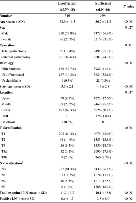
In the “insufficient” group, the mean age was 58.9 years (standard deviation [SD] 11.3 years; range, 31-85 years), whereas for the “sufficient” group, the mean age was 56.2 years (SD 11.8 years; range, 19-91 years). Patient distribution was 77.8% male in the “insufficient” group and 66.8% male in the “sufficient” group. The type of operation, tumor histology, size, and location were significantly different between the two groups (Table 1). There were also significant differences between the two groups in the distribution of T and N classification, the number of total retrieved LNs, and the number of metastatic LNs.
- 9 -
Table 1. Comparison of clinicopathologic characteristics Insufficient (eLN§≤15) Sufficient (eLN≥16) P value Number 316 9694 Age (mean ± SD*) 58.9 ± 11.3 56.2 ± 11.8 <0.001 Sex 0.027 Male 230 (77.8%) 6478 (66.8%) Female 86 (27.2%) 3216 (33.2%) Operation 0.001 Total gastrectomy 55 (17.4%) 2491 (25.7%) Subtotal gastrectomy 261 (82.6%) 7203 (74.3%) Histology <0.001 Differentiated 188 (59.7%) 3982 (41.1%) Undifferentiated 127 (40.3%) 5684 (58.6%) Unclassifiable 1 (0.3%) 28 (0.3%) Size (cm, mean ± SD) 3.1 ± 2.1 4.3 ± 2.8 <0.001 Location 0.047 Upper 29 (9.2%) 1221 (12.6%) Middle 89 (28.2%) 2469 (25.5%) Lower 197 (62.3%) 5826 (60.1%) UML 0 178 (1.8%) Unknown 1 (0.3%) 0 T classification† <0.001 T1 203 (64.2%) 4075 (42.0%) T2 46 (14.6%) 1333 (13.8%) T3 26 (8.2%) 1330 (13.7%) T4a 32 (1.2%) 2696 (27.8%) T4b 9 (2.8%) 260 (2.7%) N classification‡ <0.001 N0 257 (81.3%) 5439 (56.1%) N1 37 (11.7%) 1274 (13.1%) N2 16 (5.1%) 1213 (12.5%) N3 6 (1.9%) 1768 (18.2%)
Total examined LN (mean ± SD) 11.9 ± 3.2 40.1 ± 15.0 <0.001
- 10 -
SD* indicates standard deviation; T† and N‡ classification based on the 7th edition of the UICC/AJCC TNM staging system; eLN§, examined lymph node.
- 11 - 3. Survival analysis
When overall survival curves were compared, there was no statistically significant difference between the “insufficient” and “sufficient” groups (81.8% vs. 75.3% in 5-year survival rates, respectively, log-rank P=0.09). When the patients were categorized using the most recent TNM staging classification, the “insufficient” group had significantly worse survival than the “sufficient” group only in T1 classification (P<0.001, Figure 2A). For N classification, the “insufficient” group showed significantly worse survival only in N0 classification (P=0.01, Figure 2B). Although worse survival was noted in the “insufficient” group in T1 and N0 classifications, this group included more node negative cases and early gastric cancer than the “sufficient” group in T1 and N0 classification [93.6% vs. 89.2%, respectively, of node negative cases (P=0.048), and 73.9% vs. 66.9%, respectively, of early gastric cancer, P=0.018]. Significant survival difference was also noted between the two groups in stage I subgroup but not in the other subgroups (Figure 2C, P<0.001).
- 13 -
Figure 2. Survival curves of the “insufficient” and “sufficient” groups. A. T Classification. Left panels from up to down: T1, T2, T3, T4a, and T4b; B. N Classification. Middle panels from up to down: N0, N1, N2, and N3 C. Stage Classification. Right panels from up to down: I, II, and III. P values from the log-rank test are given.
- 14 -
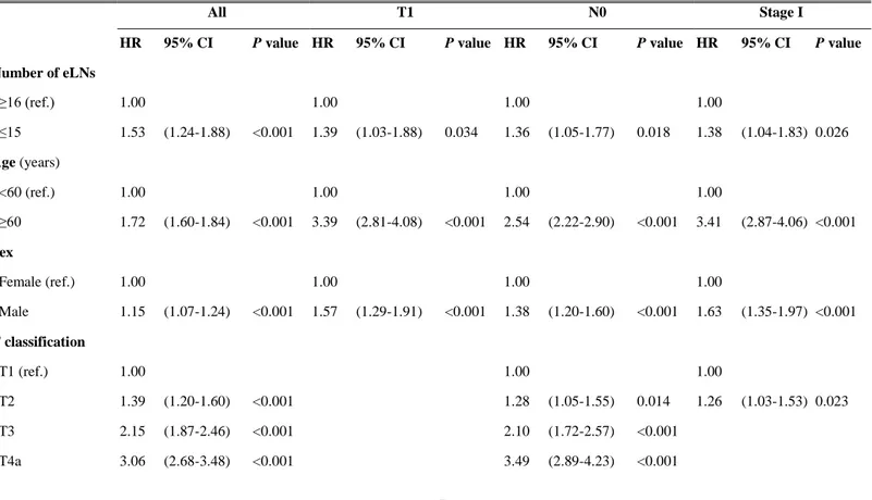
4. Multivariate analyses of factors influencing survival
For all patients analyzed, an insufficient number of examined LNs, age over 60 years, male sex, higher T and higher N classification, and upper tumor location were identified as significant risk factors for survival (Table 2). Upon further analysis, we determined that in patients with T1 and N0 classification, examination of fewer than 15 LNs was a risk factor of poor survival. In stage I, an insufficient number of examined LNs, age over 60 years, and male sex correlated with worse prognoses. However, in the other subgroups, fewer than 15 examined LNs was not a risk factor for poor survival (data not shown).
- 15 -
Table 2. Multivariate survival analysis. Before conducting subgroup analyses, interaction tests between number of examined LNs and each T classification, N classification and TNM stage were performed. Interactions were present between number of eLNs and T classification and TNM stage, but not N classification. Hence, over-interpretation of the results of subgroup analyses in N classification should be avoided.
All T1 N0 Stage I
HR 95% CI P value HR 95% CI P value HR 95% CI P value HR 95% CI P value
Number of eLNs ≥16 (ref.) 1.00 1.00 1.00 1.00 ≤15 1.53 (1.24-1.88) <0.001 1.39 (1.03-1.88) 0.034 1.36 (1.05-1.77) 0.018 1.38 (1.04-1.83) 0.026 Age (years) <60 (ref.) 1.00 1.00 1.00 1.00 ≥60 1.72 (1.60-1.84) <0.001 3.39 (2.81-4.08) <0.001 2.54 (2.22-2.90) <0.001 3.41 (2.87-4.06) <0.001 Sex Female (ref.) 1.00 1.00 1.00 1.00 Male 1.15 (1.07-1.24) <0.001 1.57 (1.29-1.91) <0.001 1.38 (1.20-1.60) <0.001 1.63 (1.35-1.97) <0.001 T classification T1 (ref.) 1.00 1.00 1.00 T2 1.39 (1.20-1.60) <0.001 1.28 (1.05-1.55) 0.014 1.26 (1.03-1.53) 0.023 T3 2.15 (1.87-2.46) <0.001 2.10 (1.72-2.57) <0.001 T4a 3.06 (2.68-3.48) <0.001 3.49 (2.89-4.23) <0.001
- 16 - T4b 5.66 (4.73-6-79) <0.001 6.64 (4.47-9.87) <0.001 N classification N0 (ref.) 1.00 1.00 1.00 N1 1.39 (1.23-1.57) <0.001 1.43 (1.05-1.96) 0.025 1.42 (1.04-1.95) 0.027 N2 1.76 (1.56-1.98) <0.001 1.97 (1.27-3.06) 0.003 N3 3.63 (3.26-4.03) <0.001 6.60 (4.62-9.43) <0.001 Size* (cm) <4 (ref.) 1.00 1.00 1.00 1.00 ≥4 1.10 (1.00-1.21) 0.047 0.84 (0.68-1.05) 0.126 1.06 (0.91-1.24) 0.420 0.97 (0.80-1.18) 0.784 Histology* Diff. (ref.) 1.00 1.00 1.00 1.00 Undiff. 1.07 (0.99-1.16) 0.074 0.92 (0.77-1.11) 0.383 1.06 (0.93-1.21) 0.405 0.87 (0.74-1.03) 0.105 Location* Lower 1/3(ref.) 1.00 1.00 1.00 1.00 Upper 2/3 1.15 (1.07-1.24) <0.001 1.07 (0.89-1.28) 0.464 1.16 (1.01-1.32) 0.030 1.10 (0.93-1.29) 0.269 Whole 1.83 (1.52-2.19) <0.001 2.52 (0.93-6.83) 0.068 2.28 (1.31-3.99) 0.004 2.07 (0.77-5.59) 0.150
* Multivariate analyses was done for only 9922 patients who have complete information of the above variables. Twenty nine patients with unclassifiable histology, 58 patients without information of tumor size and 1 patient without that of tumor location were excluded from the analyses. eLN* indicates examined lymph node; ref.†, reference; Diff.‡, differentiated; Undiff.§, undifferentiated; HR‖, hazard ratio; CI¶, confidence interval.
- 17 -
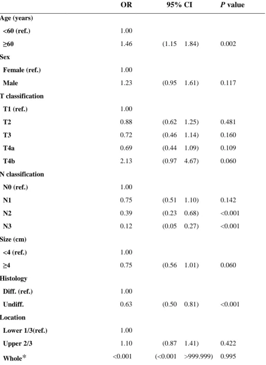
5. Binary logistic regression analyses of factors influencing inadequate evaluation of LNs
In binary logistic regression analyses to find out predictors of insufficient eLNs, older age (60 or more compared to less than 60), earlier N classification (N0, N1 and N2 classification compared to N3 classification) and differentiated type of histology (compared to undifferentiated type) were the risk factors for inadequate LN assessment. (Table 3)
- 18 -
Table 3. Binary logistic regression analyses of factors for influencing inadequate evaluation of LNs OR 95% CI P value Age (years) <60 (ref.) 1.00 ≥60 1.46 (1.15 1.84) 0.002 Sex Female (ref.) 1.00 Male 1.23 (0.95 1.61) 0.117 T classification T1 (ref.) 1.00 T2 0.88 (0.62 1.25) 0.481 T3 0.72 (0.46 1.14) 0.160 T4a 0.69 (0.44 1.09) 0.109 T4b 2.13 (0.97 4.67) 0.060 N classification N0 (ref.) 1.00 N1 0.75 (0.51 1.10) 0.142 N2 0.39 (0.23 0.68) <0.001 N3 0.12 (0.05 0.27) <0.001 Size (cm) <4 (ref.) 1.00 ≥4 0.75 (0.56 1.01) 0.060 Histology Diff. (ref.) 1.00 Undiff. 0.63 (0.50 0.81) <0.001 Location Lower 1/3(ref.) 1.00 Upper 2/3 1.10 (0.87 1.41) 0.422 Whole
*
<0.001 (<0.001 >999.999) 0.995ref indicates reference; Diff, differentiated; Undiff, undifferentiated; OR, odds ratio; CI, confidence interval.
- 19 - IV. DISCUSSION
In this study, we found that the majority of patients with fewer than 15 examined LNs for staging were T1 (64.2%) or N0 (81.3%) classification. Moreover, we found that these same patients and stage I patients had significantly worse prognoses after standard curative lymphadenectomy than patients who had at least 16 examined LNs. In multivariate analyses, fewer than 15 examined LNs was an independent prognostic factor that correlated with worse prognoses. Subgroup analyses also demonstrated that an insufficient number of examined LNs indicated a poor prognosis in T1 and N0 classification with stage I disease.
The prognostic impact of the number of examined LNs has been suggested primarily in patients with advanced gastric cancer. For these patients, a greater number of examined LNs correlated with a better prognosis.11, 15 Our previous study on the number of examined LNs in patients with locally advanced gastric cancer showed a significant impact in survival rates when a higher number of LNs were examined.15 Few studies focus on the influence of an insufficient number of examined LNs but it is known that for staging tumors, a low number of examined LNs correlates with poor survival.10, 15, 18, 22 These data suggest that inappropriate staging may lead to inaccurate diagnoses in potentially curative disease and loss of opportunity for adjuvant treatment23, usually in patients with node negative or early gastric cancer.10, 18, 19 The Surveillance, Epidemiology, and End Results (SEER) database study reported in earlier stage tumors, the
- 20 -
survival rate increased when patients had an adequate number of examined LNs.10, 19 Our results from this study also demonstrate that in patients with fewer than 15 examined LNs, there is a lower survival rate among the T1, N0 classification, and stage I. To our knowledge, these studies including ours are the largest studies of gastric cancer patients that correlate the number of examined LNs with the rate of survival, although the proportion of adequately assessed patients and median number of examined LNs among studies were far different.
The main reasons for examination of an insufficient number of LNs after curative gastrectomy are inaccurate LN dissection or retrieval.8, 24-32 These inaccuracies are most likely determined by a low operation volume of surgeons and decreased diligence of pathologists.18, 23, 33, 34 In addition, they may be closely related to host factors such as patient clinicopathologic status and immunologic response.20, 35, 36 In this study, older age, differentiated type of histology and earlier N classification were the predictors of inadequate LN assessment. In addition, patient’s comorbidities, intrinsic number of LNs, lympho-vascular invasion status and preoperative endoscopic procedures might play a role in inadequate LN dissection and assessment.
In a randomized Dutch gastric cancer group trial, LN retrieval from the specimen was suggested to be responsible for the differences in the number of examined LNs, rather than the extent of lymphadenectomy.8 In addition, pathologists can be affected by the suggested minimum number of LNs required
- 21 -
for examination.10 Presumably, a preoperative diagnosis of early cancer or a less aggressive tumor can lead doctors to biased decisions. Accordingly, these factors could make surgeons and pathologists reluctant to perform strict lymphadenectomy and retrieval. It is less likely that smaller metastatic or non-metastatic LNs might result in the failure to examine an adequate number of LNs.27 Smaller LNs have been reported to correlate with decreased immune response in gastrointestinal cancer.37-40 Older age, male, and differentiated-type histology were reported to trigger weaker immune responses. Our results show that these host factors were more likely to be observed in patients that had fewer than 15 examined LNs.
In the TNM staging system, there have been no validation studies to justify the published and suggested number of LNs that should be examined for proper staging in node negative gastric cancer. Ours is the first comprehensive study with large number of patients who underwent curative gastrectomy with extended lymph node dissection. Due to the high volume of surgery for gastric cancer, surgeons and pathologists removed and examined a much higher number of LNs with consistent results.
Our study, however, has some limitations. First, survival of patients with advanced gastric cancer could not be properly evaluated. Thus, we cannot suggest a proper staging strategy for LN positive tumors with fewer than examined 15 LNs because our sample size was too small to analyze. Second, our results are based on retrospective analyses in a single institution over a long
- 22 -
duration (1987 to 2007). Third, we also could not analyze cancer-specific survival rates to determine the relationship between the number of examined LNs and cancer recurrence after standard treatment. Finally, these results may not be directly applicable to Western institutions which more than half of non-metastatic gastric cancer patients were treated with inadequate LN assessment. Based on our results, the definition of node negative gastric cancer should be re-evaluated to take into account the number of examined LNs. Our results suggest that examination of fewer than 15 LNs may cause inaccurate staging and treatment, thus affecting survival rates. Thus, if the number of examined LNs is inadequate after standard curative resection of gastric cancer, more thorough re-assessment such as fat clearance should be performed. Otherwise, these patients should be considered a high risk group of stage migration and worse survival compared to those with adequate LN evaluation.
- 23 - V. CONCLUSION
In conclusion, future studies should further evaluate the effects of examining insufficient numbers of LNs to clarify the minimally required and optimal number that will contribute to proper TNM staging and patient survival.
- 24 - REFERENCES
1. Seto Y, Nagawa H, Muto T. Impact of lymph node metastasis on survival with early gastric cancer. World J Surg 1997;21:186-89; discussion 90.
2. Siewert JR, Bottcher K, Stein HJ, Roder JD. Relevant prognostic factors in gastric cancer: ten-year results of the German Gastric Cancer Study. Ann Surg 1998;228:449-61.
3. Wu CW, Hsieh MC, Lo SS, Tsay SH, Lui WY, P'Eng F K. Relation of number of positive lymph nodes to the prognosis of patients with primary gastric adenocarcinoma. Gut 1996;38:525-7.
4. Bozzetti F, Bonfanti G, Morabito A, Bufalino R, Menotti V, Andreola S, et al. A multifactorial approach for the prognosis of patients with carcinoma of the stomach after curative resection. Surg Gynecol Obstet 1986;162:229-34.
5. Sobin L, Wittekind C. International Union Against Cancer(UICC). TNM classification of malignant tumors. 5th edition. New York. John Wiley & Sons, Inc. 1997.
6. Edge SB, Byrd DR, Compton CC, Fritz AG, Greene FL, A. T. American Joint Committee on Cancer(AJCC). AJCC Cancer Staging Manual. 7th edition. New York. Springer. 2010.
7. Bouvier AM, Haas O, Piard F, Roignot P, Bonithon-Kopp C, Faivre J. How many nodes must be examined to accurately stage gastric carcinomas? Results from a population based study. Cancer 2002;94:2862-6.
8. Bunt AMG, Hermans J, vanDeVelde CJH, Sasako M, Hoefsloot FAM, Fleuren G, et al. Lymph node retrieval in a randomized trial on western-type versus Japanese-western-type surgery in gastric cancer. J Clin Oncol 1996;14:2289-94.
9. Coburn NG. Lymph nodes and gastric cancer. J Surg Oncol 2009;99:199-206.
10. Coburn NG, Swallow CJ, Kiss A, Law C. Significant regional variation in adequacy of lymph node assessment and survival in gastric cancer. Cancer 2006;107:2143-51.
- 25 -
11. Lee HK, Yang HK, Kim WH, Lee KU, Choe KJ, Kim JP. Influence of the number of lymph nodes examined on staging of gastric cancer. Br J Surg 2001;88:1408-12.
12. Liu C, Lu Y, Jun Z, Zhang R, Yao F, Lu P, et al. Impact of total retrieved lymph nodes on staging and survival of patients with gastric cancer invading the subserosa. Surgical oncology 2009;18:379-84.
13. Noda N, Sasako M, Yamaguchi N, Nakanishi Y. Ignoring small lymph nodes can be a major cause of staging error in gastric cancer. Br J Surg 1998;85:831-4.
14. Saito H, Fukumoto Y, Osaki T, Fukuda K, Tatebe S, Tsujitani S, et al. Prognostic significance of level and number of lymph node metastases in patients with gastric cancer. Ann Surg Oncol 2007;14:1688-93. 15. Shen JY, Kim S, Cheong JH, Kim YI, Hyung WJ, Choi WH, et al. The
impact of total retrieved lymph nodes on staging and survival of patients with pT3 gastric cancer. Cancer 2007;110:745-51.
16. Wagner PK, Ramaswamy A, Ruschoff J, Schmitzmoormann P, Rothmund M. Lymph-Node Counts in the Upper Abdomen - Anatomical Basis for Lymphadenectomy in Gastric-Cancer. British Journal of Surgery 1991;78:825-7.
17. Wittekind C, Greene F, Henson DE, Hutter RVP, Sobin LH. TNM Supplement: A Commentary on Uniform Use, 3rd edition. John Wiley & Sons Inc. 2003.
18. Smith DD, Schwarz RR, Schwarz RE. Impact of total lymph node count on staging and survival after gastrectomy for gastric cancer: data from a large US-population database. Journal of clinical oncology : official journal of the American Society of Clinical Oncology 2005;23:7114-24.
19. Dudeja V, Habermann E, Abraham A, Zhong W, Parsons HM, Tseng JE, et al. Is There a Role for Surgery Alone With Adequate Nodal Evaluation in Gastric Adenocarcinoma? Gastroenterology 2011;140:S997-S.
20. Baxter NN, Tuttle TM. Inadequacy of lymph node staging in gastric cancer patients: a population-based study. Ann Surg Oncol 2005;12:981-7.
- 26 -
21. Japanese Gastric Cancer A. Japanese Classification of Gastric Carcinoma - 2nd English Edition. Gastric Cancer. 1998;1:10-24.
22. Hundahl SA, Phillips JL, Menck HR. The National Cancer Data Base Report on poor survival of U.S. gastric carcinoma patients treated with gastrectomy: Fifth Edition American Joint Committee on Cancer staging, proximal disease, and the "different disease" hypothesis. Cancer 2000;88:921-32.
23. Al-Refaie WB, Gay G, Virnig BA, Tseng JF, Stewart A, Vickers SM, et al. Variations in gastric cancer care: a trend beyond racial disparities. Cancer 2010;116:465-75.
24. Bonenkamp JJ, Songun I, Hermans J, Sasako M, Welvaart K, Plukker JTM, et al. Randomized Comparison of Morbidity after D1 and D2 Dissection for Gastric-Cancer in 996 Dutch Patients. Lancet 1995;345:745-8.
25. Roviello F, Rossi S, Marrelli D, Pedrazzani C, Corso G, Vindigni C, et al. Number of lymph node metastases and its prognostic significance in early gastric cancer: A multicenter Italian study. J Surg Oncol 2006;94:275-80.
26. Siewert JR, Bottcher K, Roder JD, Busch R, Hermanek P, Meyer HJ. Prognostic relevance of systematic lymph node dissection in gastric carcinoma. German Gastric Carcinoma Study Group. Br J Surg 1993;80:1015-8.
27. Noguchi Y, Imada T, Matsumoto A, Coit DG, Brennan MF. Radical surgery for gastric cancer. A review of the Japanese experience. Cancer 1989;64:2053-62.
28. Ichikura T, Tomimatsu S, Okusa Y, Uefuji K, Tamakuma S. Comparison of the prognostic significance between the number of metastatic lymph nodes and nodal stage based on their location in patients with gastric cancer. Journal of clinical oncology : official journal of the American Society of Clinical Oncology 1993;11:1894-900.
29. Hermanek P, Altendorf-Hofmann A, Mansmann U, Dworak O, Wittekind C, Hohenberger W. Improvements in staging of gastric carcinoma from using the new edition of TNM classification. Eur J Surg Oncol 1998;24:536-41.
- 27 -
30. Cheong JH, Hyung WJ, Shen JG, Song C, Kim J, Choi SH, et al. The N ratio predicts recurrence and poor prognosis in patients with node-positive early gastric cancer. Ann Surg Oncol 2006;13:377-85.
31. Coburn NG, Mahar AL, Qureshi AP, Ottensmeyer CA, Pollett A, Wright FC, et al. A Descriptive Analysis of Gastric Cancer Specimen Processing Techniques. J Surg Oncol 2011;103:248-56.
32. Lemmens VE, van Lijnschoten I, Janssen-Heijnen ML, Rutten HJ, Verheij CD, Coebergh JW. Pathology practice patterns affect lymph node evaluation and outcome of colon cancer: a population-based study. Annals of oncology : official journal of the European Society for Medical Oncology / ESMO 2006;17:1803-9.
33. Hannan EL, Radzyner M, Rubin D, Dougherty J, Brennan MF. The influence of hospital and surgeon volume on in-hospital mortality for colectomy, gastrectomy, and lung lobectomy in patients with cancer. Surgery 2002;131:6-15.
34. Callahan MA, Christos PJ, Gold HT, Mushlin AI, Daly JM. Influence of surgical subspecialty training on in-hospital mortality for gastrectomy and colectomy patients. Ann Surg 2003;238:629-36.
35. Dhar DK, Kubota H, Tachibana M, Kotoh T, Tabara H, Masunaga R, et al. Body mass index determines the success of lymph node dissection and predicts the outcome of gastric carcinoma patients. Oncology-Basel 2000;59:18-23.
36. Dudeja V, Habermann EB, Zhong W, Tuttle TM, Vickers SM, Jensen EH, et al. Guideline recommended gastric cancer care in the elderly: insights into the applicability of cancer trials to real world. Ann Surg Oncol 2011;18:26-33.
37. Kim J, Huynh R, Abraham I, Kim E, Kumar RR. Number of lymph nodes examined and its impact on colorectal cancer staging. The American surgeon 2006;72:902-5.
38. Caplin S, Cerottini JP, Bosman FT, Constanda MT, Givel JC. For patients with Dukes' B (TNM Stage II) colorectal carcinoma, examination of six or fewer lymph nodes is related to poor prognosis. Cancer 1998;83:666-72.
- 28 -
Number of lymph nodes examined and prognosis of TNM stage II colorectal cancer. European Journal of Cancer 2005;41:272-9.
40. Wong SL, Ji H, Hollenbeck BK, Morris AM, Baser O, Birkmeyer JD. Hospital lymph node examination rates and survival after resection for colon cancer. JAMA : the journal of the American Medical Association 2007;298:2149-54.
- 29 -
ABSTRACT (IN KOREAN)
위암의 근치적 절제술 후 불충분한 전체 획득
림프절 수가 갖는 임상적 의미
<지도교수 노 성 훈>
연세대학교 대학원 의학과
손 태 일
목적: 최근 개정된 위암의 TNM 병기 분류법에서는 위 절제술 후 전이 림프절이 없을 경우 검사된 림프절의 개수에 상관없이 N0 병기로 구분할 수 있다고 새롭게 정의하였다. 이 연구는 위암의 근치적 수술 후 검사된 림프절의 개수가 권고하는 경우보다 적은 경우 환자의 예후에 미치는 영향에 대해서 알아보고자 계획하였다. 방법: 1987년부터 2007년까지 연세대학교 세브란스 병원에서 위암을 진단으로 근치적 위 절제술을 받은 10,010명의 환자의 임상 병리학적 특성과 예후를 후향적으로 분석하였다. 이 환자들을 검사 림프절 수가 부족한 그룹 (15개 이하), 검사 림프절 수가 충분한 그룹 (16개 이상)으로 나누어 7판 TNM 병기 분류법에 따라 각 병기 내에서의 예후의 차이 및 이 차이를 나타내는 원인에 대한 분석을 시행하였다. 결과: 대상 환자 중 316명 (3.2%)의 환자에서 검사된 림프절의 개수가 15개 이하였다. T1, N0, Stage I에 속한 환자들에서 검사 림프절의 수가 부족할 경우 그렇지 않은 경우 보다 의미 있게 나쁜 예후를 보였다. 다변량 분석을 시행한 결과 위의 병기 내에서 부족한 검사 림프절의 개수는 환자의 예후를 불량하게 할 수 있는 독립적인 요인임을 확인하였다. 결론: 위암의 위 절제술 후 림프절 병기에서 N0로 분류하기 위해서는 검사된 림프절의 개수가 다른 병기와 마찬가지로 충분 (16개 이상)하여야 한다. --- 핵심되는 말: 림프절 전이, 위암, 병기 분류법, 예후- 30 -
PUBLICATION LIST
Son T, Hyung WJ, Lee JH, Kim YM, KIM HI, An JY, Cheong JH, Noh SH. Clinical Implication of an Insufficient Number of Examined Lymph Nodes after Curative Resection for Gastric Cancer. Cancer. In press.