Effects of vitamin A deficiency on
age-related and noise-induced
sensorineural hearing loss in mice
Dae Bo Shim
Department of Medicine
Effects of vitamin A deficiency on
age-related and noise-induced
sensorineural hearing loss in mice
Dae Bo Shim
Department of Medicine
Effects of vitamin A deficiency on
age-related and noise-induced
sensorineural hearing loss in mice
Directed by Professor Won-Sang Lee
The Doctoral Dissertation
submitted to the Department of Medicine,
the Graduate School of Yonsei University
in partial fulfillment of the requirements for the degree
of Doctor of Philosophy
Dae Bo Shim
This certifies that the Doctoral
Dissertation of Dae Bo Shim
is approved.
---
Thesis Supervisor : Won-Sang Lee
---
Thesis Committee Member#1 : Jae Young Choi
---
Thesis Committee Member#2 : Jinwoong Bok
---
Thesis Committee Member#3: Ji-Hwan Ryu
---
Thesis Committee Member#4: Mee Hyun Song
The Graduate School
Yonsei University
ACKNOWLEDGEMENTS
It would have been impossible to compile this thesis, if it had
not been for the help and advice of many people. First of all, I
would like to express my sincerest gratitude to Professor
Won-Sang Lee, who is the mentor in my doctor's life and
greatest academic teacher. He has led to the completion of this
study with affectionate advice.
I owe my deepest gratitude to Professor Jae Young Choi, who
guided me to the right way when I was wandering around in
study design. And I also feel grateful to Professor Jinwoong
Bok for his unstinting advice and substantive help to my study.
I offer my sincere gratitude to the thesis committee members,
Professor Ji-Hwan Ryu and Professor Mee Hyun Song, who
provided me with constructive criticism and encouragement.
I, especially, give my thanks to Ling Wu and Mia Ki for
encouraging me with their technical support and for helping
my efficient study proceeding.
I thank my parents and parents-in-law, who have always given
me the encouragement and invisible help for completing this
thesis.
Lastly, I would like to express my love and my sincere thanks
to my wife and children for the indescribable help during the
years of study.
December 2013
<TABLE OF CONTENTS>
ABSTRACT ··· 1
I. INTRODUCTION ··· 3
II. MATERIALS AND METHODS ··· 6
1. Vitamin A-deficient mice ··· 6
2. Retinol level measurement ··· 6
3. Noise generation ··· 7
4. Audiologic evaluation ··· 8
5. Histologic evaluation ··· 9
6. Statistical analysis ··· 10
III. RESULTS ··· 11
1. Body weight and general morbidity of vitamin A-deficient mice · 11
2. Serum retinol level of vitamin A-deficient mice ··· 11
3. The changes of hearing threshold with aging ··· 12
4. Histological comparison between VAD group and control group in
albino mice ··· 13
5. Changes in hearing thresholds after noise exposure in pigmented
mice ··· 16
6. Melanocyte recruitment in pigmented mice after VAD diet ··· 18
IV. DISCUSSION ··· 20
V. CONCLUSION ··· 24
REFERENCES ··· 25
ABSTRACT (IN KOREAN) ··· 30
LIST OF FIGURES
Figure 1.
Specially designed noise exposure cage and apparatus··· 8
Figure 2.
Body weights··· 11
Figure 3.
Serum retinol levels··· 12
Figure 4.
Changes in hearing threshold with aging··· 13
Figure 5.
Histology of Organ of Corti··· 14
Figure 6.
Histology of cochlear duct··· 15
Figure 7.
Histology of spiral ganglion··· 16
Figure 8.
Changes in hearing threshold using click-ABR after noise exposure··· 17
Figure 9.
Changes in hearing threshold using tone burst-ABR after noise exposure··· 18
1
ABSTRACT
Effects of vitamin A deficiency on age-related and noise-induced
sensorineural hearing loss in mice
Dae Bo Shim
Department of Medicine
The Graduate School, Yonsei University
(Directed by Professor Won-Sang Lee)
Vitamin A deficiency (VAD) causes a variety of pathologic phenotypes such as skin differentiation, spermatogenesis, and immune system function in human and animal subjects. VAD has also been reported to induce hearing impairment, yet its underlying mechanism is still unclear. In the present study, I evaluated the effect of VAD on hearing function using a mouse model of VAD. The hearing ability was evaluated with auditory brainstem responses (ABR) in two mouse strains: ICR (albino type) and C57BL/6 (pigmented type). For each strain, mice were divided into two groups as follows: group 1 (VAD group) was fed a purified vitamin A-free diet from 7 days after pregnancy and group 2 (control group) was fed a normal diet. Hearing thresholds was measured by ABR from 3 weeks until 20 weeks after birth. After serum levels of retinoic acid in VAD group reached less than a half of those in control group, only albino VAD group showed significant increase in hearing thresholds, compared to those of other groups (albino control group and both groups of pigmented mice) (p<.05). When identifying the histopathology in albino mice, the results were
2
closely correlated with the changes in hearing thresholds with aging. However, when noise was applied to the pigmented strain, pigmented VAD mice showed poorer recovery of auditory thresholds after noise exposure than pigmented control mice. In conclusion, hearing thresholds of VAD mice were impaired by aging and noise insult when compared to other groups, although cochlear melanocytes might play some protective roles in pigmented VAD mice. These results suggest that VAD may cause age-related hearing loss and also affect the recovery of noise-induced hearing loss in mice.
---
Key words: Vitamin A, Hearing loss, Aging, Noise, Melanocyte,
Albinism
3
Effects of vitamin A deficiency on age-related and noise-induced
sensorineural hearing loss in mice
Dae Bo Shim
Department of Medicine
The Graduate School, Yonsei University
(Directed by Professor Won-Sang Lee)
I. INTRODUCTION
Sensorineural hearing loss (SNHL) is one of the most common health problems, which accounts for 10% of general population.1 Age-related hearing loss (ARHL), or presbyacusis, is the most common type of progressive SNHL. The incidence of ARHL is 30-35% of adults between the ages of 65 and 75 years.2 Noise-induced hearing loss (NIHL) is another significant socio-economic problem in industrial societies. Exposure to high intensity noise [at least 130 dB sound pressure level (SPL)] also induces direct mechanical destruction of the sensory hair cells and adjacent supporting cells in the organ of Corti. The prevalence of NIHL is internationally estimated from about 7% of the population in Western countries to 21% in developing nations.3
Vitamin A (Retinol) is essential for a diversity of physiological process, including vision, embryonic development, skin differentiation, spermatogenesis, and immune system function.4-6 The deficiency of vitamin A is caused by the lack of intake, malabsorption, and increased consumption in the body. As the
4
World Health Organization estimated, vitamin A deficiency (VAD) has an impact on more than 20% of preschool children and 10% of pregnant women in many developing countries, such as Africa and southern and central Asia.7 Cross-sectional studies showed that vitamin A is related to the hearing loss.1,8-10 Recently, Michikawa et al. found that increased serum levels of retinol and provitamin A carotenoids were clearly associated with a decreased prevalence of hearing impairment in Japanese community.1 In addition, some authors reported that VAD increases the sensitivity of the inner ear to noise, which eventually increases the probability of NIHL.11 However, several published animal studies regarding the effect of vitamin A on hearing remained controversial. There were no morphological changes induced by VAD condition in the cochlea of guinea pig, and vitamin A did not play an important role for development, metabolism, and function in the rat cochlea.12,13 In contrast, Chole reported binding sites for vitamin A in the stria vascularis and high vitamin A levels in the whole guinea pig cochlea.11,13 Also, VAD induced a reduction in hearing ability in the specific animal group.12 The discordance between the findings on roles of vitamin A on auditory functions seems to stem from the difference of genetic backgrounds even in the same animal species as well as the difference of methodology to make a mouse model of VAD.
Melanocytes, which are present in the cochlea as well as skin, produce the melanin pigment that determines skin color. One temporal bone histology study has suggested that there may be concordance in the degree of skin and cochlear
5
pigmentation.14 These cochlear melanocytes are essential functioning and structural components in stria vascularis. In the inner ear, the mechanisms in which melanocytes play a protective role against various types of hearing loss such as ARHL and NIHL have been proposed, and they include calcium buffering, heavy metal scavenging and antioxidant activity.15-19 Some studies referred that melanocyte deficiencies result in a variety of hearing impairments in the mammals.20,21 In addition, in the studies with human subjects, black individuals showed lower rates of hearing loss than white people, suggesting a protective role of cochlear melanocytes against hearing loss.22
The aim of this study is to identify the effect of VAD on the development of ARHL and NIHL through the evaluation of the changes of hearing thresholds and histopathology in pigmented and albino type mouse models of VAD.
6 II. MATERIALS AND METHODS
1. Vitamin A-deficient mice
ICR and C57BL/6 mice were purchased from Orient Bio Inc. (Seongnam, Korea). They were bred and handled according to protocols approved by the Institutional Animal Ethics Committee of Yonsei University. Pregnant female mice of two strains were arbitrarily distributed into two groups. One (VAD) group received a chemically defined diet that lacked vitamin A (the modified AIN-93M feed without vitamin A, Purina Mills Inc., St Louis, MO, USA) and the other (control) group was fed a control diet containing 16 mg/kg of vitamin A palmitate (AIN-93M feed, Feedlab Inc., Guri, Korea). These diets started from 7-10 days of gestation. Both groups had ad libitum access to food. The pups were weaned at 3 weeks of age and maintained on the same diet for at least until 8 and 20 weeks of age before histologic analysis was performed. Male mice were used for all experiments. The body weight and general morbidity of all mice were checked every week until 20 weeks of age.
2. Retinol level measurement
The serum retinol levels was measured at the ages of 4, 8, and 20 weeks. Mice were anesthetized at the ages of 4, 8, and 20 weeks by intraperitoneal injection of 30 mg/kg Zoletil (Virbac S.A., Carros, France) and 10 mg/kg Rompun (Bayer Healthcare Korea, Seoul, Korea). Whole blood was obtained from the
7
right ventricle of the heart, which was exposed after thoracotomy.
Hepatic levels of retinol were measured using high-performance liquid chromatographic (HPLC; HP1100 series, Hewlett Packard Co., Waldbronn, Germany) methods that allow for the determination of retinol. All extraction and HPLC procedures were carried out under reduced light and under N2 to prevent oxidation of the compounds. Low limit of detection for serum retinol was 0.03 μmol/L.
3. Noise generation
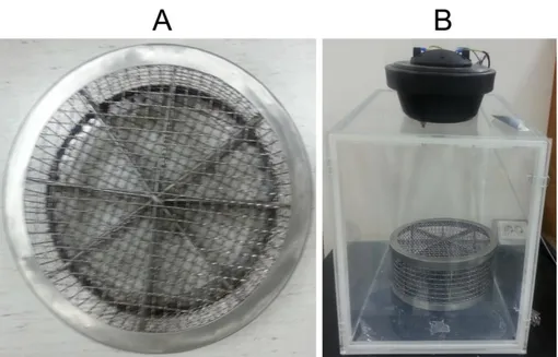
Noise exposure and auditory brainstem response (ABR) testing were performed in a foam-lined, single-walled soundproof room (TCA-500S, SONTEK, Gyeonggido, Korea). The noise exposure apparatus was specially designed with an acrylic frame (53 cm x 35 cm x 53 cm x 2 cm [length x width x height x thickness]; Seoul, Korea) with a speaker on top. White noise (300-10,000 Hz) was generated by a personal computer and amplifier (R150-plus, Inter M, Seoul, Korea) and delivered through the speaker (DH1A, Electro-Voice, Burnsville, MN, USA). C57BL/6 (pigmented) mice were exposed to 95 dB peak equivalent (PE) SPL white noise for only 3 hours to make transient thresholds shift model. To avoid inappropriate exposure to noise caused by the mice gathering together and hiding their heads during noise stimulation, a specially designed pie-shaped wire cage with 8 separated compartments was used, as described in a previous study (Fig. 1).23 Each separated partition contained a different mouse group.
8
Figure 1. Specially designed noise exposure cage and apparatus. A pie-shaped,
wire-mesh noise exposure cage with eight compartments (A). Noise exposure apparatus consisted of an acryl frame with the speaker attached on top of the frame (B).
4. Audiologic evaluation
The hearing level of each mouse was determined by measuring ABR thresholds using auditory evoked potentials workstation (Tucker-Davis Technologies, Alachua, FL, USA). Mice were anesthetized by intraperitoneal injection of Zoletil (30 mg/kg) and Rompun (10 mg/kg), and each ear of each mouse was stimulated with an ear probe sealed into the ear canal. Body temperature of the animal was maintained at 38℃ with an isothermic heating water-pad. Click and
9
tone-burst (4k, 8k, 16k, and 32kHz) stimuli were applied and the threshold was defined as the lowest intensity of stimulation that yielded a repeatable waveform based on an identifiable ABR wave V. The ABR was recorded every 1 or 2 weeks from 3 weeks of age in each mouse for evaluation of hearing thresholds with aging. For evaluation of NIHL, ABR was performed before the noise insult to confirm normal hearing function, and immediately after the noise exposure at 1, 3, 5, 10, and 30 days following the noise insult to determine the magnitude of threshold shift. Hearing thresholds are shown as mean ± SEM.
5. Histologic evaluation
At 8 and 20 weeks of age following ABR measurements, some mice were deeply anesthetized by intraperitoneal injections of 30 mg/kg Zoletil and 10 mg/kg Rompun, and both cochleae were removed from each mouse. Stapes were removed and a small hole was made in the cochlear apex with a fine pick. Fixative (4% formalin and 1% glutaraldehyde in 0.1M sodium phosphate buffer) was infused through this hole and the cochleae were immersed in fixative for 48 h at 4℃. Cochleae were kept in 10% EDTA in phosphate-buffered saline (PBS) for 3 days, and half of removed cochleae were dehydrated and embedded in paraffin. The paraffin blocks were sectioned at 5 ㎛ along the mid-modiolar axis. The sections were observed using hematoxylin-eosin staining. The others were washed by PBS for the fluorescent staining. After washing with PBS, the outer bony wall of each cochlea and the
10
tectorial membrane were gently removed and the organ of Corti was harvested with fine forceps beginning from the apex. The organ of Corti was stained with a fluorescent isothiocyanate (FITC)-conjugated phalloidin probe (Invitrogen, Carlsbad, CA, USA), washed and examined at high power magnification (x 600) under a fluorescence microscope and a confocal microscope to evaluate the damaged hair cells. Only the basal region could not be completely examined.
6. Statistical analysis
All values were presented as mean ± SEM. The serum retinol concentrations were logarithmically transformed to improve normality and to compensate for unequal variance and were analyzed by 2-way ANOVA followed by post hoc Bonferroni tests. 2-way ANOVA was performed to compare body weights and the hearing parameters between the age and diet groups. Post hoc analyses were performed with the Bonferroni test when comparing VAD group to the other groups. In addition, independent sample t tests were applied to determine between the time points when significant differences of auditory thresholds the two groups were seen in the NIHL model. The statistical analysis was carried out with SPSS (version 13.0, SPSS Inc., Chicago, IL, USA) software. Statistical significance was assumed for P<0.05.
11 III. RESULTS
1. Body weight and general morbidity of Vitamin A-deficient mice
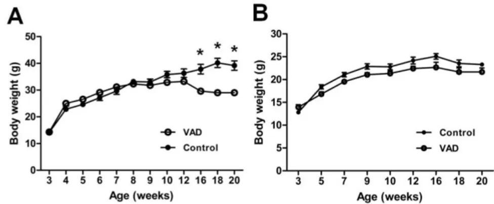
There were no significant differences in the growth pattern of the two groups in both albino and C57BL/C mice until 12 weeks, whereas the weight of VAD group in only albino mice was reduced significantly compared to that of the control group after 16 weeks (Fig. 2A and 2B). VAD mice showed no general morbidity at 8 weeks; however, on gross examination, the livers were yellowish and slightly enlarged in two of the 20-week-old VAD mice. It might suggest that the weight loss of VAD group was caused by the decreased function of immune system in intestine and liver.24,25
Figure 2. Body weight of each group in (A) albino mice and (B) pigmented
mice. (A) Mean body weights of control and VAD mice. Values are means ± SEM, n=15. Asterisks indicate difference from control mice at that age: *P<0.001. VAD; vitamin A deficient group.
2. Serum retinol level of vitamin A-deficient mice
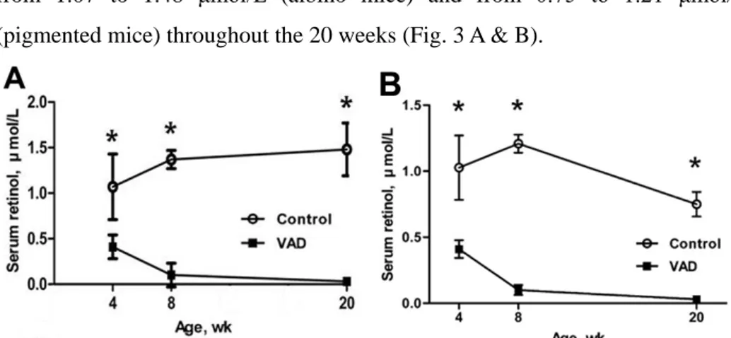
The serum retinol levels of VAD mice in albino and pigmented mice were 0.41±0.13 μmol/L and 0.41±0.07 μmol/L at 4 weeks, respectively. They decreased to 0.10±0.13 μmol/L and 0.10±0.04 μmol/L at 8 weeks, and almost below the detection limit (0.03 μmol/L) at 20 weeks, whereas those of control
12
mice in both mice strains were maintained in a relatively stable manner, ranging from 1.07 to 1.48 μmol/L (albino mice) and from 0.75 to 1.21 μmol/L (pigmented mice) throughout the 20 weeks (Fig. 3 A & B).
Figure 3. Serum retinol concentrations of control and VAD mice at 4, 8, and 20
weeks of age in albino (A) and pigmented mice (B). Values are means ± SEM,
n=4 except for 8 weeks, n=10. Asterisks indicate difference from control mice
at that age: *P<0.01.
3. The changes of hearing thresholds with aging
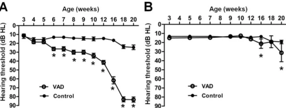
There was no statistically significant change of hearing thresholds until 20 weeks in albino control mice. However, albino VAD mice showed hearing loss after 6 weeks, and then hearing thresholds were rapidly increased after 12 weeks. The hearing levels eventually reached a status of deafness (> 80dB HL) in albino VAD mice at 18 weeks (Fig. 4A). In contrast, there was no significant change of hearing thresholds until 12 weeks in both control and VAD groups of pigmented mice. However, the hearing threshold of pigmented VAD mice was slightly increased only at 16 weeks and 20 weeks when compared to hearing threshold of pigmented control mice, and there was no deafness in both groups of pigmented mice (Fig. 4B).
13
Figure 4. Changes in hearing thresholds measured using auditory brainstem
responses with aging in albino (A) and pigmented mice (B). In albino mice, there are statistically significant differences between VAD and control group after 6 weeks and premature deafness (>80dB HL) was observed in VAD group after 18 weeks. Asterisks indicate difference from control mice at that age: *P<0.01. VAD; vitamin A deficient group. Number of individuals analyzed per group: 20 albino VAD, 18 albino control, 20 pigmented VAD, and 20 pigmented control mice.
4. Histological comparison between VAD group and control group in albino mice
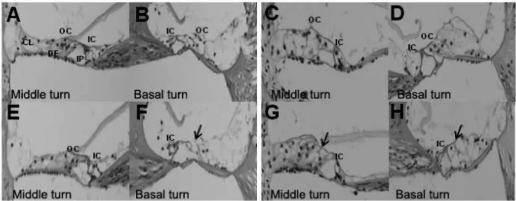
The histologic changes of the cochlea were analyzed in albino mice that showed hearing loss in VAD group. VAD mice showed damaged outer hair cells only in the basal turn at 8 weeks. These phenomena were more obvious at 20 weeks, and the damaged outer hair cells were observed in the middle turn as well as in the basal turn (Fig. 5 A-H).
14
Figure 5. Photographs of H&E stain of cochlea in control group (1st row; A-D)
and VAD group (2nd row; E-H) at 8 weeks (A,B,E and F) and 20 weeks (C,D,G and H) by light microscope (400x). Although only cochlear basal turn in VAD group (F) show some damaged outer hair cells at 8 weeks, old-aged VAD mice (20 weeks) show damages of outer hair cells in middle turn as well as basal turn (G and H). Black arrows indicate damaged outer hair cells. VAD; vitamin A deficient group.
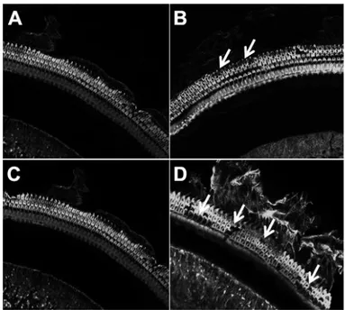
When fluorescence staining of the cochlea was performed, damaged outer hair cells were observed as blanket spaces in contrast to the normal greenish fluorescent stereocilia. 20-week-old mice were more severely damaged in both middle and basal turns compared to the young-aged mice (8 weeks) (Fig. 6 A-D). However, the findings in outer hair cells were not closely correlated with the changes of hearing thresholds with aging because there were no loss of inner hair cells and the extent of damaged outer hair cells was not severe compared to that of hearing loss. These findings suggest that other defects might also be involved such as loss of spiral ganglion neurons.
15
Figure 6. Photograph of FITC phallodin fluolescence stain of cochlea in control
group (A and C) and VAD group (B and D) at 8 weeks (A and B) and 20 weeks (C and D). Cochlear basal turn in VAD group (B and D) shows some stain-free areas with stereocilia loss, and defect is more severe in old-aged (20 weeks) mice (D), whereas the three rows of outer hair cell in control group show intact integrity (A and C). These findings were examined at high power magnification (x600) under a fluorescence confocal microscope to evaluate the damaged hair cells. White arrows indicate stereocilia loss. VAD; vitamin A deficient group.
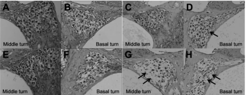
The VAD group had a greater neuronal loss than the control group in 20 weeks of age, while there was no difference of neuronal damage between the two groups in 8 weeks of age (Fig. 7 A-H). These findings suggested that multiple defects are involved in the increased hearing thresholds with aging.
16
Figure 7. Photomicrographs of spiral ganglion in control group (1st row; A-D)
and VAD group (2nd row; E-H) at 8 weeks (A,B,E and F) and 20 weeks (C,D,G and H) by light microscope (400x). A greater loss of neurons in middle turn as well as basal turn (G and H) was observed in old-aged VAD mice (20 weeks), whereas old-aged control group (C and D) showed smaller loss of neurons only in the basal turn. Black arrows indicate neuronal loss in spiral ganglion. VAD; vitamin A deficient group.
5. Changes in hearing thresholds after noise exposure in pigmented mice
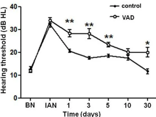
Since there was no change in the hearing thresholds with aging even under VAD condition in pigmented mice, noise trauma was given to VAD and control groups of pigmented mice in order to induce a transient threshold shift (TTS). When analyzing the data of click ABR, I observed that the hearing thresholds of control mice were increased immediately after noise damage. Then, the hearing loss was gradually recovered and eventually restored completely after 30 days. Although there was no significant difference immediately after noise exposure, a delay of hearing recovery was observed in VAD mice. In addition, VAD mice showed some threshold shift (about 10dB) compared with control mice after 30 days (Fig. 8).
17
Figure 8. Changes in hearing thresholds in pigmented mice measured using
auditory brainstem responses (in response to click stimuli) after 3 hours of exposure to noise (95 dB SPL). There is statistically significant differences of hearing restoration between VAD group and control group 1 day after noise exposure and hearing threshold shift of 10-20 dB nHL remained after 30 days in VAD group only. Asterisks indicate difference from control mice at that age: *P<0.05, **P<0.01. BN; before noise, IAN; Immediately after noise, VAD; vitamin A deficient group.
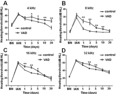
When analyzed by frequency, control mice showed complete recovery in all frequencies except 32 kHz. However, VAD mice showed delayed hearing restoration in 8, 16, and 32 kHz, and there was incomplete recovery of hearing thresholds in 4 kHz. Considering these changes in hearing thresholds in 4 different frequencies, there was no statistically significant difference of hearing thresholds between the two groups except in the low frequency (4 kHz) when measured 30 days after noise exposure. In all frequencies, the hearing restoration of VAD group was delayed from 1 day after noise exposure compared to that of control group (Fig. 9A-D).
18
Figure 9. Changes in hearing thresholds in pigmented mice at different
frequencies measured using auditory brainstem responses (in response to tone burst stimuli) after 3 hours of exposure to noise. There is statistically significant differences of hearing restoration between VAD and control group from one day after noise exposure. Asterisks indicate difference from control mice at that age: *P<0.05, **P<0.01. BN; before noise exposure, IAN; Immediately after noise exposure, VAD; vitamin A deficient group.
6. Melanocyte recruitment in pigmented mice after VAD diet
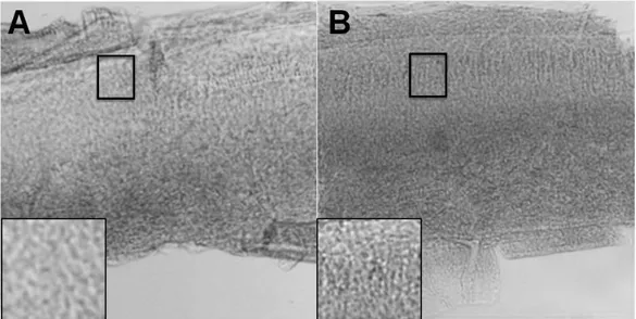
Because I made the hypothesis that cochlear melanocytes play a protective role against hearing loss, I compared the proportions of cochlear melanocytes between the two groups in pigmented mice. The melanosomes containing melanin granules increased in VAD group compared to the control group in pigmented mice (Fig. 10).
19
Figure 10. The density of melanosomes containing melanin granules in control
(A) and VAD group (B) in basal turn of pigmented mice. The granules increased in VAD group (B) compared to the control group (A). Insets: magnification of the stria vascularis to compare the density between the two groups.
20 IV. DISCUSSION
In this study, I showed that VAD diet induced severe hearing loss with age in albino type (ICR) mice through the evaluation of changes in hearing thresholds and results of histopathology. At 8 weeks of age, which is early stage of increasing auditory thresholds, the damaged outer hair cells were already observed in the basal turn, which eventually extended to the middle turn with time in albino VAD mice. Old aged (20 weeks) VAD group showed a greater loss of neurons in the middle and basal turns compared to the control group in albino mice. These findings were closely correlated with the changes in hearing thresholds with aging. However, the results of this study that VAD mice develop hearing loss with aging run counter to some of the previous studies of VAD mouse models. That is, some studies reported no significant morphologic changes related to hearing loss after VAD diet.12,13 The difference of results between this study and the previous studies might stem from two factors. First, there could be some methodological problems in making a VAD animal model. The animals were fed the VAD diet after birth in the previous studies dealing with the hearing in VAD animal model. In contrast, I started feeding VAD diet to the animal models from 7-10 days of gestation. To decrease the plasma retinol level, a vitamin A-free diet needs to last until the hepatic reserves are depleted. However, the previous studies did not fulfill these requirements of complete VAD condition. Second, there could be the difference of genetic background between different animal species used in the previous studies and
21
my study. The previous studies used different animals such as guinea pig and rat, while I used mice. The hearing thresholds were steeply increased after 12 weeks of ages in albino VAD mice. This result needs to be interpreted carefully because the general morbidity is changed after 12 weeks of age by impaired immunity of internal organs such as intestine.26,27
I identified that albino VAD mice show profound premature deafness and both groups of pigmented (C57BL/6) mice did not show hearing loss with aging, although there were significant differences of hearing thresholds between the two groups in pigmented mice at 16 and 20 weeks. These differences of hearing thresholds in pigmented VAD mice at 16 and 20 weeks were caused by middle ear effusion, which were identified using 2.7 mm endoscope. Only albino VAD mice showed progressive increases of hearing thresholds after 6-week of age, compared with the other mice groups (albino control mice, pigmented VAD mice, and pigmented control mice). This difference between the two strains might indicate the role of cochlear melanocytes. In addition, such damage might be caused by the absence of intrinsic and extrinsic antioxidants. That is, the lack of a protective role of vitamin A as well as cochlear melanocyte could result in hearing loss. Pigmented VAD mice also showed increased melanocytes in stria vascularis compared to pigmented wild-type mice. Such increases might provide an explanation to the reason why the pigmented VAD mice did not become deaf prematurely under the condition of VAD.
22
control and VAD groups after noise damage in pigmented mice. The differences were similar to the result of a previous study that VAD increased the sensitivity of the inner ear to noise insult.11 This might be interpreted that the cochlear melanocytes no longer play a protective role against noise damage. In other words, VAD group could no longer scavenge oxidative stress because of previously increased cochlear melanocytes before noise insult, while cochlear melanocytes increase and then may play a protective role against oxidative stress after noise exposure in control group.
Furthermore, my study presented that pigmented VAD mice had poorer hearing restoration after noise exposure compared to pigmented control mice, and the delayed hearing recovery was shown especially in low frequency (4 kHz). Pigmented VAD mice showed increased cochlear melanocytes before noise exposure, which could imply that the increased melanocytes play a protective role against ARHL. However, there was no more increase in the number of melanocytes in pigmented VAD mice after noise damage. It was hypothesized that there is a limitation of the increase in the number of melanocytes in a cochlea, which might cause reduction of protective role of melanocytes in pigmented VAD mice after noise insults.
The protective role of cochlear melanocytes against hearing loss has been revealed in various published studies.20-22,28-30 Epidemiologic studies of hearing loss in adults have demonstrated that the odds of hearing loss are substantially lower in black than in white individuals.22 In the epidemiologic studies, it was
23
hypothesized that skin pigmentation as a marker of melanocytic functioning mediates this observed association and that skin pigmentation is associated with hearing loss independent of race/ethnicity. In addition, other studies suggested that increased melanin in the inner ear may subsequently protect the cochlea against age-related cellular declines and hearing loss in darker skinned individuals.31,32 Nevertheless, the association of coat color, cochlear pigmentation, and hearing loss in mice and other animal models remains unclear.18,30,32 However, many of these prior experiments have been confounded by comparing animals of different genetic backgrounds, and recent work using coisogenic mouse strains has demonstrated that the absence of strial melanin is strongly associated with marginal cell loss and endocochlear potential decline.32 Interestingly in my study, pigmented VAD mice did not show enough protection against NIHL, while pigmented control mice showed a normal hearing recovery after noise damage. It could be strong evidence that extrinsic vitamin A plays an important role in the prevention of hearing loss.
VAD is still common in developing countries; it is estimated that there are more than 100 million preschool children with VAD throughout the world.33 The prevalence of subclinical VAD extends even further and is observed even in developed countries.34 In addition, preschool children with VAD in southern Asia have high prevalence in hearing loss, which can be prevented with high dose vitamin A supplementation.9,10 Therefore, the results of my study, showing that VAD has an impact on hearing loss, might have a significant meaning for
24
the prevention and treatment of hearing loss.
V. CONCLUSIONS
My data demonstrated that VAD with albinism led to the premature ARHL, and VAD with pigmentation showed poorer recovery after noise exposure when compared to the control group. In addition, cochlear melanocytes had a protective role by increasing melanin level under VAD state. These findings were associated with the serum retinol level in both control and VAD groups and might imply that vitamin A has a protective role against stressful insults such as aging and noise exposure. My results also suggest that patients with pigmented disorder may as well avoid conditions of noise exposure under conditions of VAD. Since my results were derived from only morphological and functional aspects, further research is needed to investigate the specific mechanism in the protective role of vitamin A against hearing loss.
25
REFERENCES
1. Michikawa T, Nishiwaki Y, Kikuchi Yet al. Serum levels of retinol and other antioxidants for hearing impairment among Japanese older adults. The journals of gerontology Series A, Biological sciences and medical sciences 2009; 64:910-5.
2. National Institute of Aging. Hearing Loss. Age Page.
www.niapublications.org/agepages /hearing.asp. Bethesda: U.S. Department of Health and Human Services, National Institutes of Health; September 2002; Reprinted August 2005.
3. Nelson DI, Nelson RY, Concha-Barrientos M, Fingerhut M. The
global burden of occupational noise-induced hearing loss. American journal of industrial medicine 2005; 48:446-58.
4. Wald G. The molecular basis of visual excitation. Nature 1968; 219:800-7.
5. De Luca LM. Retinoids and their receptors in differentiation, embryogenesis, and neoplasia. FASEB J 1991; 5:2924-33.
6. Smith SM, Levy NS, Hayes CE. Impaired immunity in vitamin
A-deficient mice. The Journal of nutrition 1987; 117:857-65.
7. World Health Organization. 2009. Global Prevalence of Vitamin A Deficiency in Populations at Risk 1995-2005: WHO Global Database on Vitamin A Deficiency. World Health Organization, Geneva, Switzerland.,
26
2009.
8. Gopinath B, Flood VM, McMahon CMet al. Dietary antioxidant intake
is associated with the prevalence but not incidence of age-related hearing loss. The journal of nutrition, health & aging 2011; 15:896-900.
9. Schmitz J, West KP, Jr., Khatry SKet al. Vitamin A supplementation in preschool children and risk of hearing loss as adolescents and young adults in rural Nepal: randomised trial cohort follow-up study. Bmj 2012; 344:d7962.
10. Wieringa FT, Dijkhuizen MA, Berger J. Vitamin A supplementation in
children and hearing loss. Bmj 2012; 344:d7603.
11. Biesalski HK, Wellner U, Weiser H. Vitamin A deficiency increases noise susceptibility in guinea pigs. The Journal of nutrition 1990; 120:726-37. 12. Chole RA, Quick CA. Estimate of vitamin A in the guinea pig cochlea.
The Annals of otology, rhinology, and laryngology 1978; 87:380-2.
13. Bichler E, Wieser M. The influence of a chronic vitamin A deficiency on the rat cochlea. Archives of oto-rhino-laryngology 1982; 234:175-9. 14. Wolff D. Melanin in the inner ear. Archives of otolaryngology 1931;
14:195.
15. Bush WD, Simon JD. Quantification of Ca(2+) binding to melanin supports the hypothesis that melanosomes serve a functional role in regulating calcium homeostasis. Pigment cell research / sponsored by the European Society for Pigment Cell Research and the International Pigment Cell Society
27
2007; 20:134-9.
16. Drager UC. Calcium binding in pigmented and albino eyes.
Proceedings of the National Academy of Sciences of the United States of America 1985; 82:6716-20.
17. Seagle BL, Rezai KA, Gasyna EM, Kobori Y, Rezaei KA, Norris JR,
Jr. Time-resolved detection of melanin free radicals quenching reactive oxygen species. Journal of the American Chemical Society 2005; 127:11220-1.
18. Conlee JW, Abdul-Baqi KJ, McCandless GA, Creel DJ. Effects of aging on normal hearing loss and noise-induced threshold shift in albino and pigmented guinea pigs. Acta oto-laryngologica 1988; 106:64-70.
19. Ohlemiller KK, Gagnon PM. Genetic dependence of cochlear cells and
structures injured by noise. Hearing research 2007; 224:34-50.
20. Steel KP, Barkway C. Another role for melanocytes: their importance for normal stria vascularis development in the mammalian inner ear. Development 1989; 107:453-63.
21. Motohashi H, Hozawa K, Oshima T, Takeuchi T, Takasaka T.
Dysgenesis of melanocytes and cochlear dysfunction in mutant
microphthalmia (mi) mice. Hearing research 1994; 80:10-20.
22. Lin FR, Maas P, Chien W, Carey JP, Ferrucci L, Thorpe R. Association of skin color, race/ethnicity, and hearing loss among adults in the USA. Journal of the Association for Research in Otolaryngology : JARO
28
2012; 13:109-17.
23. White CH, Ohmen JD, Sheth Set al. Genome-wide screening for genetic loci associated with noise-induced hearing loss. Mammalian genome : official journal of the International Mammalian Genome Society 2009; 20:207-13.
24. Yang Y, Yuan Y, Tao Y, Wang W. Effects of vitamin A deficiency on
mucosal immunity and response to intestinal infection in rats. Nutrition 2011; 27:227-32.
25. Venu M, Martin E, Saeian K, Gawrieh S. High prevalence of vitamin
A deficiency and vitamin D deficiency in patients evaluated for liver transplantation. Liver transplantation : official publication of the American Association for the Study of Liver Diseases and the International Liver Transplantation Society 2013; 19:627-33.
26. Iwata M, Hirakiyama A, Eshima Y, Kagechika H, Kato C, Song SY. Retinoic acid imprints gut-homing specificity on T cells. Immunity 2004; 21:527-38.
27. Chang SY, Cha HR, Chang JHet al. Lack of retinoic acid leads to increased langerin-expressing dendritic cells in gut-associated lymphoid tissues. Gastroenterology 2010; 138:1468-78.
28. Tachibana M, Hara Y, Vyas Det al. Cochlear disorder associated with melanocyte anomaly in mice with a transgenic insertional mutation. Molecular and cellular neurosciences 1992; 3:433-45.
29
29. Hong CK, Lee MH, Jeong KH, Cha CI, Yeo SG. Clinical analysis of
hearing levels in vitiligo patients. European journal of dermatology : EJD 2009; 19:50-6.
30. Bartels S, Ito S, Trune DR, Nuttall AL. Noise-induced hearing loss: the effect of melanin in the stria vascularis. Hearing research 2001; 154:116-23.
31. Hood JD, Poole JP, Freedman L. The influence of eye colour upon temporary threshold shift. Audiology : official organ of the International Society of Audiology 1976; 15:449-64.
32. Ohlemiller KK. Mechanisms and genes in human strial presbycusis from animal models. Brain research 2009; 1277:70-83.
33. West KP, Jr. Extent of vitamin A deficiency among preschool children and women of reproductive age. The Journal of nutrition 2002; 132:2857S-66S.
34. Kim YN, Giraud DW, Cho YO, Driskell JA. Vitamin A inadequacy observed in a group of 2- to 6-year-old children living in Kwangju, Republic of Korea. International journal for vitamin and nutrition research Internationale Zeitschrift fur Vitamin- und Ernahrungsforschung Journal international de vitaminologie et de nutrition 2007; 77:311-9.
30
ABSTRACT(IN KOREAN)
쥐에서 비타민A 결핍이 노화 관련 및
소음 유발 감각신경성 난청에 미치는 영향
< 지도교수 이 원 상 >
연세대학교 대학원 의학과
심 대 보
비타민A 결핍은 사람 및 동물에서 피부 분화, 생식 및 면역 체계 기능 등에서 다양한 병리적 표현형을 일으킨다. 비록 많은 연구자들이 비타민A 결핍이 사람과 동물에서 청력을 감소시킨다고 보고하였으나, 내이에 대한 비타민A 결핍의 영향은 아직 명확하지 않은 상태이다. 따라서 본 연구자는 비타민A 결핍 마우스 모델을 만들어 청력의 변화 및 내이의 조직학적 변화를 관찰함으로써 비타민A 결핍이 내이에 미치는 영향을 확인하고자 하였다. 두 계통의 마우스(ICR; 흰쥐와 C57BL/6, 색소침착쥐)에서 청성 뇌간 반응검사를 시행하여 분석하였다. 각 계통에서, 마우스는 임의의 두 군으로 나누어 임신 7일째부터 한 군에게는 비타민A 결핍 사료를, 다른 군에게는 대조군 사료를 투여하였다. 청력 역치는 생 후 20주까지 청성 뇌간 반응검사를 이용하여 측정하였다. 비타민A 결핍 군의 혈중 비타민A 농도가 대조군의 절반 이하에 도달한 이후, 흰쥐 비타민A31 결핍 군에서만 청력 역치가 다른 세 군들(흰쥐 대조군 및 색소침착쥐 두 군)에 비해 유의하게 증가하였다(p<0.05). 또한, 흰쥐에서 조직학적 소견을 확인하였을 때, 조직학적 소견이 노화에 따른 난청의 정도와 밀접한 연관성을 가지는 것을 확인하였다. 그러나, 색소침착쥐에 소음을 주었을 때, 소음 노출 후에는 색소침착 비타민A 결핍 군에서 대조군보다 청력의 회복이 지연되는 것을 관찰하였다. 따라서 본 연구자들은 비록 와우 멜라닌 세포들이 색소침착 비타민A 결핍 마우스에서는 일부 난청에 대한 보호 역할을 하지만, 비타민A 결핍 마우스의 청력 역치는 대조군에 비해 노화 및 소음 손상에 의해 악화될 수 있음을 확인하였다. 이는 비타민A 결핍이 쥐에서 노인성 난청의 발생 및 소음성 난청의 회복에 영향을 줄 수 있음을 의미한다.
