ISSN 2234-3806 • eISSN 2234-3814
http://dx.doi.org/10.3343/alm.2016.36.5.475
Frequency and Clinical Characteristics of
Intrachromosomal Amplification of Chromosome 21 in
Korean Childhood B-lineage Acute Lymphoblastic
Leukemia
Jieun Kim, M.D.1, Chuhl Joo Lyu, M.D.2, Saeam Shin, M.D.3, Seung-Tae Lee, M.D.1, and Jong Rak Choi, M.D.1
Department of Laboratory Medicine1, Yonsei University College of Medicine, Seoul; Department of Pediatrics2, Yonsei Cancer Research Center, Yonsei
University College of Medicine, Seoul; Department of Laboratory Medicine3, Hallym University College of Medicine, Kangnam Sacred Heart Hospital, Seoul,
Korea
Background: Intrachromosomal amplification of chromosome 21 (iAMP21) is known to
be associated with poor prognosis in B-cell ALL (B-ALL). To determine the frequency and clinical characteristics of iAMP21 in Korean B-ALL patients, we performed FISH and mul-tiplex ligation-dependent probe amplification (MLPA) analyses.
Methods: A total of 102 childhood B-ALL patients were screened with ETV6-RUNX1 FISH
probes (Abbott Molecular, USA). The presence of an iAMP21 was confirmed by using MLPA P327 iAMP21-ERG probemix (MRC Holland, The Netherlands).
Results: iAMP21 was detected in one of the screened B-ALL patients (1/102 patients,
1.0%) who presented the ALL immunophenotype and complex karyotype at initial diagno-sis. The patient relapsed twice after bone marrow transplantation. MLPA showed 12.5-Mb and 4.28-Mb regions of amplification and deletion, respectively.
Conclusions: The frequency of iAMP21 is considerable in Korean pediatric patients. Our
report suggests that iAMP21 in childhood B-ALL has very unfavorable impact on patient’s prognosis. Additional methods such as MLPA analysis is essential to rule out patients with equivocal interphase FISH results.
Key Words: iAMP21, RUNX1, ERG, FISH, MLPA, Acute lymphoblastic leukemia, Korean,
Childhood
Received: December 1, 2015 Revision received: March 30, 2016 Accepted: May 11, 2016
Corresponding author: Seung-Tae Lee Department of Laboratory Medicine, Yonsei University College of Medicine, 50-1 Yonsei-ro, Seodaemun-gu, Seoul 03722, Korea
Tel: +82-2-2228-2450 Fax: +82-2-364-1583 E-mail: [email protected]
Co-corresponding author: Saeam Shin Department of Laboratory Medicine, Hallym University College of Medicine, Kangnam Sacred Heart Hospital, 1 Singil-ro, Yeongdeungpo-gu, Seoul 07441, Korea Tel: +82-2-829-5695
Fax: +82-2-847-2403 E-mail: [email protected]
© The Korean Society for Laboratory Medicine This is an Open Access article distributed under the terms of the Creative Commons Attribution Non-Commercial License (http://creativecom-mons.org/licenses/by-nc/4.0) which permits unrestricted non-commercial use, distribution, and reproduction in any medium, provided the original work is properly cited.
INTRODUCTION
The current treatment strategies for B-cell ALL (B-ALL) are based on several factors, including patient-related parameters as well as intrinsic characteristics of particular disease subtypes. Abnormalities with the most significant effects on the manage-ment of B-ALL are t(12;21)(p13;q22)/ETV6-RUNX1 and high
hyperdiploidy for good risk management, and t(9;22)(q34;q11)/ BCR-ABL1, t(4;11)(q21;q23)/MLL-AFF1, and near-haploidy/low hypodiploidy for high-risk stratification. However, a substantial proportion of childhood leukemias remain genetically undefined or the correlation of genetic abnormalities with clinical outcome is not fully understood [1]. The detection of chromosomal ab-normalities by conventional cytogenetic analysis is an essential
approach for the diagnosis, and its utility has been expanded by the development of FISH [2]. The use of FISH has enabled the identification of a number of alterations in patients with failed cytogenetic results and has led to the detection of cryptic abnor-malities that cannot be determined [3].
Intrachromosomal amplification of chromosome 21 (iAMP21), discovered during routine screening for the presence of the ETV6-RUNX1 fusion by FISH analysis [3], is usually defined as three or more additional copies of RUNX1 signals on a single abnormal chromosome 21 (a total of five or more RUNX1 sig-nals per cell) [4]. This chromosomal abnormality is found in 1.5-2% of ALL cases [5] and is characterized by a low white blood cell count and a pre-B/common immunophenotype in children and adolescent populations [2]. A significant finding was that patients with iAMP21 had an inferior outcome when treated with standard therapy and an improved outcome on in-tensive therapy [6, 7]. Therefore, an accurate detection of this abnormality is very important for the choice of treatment.
The morphology of the abnormal chromosome 21 is highly variable among cases and a variety of cytogenetic nomencla-tures such as add(21), dup(21), and der(21) were used to de-scribe the iAMP21 abnormalities, with the loss of chromosome 21 and an associated gain of a marker chromosome being the most common [8]. Furthermore, in cases where metaphase chromosomes are absent, other methods should be applied to distinguish iAMP21 from high hyperdiploidy with extra copies of chromosome 21. These include a subtelomeric FISH probe for chromosome 21 to demonstrate that the number of RUNX1 sig-nals is greater than the number of chromosomes 21 [4], array comparative genomic hybridization, single nucleotide polymor-phism array, and multiplex ligation-dependent probe amplifica-tion (MLPA) [9].
Because little data are available on the frequency and clinical characteristics of iAMP21 in Korean pediatric B-ALL patients, we comprehensively analyzed FISH data and further performed MLPA analysis to confirm suspected and equivocal cases.
METHODS
1. Patient selection and FISH analysis
A total of 102 pediatric patients diagnosed with B-ALL between February 2009 and June 2015 were retrospectively investigated in this study. Of the 102 pediatric B-ALL patients, 65 were men (63.7%) and 37 were women (36.3%), and the median age at diagnosis was 5 yr (range, 0 to 19 yr) (Table 1). Written in-formed consent was obtained from the patients or patients’
par-ents for genetic analysis.
The chromosomal abnormalities were comprehensively ana-lyzed on the basis of G-banding karyotype, FISH, and multiplex reverse transcription-PCR (RT-PCR). The screening of iAMP21 was performed by using FISH probe, ETV6-RUNX1 (TEL-AML1) dual color extra signal probe (Abbott Molecular, Abbott Park, IL, USA). For interphase FISH analysis, the slides were subjected to hybridization according to the manufacturer’s instructions. The slides were counterstained with 4,6-diamidino-2-phenylin-dole. The slides were viewed at a magnification of 400×, and the number of interphase cells with five or more RUNX1 signals was counted; a total of 300 cells were counted for each sample.
2. Detection of recurrent translocations
Total RNA was extracted from EDTA-anticoagulated blood sam-ples by using the QIAamp RNA Blood Mini kit (Qiagen, Hilden, Germany). cDNA was synthesized by using the Transcriptor First Strand cDNA Synthesis kit (Roche Diagnostics Corp., Indi-anapolis, IN, USA). To determine recurrent translocations, RT-PCR was performed by using the HemaVision kit (DNA Technol-ogy, Aarhus, Denmark) according to the manufacturer’s
instruc-Table 1. Patient characteristics
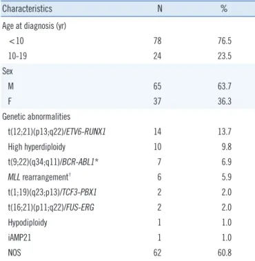
Characteristics N %
Age at diagnosis (yr)
<10 78 76.5 10-19 24 23.5 Sex M 65 63.7 F 37 36.3 Genetic abnormalities t(12;21)(p13;q22)/ETV6-RUNX1 14 13.7 High hyperdiploidy 10 9.8 t(9;22)(q34;q11)/BCR-ABL1* 7 6.9 MLL rearrangement† 6 5.9 t(1;19)(q23;p13)/TCF3-PBX1 2 2.0 t(16;21)(p11;q22)/FUS-ERG 2 2.0 Hypodiploidy 1 1.0 iAMP21 1 1.0 NOS 62 60.8
*Five patients with e1a2, one with b2a2, and one with b3a2 fusion tran-scripts were included; †Three patients with t(4;11)(q21;q23)/MLL-AFF1, two
with t(11;19)(q23;p13.3)/MLL-MLLT1, and one with t(1;11)(p32;q23)/MLL-EPS15 were included.
Abbreviations: M, male; F, female; iAMP21, intrachromosomal amplification of chromosome 21; NOS, not otherwise specified.
tions. Recurrent genetic abnormalities were detected in 40 (39.2%) patients, including ETV6-RUNX1 (14 patients, 13.7%), high hyperdiploidy (10 patients, 9.8%), BCR-ABL1 (7 patients, 6.9%), MLL rearrangement (6 patients, 5.9%), TCF3-PBX1 (2 patients, 2.0%), FUS-ERG (2 patients, 1.2%), and hypodiploidy (1 patient, 1.0%) in the order of decreasing frequency.
3. DNA extraction and MLPA analysis
DNA was extracted from bone marrow samples of patients and controls by using the QIAamp DNA Blood Mini kit (Qiagen) ac-cording to standard procedures. The presence of iAMP21 was retrospectively evaluated in three patients by using SALSA MLPA P327-B1 iAMP21-ERG probemix (MRC Holland, Amster-dam, The Netherlands), including 46 probes encompassing a region around 21q11.2 to 21q22.3, according to the manufac-turer’s instructions. Hybridized probes were amplified on a C1000 thermal cycler (Bio-Rad, Hercules, CA, USA). Amplicons were separated on an ABI 3730 capillary sequencer (Applied Biosystems, Foster City, CA, USA). The data were analyzed by using GeneMarker software (Softgenetics, State College, PA,
USA). The relative fluorescence peak ratios were calculated from patients’ peak heights divided by control’s peak heights. Relative ratios between 0.75 and 1.3 were considered normal, whereas those below 0.75 and above 1.3 indicated loss or gain of genomic material.
RESULTS
1. FISH and MLPA results
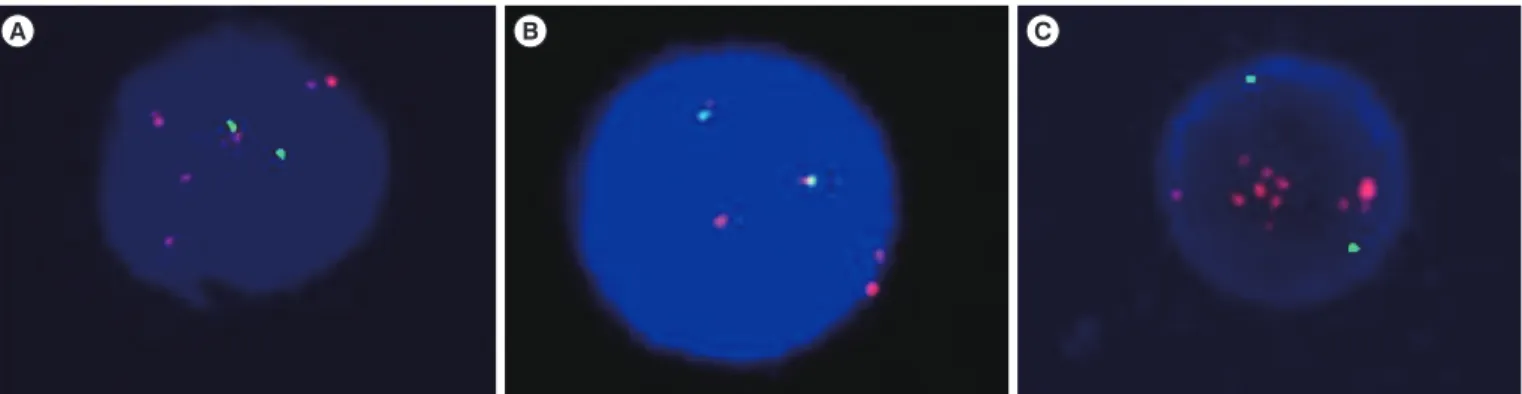
FISH screening revealed three patients with more than five RUNX1 signals (Fig. 1). The clinical characteristics of the three patients are summarized in Table 2. MLPA analysis revealed amplification of chromosome 21 in only patient C (Fig. 2). The 32 probes encompassing a 12.5-Mb region around 21q21.3 to 21q22.3, which include NCAM2, BACH1, TIAM1, OLIG2, KCNE2, RUNX1, SIM2, HLCS, DYRK1A, KCNJ6, ERG, ETS2, PSMG1, TMPRSS2, and PIPK4, showed amplification (Fig. 3). The average relative ratio of probes in amplified regions was 2.82 (range, 1.87-3.50), indicating the presence of more than five allele copies compared with the two alleles in normal
con-Table 2. Clinical presentation at diagnosis of the three patients with RUNX1 signal increment by FISH Age (yr)/Sex
Initial CBC
Immunophenotype abnormalitiesGenetic interphase FISH RUNX1 gain by
(%) Karyotype WBC (×109/L)/Hb (g/dL)/ Platelet (×109/L ) A 6/M 4.48/8.7/117 CD10, CD13, CD19, CD33, CD34, CD45, CD79a, and TdT ETV6-RUNX1 17.28 46,XY[20] B 18/F 71.23/4.1/29 CD10, CD13, CD19, CD20, CD22, CD33,
CD38, CD45, CD79a, HLA-DR, and TdT
ETV6-RUNX1 4.88 46,XX[17]
C 9/M 9.16/6.7/15 CD7, CD10, CD19, CD34, CD45, CD79a,
and TdT iAMP21* 34.8%* 44,XY,add(4)(p15.2),-5,i(7)(p10), -21,-22,+mar[16]/45,idem,+7[4]
*iAMP21 in patient C was detected in the second bone marrow study after 1 week of induction chemotherapy.
Abbreviations: M, male; F, female; CBC, complete blood count; WBC, white blood cell count; TdT, terminal deoxynucleotidyl transferase; iAMP21, intrachro-mosomal amplification of chromosome 21.
Fig. 1. FISH analysis using ETV6-RUNX1 dual fusion probe revealed increments of the RUNX1 signal in three patients (A, B, and C). The RUNX1 signals are indicated in orange, and the ETV6 signals are indicated in green.
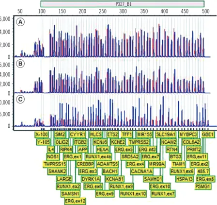
Fig. 2. Multiplex ligation-dependent probe amplification analysis of three patients (A, B, and C). The control peaks are indicated in red, and the patients’ peaks are indicated in blue; amplification of multiple probe sites on 21q21.1-21q22.3 was observed in patient C.
Fig. 3. Schematic representation of regions of amplification and deletion on chromosome 21. The x-axis in the graph indicates the genomic position of the multiplex ligation-dependent probe amplification (MLPA) probe site mapped to the human reference genome hg19. The y-axis indicates ratios of the patients’ fluorescence peak height relative to the controls’ peak height, as determined by MLPA analysis. Regions of amplification are highlighted in red and regions of deletion are highlighted in blue.
6,000 4,000 2,000 0 6,000 4,000 2,000 0 0,000 5,000 0 A B C 50 100 150 200 250 300 350 400 450 500 Position 15,000,000 20,000,000 25,000,000 30,000,000 35,000,000 40,000,000 45,000,000 50,000,000 P327_B1 3.0 2.0 1.0 0 Ratio
trols. MIR99A on the proximal site of the amplified region and TFF1, ITGB2, SLC19A1, COL6A2, and PRMT2 on the distal site of the amplified region showed deletion signals. The average relative ratio of probes in the deleted regions was 0.62 (range, 0.53-0.72).
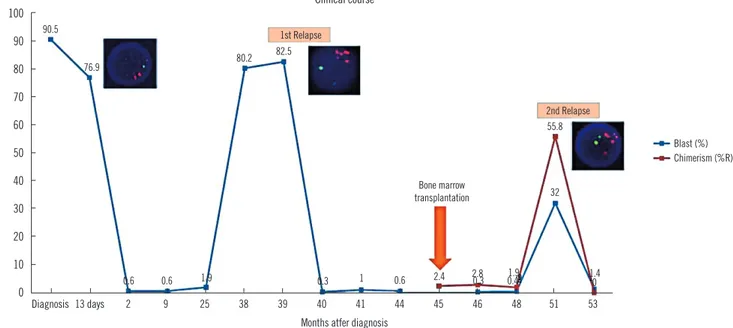
2. Clinical presentation and course of the patient with
iAMP21
A 9-yr-old boy, the patient C, was admitted because of general weakness for three weeks. At initial diagnosis, he showed the complex karyotype 44,XY,add(4)(p15.2),-5,i(7)(p10),-21, -22,+mar[16]/45,idem,+7[4]. At follow-up one week after the initiation of high-risk chemotherapy comprising the drugs vin-cristine, prednisolone, and L-asparaginase, a bone marrow ex-amination revealed 76.9% residual blasts in the observed mar-row cells and the FISH analysis revealed a 34.8% RUNX1 signal amplification of interphase cells. Chemotherapy with vincristine was maintained for three years after diagnosis; however, a bone marrow examination showed relapse of B-ALL, and the amplifi-cation of the RUNX1 signal was also observed. High-risk che-motherapy with vincristine was reinitiated to treat the relapsed B-ALL and bone marrow transplantation from a sibling donor was performed after conditioning chemotherapy. The patient re-mained in complete remission for eight months; however, a fol-low-up bone marrow examination revealed a second relapse of B-ALL. After completion of the clofarabine-based reinduction
therapy, a complete hematological remission was achieved. The clinical course of the patient is summarized in Fig. 4.
DISCUSSION
iAMP21 is a primary cytogenetic change, which remains con-stant in structure between diagnosis and relapse [10]. The ALL patients with iAMP21 showed inferior outcomes than those from the other ALL subgroups, including t(9;22), low hypoploidy, and MLL translocation [6]. Our patient also showed an aggressive course, with the first relapse occurring after chemotherapy and the second relapse after bone marrow transplantation, suggest-ing that one of the effects of iAMP21 is a poor prognosis.
Previous studies reported a common region of amplification spanning 6.6 Mb of chromosome 21 that almost always con-tained RUNX1; however, other involved regions varied among patients [8, 11]. In addition, a 3.3-Mb deleted region in the telo-mere was found in a large proportion of patients [10]. The am-plified regions of our patient overlapped with the commonly re-ported amplified regions, including RUNX1 and ERG [8]. The mechanism of this copy number alteration has been explained as chromothripsis following breakage-fusion-bridge cycles, which is often combined with deletions of RB1, ETV6, CDKN2A/ B, and IKZF1 [9].
Currently, the recommended laboratory test to detect iAMP21 is FISH, which can also determine copy number, with probes
Fig. 4. Clinical course of patient C. The x-axis in the graph indicates the time (months) after diagnosis. The y-axis indicates the percent of blast count in bone marrow (blue line) and percent recipient chimerism as measured by short tandem repeat analysis (red line). RUNX1 signal amplification by FISH analysis was detected at 13 days after diagnosis, first relapse, and second relapse.
100 90 80 70 60 50 40 30 20 10 0 Diagnosis 13 days 2 9 25 38 39 40 41 44 45 46 48 51 53
Months atfer diagnosis
Clinical course 90.5 76.9 0.6 0.6 1.9 0.3 1 0.6 2.4 2.80.3 1.90.4 0 32 55.8 1.4 80.2 82.5 1st Relapse 2nd Relapse Bone marrow transplantation Blast (%) Chimerism (%R)
specific for RUNX1 [12]. However, the stratification of patients with interphase cells should be made with caution, because ad-ditional RUNX1 signals are also seen in extra copies of chromo-some 21, which are characteristic of high-hyperdiploid ALL [7]. The characteristic signal patterns of iAMP21 are noted as clus-tered RUNX1 signals, with one signal, located apart, represent-ing the normal chromosome 21 [4]. However, because con-cerns are still present, a distinctive genomic profile of chromo-some 21 needs to be used to confirm the accuracy of iAMP21 diagnosis, such as by single nucleotide polymorphism array analysis [13] or FISH with the addition of a subtelomeric probe specific for chromosome 21 [11]. We suggest that MLPA is also helpful to verify iAMP21 in clinical settings.
MLPA is a rapid multiplex PCR-based technique that allows the relative quantification of multiple gene sites in a single test [14]. Although MLPA has a challenge in detection of a low per-centage of positive cells in sample, it targets very small se-quences and can distinguish between those differing by a single nucleotide.
To our knowledge, no study has reported a case of iAMP21 in Korea. The prevalence of iAMP21 in childhood leukemia in our study (1/102 patients, 1.0%) is comparable to that of previous reports [5, 7, 12]. In our report, the impact of the iAMP21 on disease course appeared to be very unfavorable. To determine the exact frequency and clinical impact of iAMP21 in Korean patients, appropriate test strategies to detect iAMP21 are war-ranted. To consider the prognostic implications of iAMP21 in patients with ALL, we suggest the inclusion of the MLPA test to detect iAMP21 in the initial diagnosis of ALL.
Authors’ Disclosures of Potential Conflicts of
Interest
No potential conflicts of interest relevant to this article were re-ported.
Acknowledgments
This research was supported by Basic Science Research Pro-gram through the National Research Foundation of Korea (NRF) funded by the Ministry of Education (NRF-2012R1A1A2043879).
REFERENCES
1. Pui C-H. Childhood leukemias. 3rd ed. Cambridge: Cambridge Univer-sity Press, 2012:880 S.
2. Harewood L, Robinson H, Harris R, Al-Obaidi MJ, Jalali GR, Martineau M, et al. Amplification of AML1 on a duplicated chromosome 21 in acute lymphoblastic leukemia: a study of 20 cases. Leukemia 2003;17: 547-53.
3. Harrison CJ, Moorman AV, Barber KE, Broadfield ZJ, Cheung KL, Harris RL, et al. Interphase molecular cytogenetic screening for chromosomal abnormalities of prognostic significance in childhood acute lymphoblas-tic leukaemia: a UK Cancer Cytogenelymphoblas-tics Group Study. Br J Haematol 2005;129:520-30.
4. Harrison CJ, Haas O, Harbott J, Biondi A, Stanulla M, Trka J, et al. De-tection of prognostically relevant genetic abnormalities in childhood B-cell precursor acute lymphoblastic leukaemia: recommendations from the Biology and Diagnosis Committee of the International Berlin-Frank-fürt-Münster study group. Br J Haematol 2010;151:132-42.
5. Reichard KK, Kang H, Robinett S. Pediatric B-lymphoblastic leukemia with RUNX1 amplification: clinicopathologic study of eight cases. Mod Pathol 2011;24:1606-11.
6. Moorman AV, Richards SM, Robinson HM, Strefford JC, Gibson BE, Kinsey SE, et al. Prognosis of children with acute lymphoblastic leuke-mia (ALL) and intrachromosomal amplification of chromosome 21 (iAMP21). Blood 2007;109:2327-30.
7. Harrison CJ. Blood Spotlight on iAMP21 acute lymphoblastic leukemia (ALL), a high-risk pediatric disease. Blood 2015;125:1383-6.
8. Strefford JC, van Delft FW, Robinson HM, Worley H, Yiannikouris O, Selzer R, et al. Complex genomic alterations and gene expression in acute lymphoblastic leukemia with intrachromosomal amplification of chromosome 21. Proc Natl Acad Sci U S A 2006;103:8167-72. 9. Harrison CJ, Moorman AV, Schwab C, Carroll AJ, Raetz EA, Devidas M,
et al. An international study of intrachromosomal amplification of chro-mosome 21 (iAMP21): cytogenetic characterization and outcome. Leu-kemia 2014;28:1015-21.
10. Rand V, Parker H, Russell LJ, Schwab C, Ensor H, Irving J, et al. Ge-nomic characterization implicates iAMP21 as a likely primary genetic event in childhood B-cell precursor acute lymphoblastic leukemia. Blood 2011;117:6848-55.
11. Robinson HM, Harrison CJ, Moorman AV, Chudoba I, Strefford JC. In-trachromosomal amplification of chromosome 21 (iAMP21) may arise from a breakage-fusion-bridge cycle. Genes Chromosomes Cancer 2007;46:318-26.
12. Harrison CJ. Cytogenetics of paediatric and adolescent acute lympho-blastic leukaemia. Br J Haematol 2009;144:147-56.
13. Dirse V, Bertasiute A, Gineikiene E, Zvirblis T, Dambrauskiene R, Ger-butavicius R, et al. A population-based single nucleotide polymorphism array analysis of genomic aberrations in younger adult acute lympho-blastic leukemia patients. Genes Chromosomes Cancer 2015;54:326-33.
14. Schouten JP, McElgunn CJ, Waaijer R, Zwijnenburg D, Diepvens F, Pals G. Relative quantification of 40 nucleic acid sequences by multiplex li-gation-dependent probe amplification. Nucleic Acids Res 2002;30:e57.