Clinical Characteristics of Idiopathic Pulmonary Fibrosis Patients
with Diabetes Mellitus: the National Survey in Korea from 2003 to
2007
Evidence suggests that diabetes mellitus (DM) is associated with idiopathic pulmonary fibrosis (IPF). According to the new IPF guidelines, high-resolution computed tomography (HRCT) is an essential means of diagnosing IPF. We investigated the relationship between IPF and DM in patients treated between 2003 and 2007. Newly diagnosed IPF patients in large university teaching hospitals in Korea were enrolled from January 2003 to December 2007. We retrospectively analyzed 1,685 patients using the interstitial lung disease (ILD) registry. In total, 299 IPF patients (17.8%) also had DM. The mean age of our subjects was 68.0 ± 9.4 yr. HRCT showed significantly more reticular and honeycomb patterns in IPF patients with DM than in IPF patients without DM (P = 0.014, P = 0.028, respectively). Furthermore, significantly higher incidences of hypertension, cardiovascular diseases, and other malignancies (except lung cancer) were found in IPF patients with DM than in IPF patients without DM. In conclusion, IPF patients with DM are more likely to have the usual interstitial pneumonia (UIP) pattern, including reticular and honeycomb patterns, on HRCT than are those without DM.
Key Words: Idiopathic Pulmonary Fibrosis; Diabetes Mellitus; High Resolution Computed Tomography
Yu Jin Kim1, Jeong-Woong Park1,
Sun Young Kyung1, Sang Pyo Lee1, Man Pyo Chung2,
Young Hwan Kim3, Jae Ho Lee3, Yong Chul Kim4,
Jong Seon Ryu5, Hong Lyeol Lee5, Choon Sik Park6,
Soo-Tak Uh6, Young Chul Lee7, Kwan Hyung Kim8,
Young Joon Chun9, Young Bum Park10,
Dong Soon Kim11, Yongjin Jegal12, Jin Hwa Lee13,
Moo Suk Park14, and Sung Hwan Jeong1
1Division of Pulmonology and Critical Care Medicine, Department
of Internal Medicine, Gachon University Gil Hospital, Incheon;
2Division of Pulmonology and Critical Care Medicine, Department
of Medicine, Samsung Medical Center, Sungkyunkwan University School of Medicine, Seoul; 3Division of Pulmonology and Critical
Care Medicine, Department of Internal Medicine, Seoul National University College of Medicine and Lung Institute, Seoul National University Hospital, Seoul and Seongnam; 4Department of
Internal Medicine, Chonnam National University Medical School, Gwangju; 5Division of Pulmonology and Critical Care Medicine,
Department of Internal Medicine, Inha University Hospital, Inha University School of Medicine, Incheon; 6Department of Internal
Medicine, Soon Chun Hyang University College of Medicine, Bucheon and Seoul; 7Department of Internal Medicine, Chonbuk
National University Medical School, Jeonju; 8Department of
Internal Medicine, The Catholic University of Korea College of Medicine, Seoul; 9Department of Internal Medicine, Keimyung
University School of Medicine, Daegu; 10Department of Internal
Medicine, Hallym University College of Medicine, Kangdong Sacred Heart Hospital, Seoul; 11Department of Pulmonary and
Critical Care Medicine, Asan Medical Center, University of Ulsan College of Medicine, Seoul; 12Department of Internal Medicine,
Ulsan University Hospital, University of Ulsan College of Medicine, Ulsan; 13Department of Internal Medicine, Ewha Women’s
University School of Medicine, Seoul; 14Department of Internal
Medicine, Younsei University College of Medicine, Seoul, Korea Received: 18 October 2011
Accepted: 19 April 2012 Address for Correspondence: Sung Hwan Jeong, MD
Division of Pulmonology and Critical Care Medicine, Department of Internal Medicine, Gachon University Gil Hospital, 21 Namdong-daero 774beon-gil, Namdong-gu, Incheon 405-760, Korea
Tel: +82.32-460-8925, Fax: +82.32-469-4320 E-mail: jsw@gilhospital.com
We are especially grateful to all members of The Korean Interstitial Lung Disease (ILD) Research Group, pulmonology specialists and radiologists at 54 university and training hospitals who helped gather the data for analysis.
http://dx.doi.org/10.3346/jkms.2012.27.7.756 • J Korean Med Sci 2012; 27: 756-760
INTRODUCTION
Idiopathic pulmonary fibrosis (IPF) is a chronic progressive
dis-ease with unclear etiology and pathophysiology (1, 2). Its prog-nosis is poor; survival time after initial diagprog-nosis is only 2.5-3.0 yr. There is currently no effective therapy known to improve
surviv-al times (3, 4). Numerous studies on the etiology of IPF have identified many possible inciting factors including sawdust, metal particles, smoking, gastroesophageal reflux, and viruses (1). Since IPF is more prevalent in the aged population, it has been hypothesized that the development and progression of IPF may be affected by age-related diseases such as diabetes mellitus (DM), metabolic syndrome, obesity, and cardiovascu-lar disease (5-7). Several studies have recently reported relation-ships between the aforementioned metabolic conditions and IPF. The prevalence of DM ranges from 10% and 32.7% in pa-tients with IPF (5-10). Our previous study found a DM preva-lence of 25.4% in patients with IPF compared with 13.4% in con-trol subjects, suggesting that DM is related to IPF (11).
In 2011, the American Thoracic Society, the European Respi-ratory Society, the Japanese RespiRespi-ratory Society, and the Latin American Thoracic Association published international evi-denced-based guidelines on the diagnosis and management of IPF (12). Those guidelines identify high-resolution computed tomography (HRCT) as an essential diagnostic method for IPF. Although the relationship between IPF and DM has been exten-sively studied, a large-scale national survey has yet to be con-ducted. Since the Korean population almost entirely comprises a single ethnicity, it is uniquely suitable for investigation of the clinical characteristics of a disease. The Korean Interstitial Lung Disease (ILD) Research Group retrospectively conducted a national multi-center survey to evaluate the prevalence of IPF and the characteristics of patients with the disease. DM was shown to be the most common concurrent disease with IPF. Here, we aimed to identify possible risk factors and to examine detailed clinical and radiological characteristics of IPF associat-ed with DM.
MATERIALS AND METHODS
The Korean ILD Study Group affiliated with The Korean Acade-my of Tuberculosis and Respiratory Diseases analyzed the med-ical records of patients who had been newly diagnosed with IPF by pulmonology specialists at 54 large university and training hospitals between January 1, 2003, and December 31, 2007. Patients with the following clinical conditions were excluded from the study: connective tissue diseases, left ventricular heart failure, pulmonary fibrosis induced by occupational or environ-mental exposure to chemicals or other known etiologies, or ad-verse drug reactions.
Patients diagnosed with IPF confirmed by lung biopsy and those who met the criteria for IPF according to the 2002 criteria of the American Thoracic Society/European Respiratory Soci-ety (ATS/ERS) were included (13).
The patients’ medical records were entered into the ILD web-based registry. IPF was diagnosed by pulmonology and radiol-ogy specialists. From the medical records, we analyzed patient
age, clinical features, smoking history, concurrent diseases in-cluding malignancies, diagnostic methods, time point of diag-nosis, pulmonary-function test results, HRCT findings, symp-toms at the time of initial diagnosis, duration of sympsymp-toms, arte-rial blood gas results, follow-up duration, and survival rate. The diagnosis of DM was established by satisfying one of the follow-ing criteria: 1) known DM patient or history of DM and 2) newly diagnosed DM patient before the diagnosis of IPF. Patients were classified as current smokers, former smokers (cessation of smok-ing for ≥ 1 yr), and non-smokers. HRCT findsmok-ings were interpret-ed as reticular, honeycomb, ground-glass, or nodular patterns. An asymptomatic patient was defined as one who experienced no clinical symptoms despite abnormal findings on thoracic radiographs.
Statistical analysis
Continuous variables were analyzed using the independent t-test, and categorical variables were analyzed using the Pearson’s chi-squared test. Survival time was estimated using the Kaplan-Meier method. Multivariate analysis was applied to determine risk factors for idiopathic pulmonary fibrosis. All statistical anal-yses were performed using SPSS Version 15 (SPSS Inc., Chicago, IL, USA). A P value of < 0.05 derived from a two-tailed test was considered statistically significant.
Ethics statement
This study protocol was approved by the Institutional Review Board of Gachon University Gil Hospital (IRB approval num-ber: 2007/3/29-GIRBA 1652), which deemed informed consent unnecessary.
RESULTS
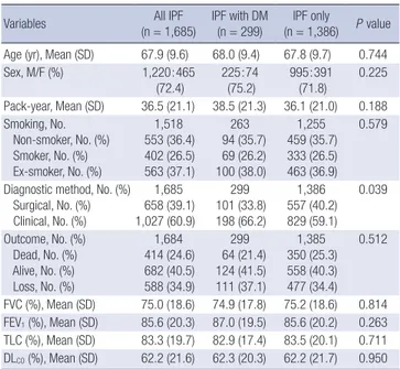
A total of 1,685 patients with IPF were included in this study. Table 1 shows the distribution of IPF patients with and without DM by clinical features. Of these, 39.1% were diagnosed with IPF by lung biopsy, and 60.9% were clinically diagnosed with IPF. Patients were classified into two groups: those without DM (group A, n = 1,386; 82.3%) and those with DM (group B, n = 299; 17.7%). The mean age and duration of cigarette use for Group B patients were 68.0 ± 9.4 yr and 38.5 yr, respectively. There were no significant differences in sex and age between the two groups (P = 0.225 and P = 0.744, respectively). There were no significant differences in arterial blood gas analysis outcome and number years of cigarette use between the two groups. Measurements of pulmonary function did not significantly differ between the time of initial diagnosis and 6 months later. There were no signif-icant differences between the two groups in clinical symptoms at the time of initial diagnosis, such as cough, sputum, hemop-tysis, and chest pain. Asymptomatic patients accounted for 4.8% of all patients. Exertional dyspnea and cough occurred
frequent-ly. Exertional dyspnea occurred in 87.6% of group A patients and 80.1% of group B patients. Coughing was present in 72.6% of group A patients and 76.6% of group B patients. The preva-lence of rheumatoid factor (RF) and antinuclear antibody (ANA) were not significantly different between the two groups. Table 2 shows radiological findings. On HRCT, reticular pat-terns were observed in 58.6% of group A patients and 67.2% of group B patients, a statistically significant difference (P = 0.014). Honeycomb lung patterns were observed in 71.4% of group A patients and 78.9% of group B patients, a statistically significant difference (P = 0.028). However, there were no significant differ-ences in ground-glass and nodular patterns between the two groups. We analyzed data on hypertension, cardiovascular dis-ease, pulmonary tuberculosis, lung cancer, and other malignant disease including stomach cancer (n = 26), bladder cancer (n = 7), colon cancer (n = 4), hematologic malignancy (n = 4), esoph-ageal cancer (n = 3), biliary duct cancer (n = 3), laryngeal can-cer (n = 2), and can-cervical cancan-cer (n = 5) (Table 3). Hypertension was present in 18.5% of group A patients and 37.5% of group B patients, and the difference was statistically significant (P = 0.000). Cardiovascular disease was present in 2.3% of group A patients and 10.7% of group B patients, a difference that was also
statistically significant (P = 0.000). However, pulmonary tuber-culosis was not significantly associated with IPF in either group. As for malignant tumors, lung cancer was not significantly asso-ciated with either group, whereas the difference in other malig-nant tumors was significant: 4.0% of group A patients and 7.0% of group B patients (P = 0.039). DM was significantly less preva-lent in patients diagnosed by lung biopsy (P = 0.039). Smoking history did not significantly differ between the two groups. Multivariate analysis was performed to determine the risk factors for group B patients (Table 4). The significant risk factors included hypertension (odds ratio [OR], 2.529; 95% confidence interval [CI], 1.920-3.325), other cardiovascular diseases (OR, 1.904; 95% CI, 1.209-3.000), and malignant tumors excluding lung cancer (OR, 1.811; 95% CI, 1.060-3.093).
Approximately 40.3% of group A patients and approximately 41.5% of group B patients survived, a statistically insignificant difference. The mortality rate was 25.3% in group A and 21.4% in group B; the difference was not statistically significant. Ap-proximately 34.4% of group A patients and apAp-proximately 37.1% of group B patients were unable to be reached for follow-up. The difference was not statistically significant. The survival rate was not significantly different between the two groups, as estimated using the Kaplan-Meier method (P = 0.207).
DISCUSSION
Although the survival time of patients with IPF is as short as 2.5 to 3 yr, there is no effective therapy that improves survival (3, 4). Table 1. Clinical charateritics of IPF patients with and without diabetes mellitus
Variables (n = 1,685)All IPF IPF with DM (n = 299) (n = 1,386)IPF only P value Age (yr), Mean (SD) 67.9 (9.6) 68.0 (9.4) 67.8 (9.7) 0.744 Sex, M/F (%) 1,220:465 (72.4) 225:74 (75.2) 995:391 (71.8) 0.225 Pack-year, Mean (SD) 36.5 (21.1) 38.5 (21.3) 36.1 (21.0) 0.188 Smoking, No. Non-smoker, No. (%) Smoker, No. (%) Ex-smoker, No. (%) 1,518 553 (36.4) 402 (26.5) 563 (37.1) 263 94 (35.7) 69 (26.2) 100 (38.0) 1,255 459 (35.7) 333 (26.5) 463 (36.9) 0.579
Diagnostic method, No. (%) Surgical, No. (%) Clinical, No. (%) 1,685 658 (39.1) 1,027 (60.9) 299 101 (33.8) 198 (66.2) 1,386 557 (40.2) 829 (59.1) 0.039 Outcome, No. (%) Dead, No. (%) Alive, No. (%) Loss, No. (%) 1,684 414 (24.6) 682 (40.5) 588 (34.9) 299 64 (21.4) 124 (41.5) 111 (37.1) 1,385 350 (25.3) 558 (40.3) 477 (34.4) 0.512 FVC (%), Mean (SD) 75.0 (18.6) 74.9 (17.8) 75.2 (18.6) 0.814 FEV1 (%), Mean (SD) 85.6 (20.3) 87.0 (19.5) 85.6 (20.2) 0.263 TLC (%), Mean (SD) 83.3 (19.7) 82.9 (17.4) 83.5 (20.1) 0.711 DLCO (%), Mean (SD) 62.2 (21.6) 62.3 (20.3) 62.2 (21.7) 0.950 FVC, forced vital capacity; FEV1, forced expiratory volume in 1 second; TLC, total lung capacity; DLCO, diffusing capacity of the lung for carbon monoxide.
Table 2. High-Resolution CT findings of IPF patients with and without diabetes mellitus Variables (n = 1,685)All IPF IPF with DM(n = 299) (n = 1,386) PIPF only value Reticular, No. (%) 1,013 201 (67.2) 812 (58.6) 0.014 Honeycombing, No. (%) 1,225 236 (78.9) 989 (71.4) 0.028 Ground grass opacity, No. (%) 963 167 (55.9) 796 (57.4) 0.447 Nodular, No. (%) 344 57 (19.1) 287 (20.7) 0.755
Table 3. Co-mobidities of IPF patients with and without diabetes mellitus Variables (n = 1,685)All IPF IPF with DM(n = 299) (n = 1,386) PIPF only value Other malignancy*, No. (%) 77 21 (7.0) 56 (4.0) 0.025 Hypertension, No. (%) 368 (21.8) 112 (37.5) 256 (18.5) 0.000 Other CVD†, No. (%) 104 (6.2) 32 (10.7) 72 (2.3) 0.000 Lung cancer, No. (%) 111 (6.6) 17 (5.7) 94 (6.8) 0.488 *Stomach ca (n = 26), bladder ca (n = 7), colorectal ca (n = 4), hematologic malig-nancy (n = 4), esophageal ca (n = 3), gallbladder ca (n = 3), larynx ca (n = 2), cervix ca (n = 3), endometrial ca (n = 2), etc.; †angina pectoris, myocardiac infartion, arrhyth-mia, etc. CVD, cardiovascular disease.
Table 4. Clinical findings independently associated with IPF patients with diabetes mellitus Variable OR 95% CI P value Diagnostic method 0.758 0.578-0.993 0.444 CT finding-reticular 1.888 1.807-3.281 0.024 Hypertension 2.527 1.920-3.325 0.000 Other CVD† 1.904 1.209-3.000 0.005 Other malignancies* 1.811 1.060-3.093 0.030 *Stomach ca (n = 26), bladder ca (n = 7), colorectal ca (n = 4), hematologic malig-nancy (n = 4), esophageal ca (n = 3), gallbladder ca (n = 3), larynx ca (n = 2), cervix ca (n = 3), endometrial ca (n = 2), etc.; †angina pectoris, myocardiac infartion, arrhyth-mia, etc. OR, odds ratio; CI, confidence interval; CVD, cardiovascular disease.
It has been reported that DM and cardiovascular diseases such as hypertension, the prevalence of which increases with age, particularly beyond 60 years, affect the progression and progno-sis of IPF (5-7). Many studies have demonstrated a correlation between DM and IPF (7-10, 14). However, few studies have in-vestigated the clinical characteristics, pulmonary function, under-lying condition, and prognosis of IPF with concurrent DM. It is important to identify the conditions related to IPF and to evalu-ate its characteristics to improve management of the disease. To the best of our knowledge, this is the first large-scale study of the characteristics of IPF associated with DM and the relation-ships between IPF and other clinical entities. In this study, 17.8% of IPF cases were associated with DM. Enomoto et al. (7) re-ported a prevalence of pulmonary fibrosis associated with DM of 32.7%, but this high percentage could be due to the fact that theirs was a single-institution study. The prevalence of DM in IPF patients has also been reported as 11.3% (9) and 10% (10). In the present study, group B patients underwent lung biopsy less frequently than did patients in group A (P = 0.039). This dif-ference may be explained by fewer lung biopsies being taken in diabetic patients due to the higher risk of complications. The 2011 IPF statement recommends that IPF be confirmed by the presence of a UIP pattern on HRCT. The UIP pattern usu-ally consists of reticular abnormalities and honeycombing, with or without traction bronchiectasis. In this study, group B patients showed typical patterns on HRCT images; reticular and honey-comb patterns were observed more frequently (P = 0.014 and P = 0.028, respectively). Carolina et al. (15) have documented that ground glass patterns are typically noted in patients with non-specific interstitial pneumonia and hyperreactive pneu-monitis, with nodular patterns less common in patients with IPF. An association between DM and decreased lung function has been shown (16). Previous studies have indicated that reac-tive oxygen species (ROS) and advanced glycation end products (AGE) produced by hyperglycemia contribute to fibrosis of vari-ous organs, such as the lungs and kidneys (17-23). In our study, typical patterns were observed more frequently in group B than in group A. This might have been secondary to damage to the lungs caused by DM or hyperglycemia. Such damage may in-duce destructive lung parenchyma, seen as reticular and hon-eycomb patterns on HRCT. The prevalence of hypertension and cardiovascular disease was significantly higher in group B than in group A (37.5% vs 18.5%, P = 0.000 for hypertension; 10.7% vs 2.3%, P = 0.000 for cardiovascular disease). This result is sim-ilar to those of previous studies. It has been reported that the prevalence of cardiovascular disease increases in patients with IPF (24), and numerous studies have supported these results (25-27). Kizer et al. (25) reported that 186 of 630 patients who underwent lung transplantation had concurrent cardiovascular disease. Hubbard et al. (27) suggested that patients with IPF have a higher risk of cardiovascular disease. Our results indicate that
cardiovascular disease is more frequently found in group B pa-tients.
In our study, malignant tumors, lung cancer excluded, were more frequently found in group B than in group A. Generally, the prevalence of lung cancer in patients with IPF was signifi-cantly higher, at 6.6% (28 patients). However, the difference be-tween group A and group B patients was not statistically signifi-cant. There was no significant difference in smoking history be-tween the two groups. This result suggests that smoking may be more closely related to lung cancer than IPF is. Clinicians must be aware of the possibility of cancer development in patients with IPF based on the finding that malignant tumors, excluding lung cancer, occurred more frequently in group B patients. In this study, multivariate analysis was performed to deter-mine the risk factors for group B patients. The risk factors for idiopathic pulmonary fibrosis were reticular patterns on CT im-ages (OR, 1.888; 95% CI, 1.87-3.281), hypertension (OR, 2.527; 95% CI, 1.920-3.325), cardiovascular disease (OR, 1.904; 95% CI, 1.209-3.000), and malignant tumors other than lung cancer (OR, 1.811; 95% CI, 1.060-3.093). The survival rate was not significant-ly different between the two groups (P = 0.207).
In this study, there were no significant differences between the groups in age, smoking history, and lung cancer. These factors were regarded as risk factors for IPF in previous studies. These results differ from those of previous studies that did not include IPF patients as controls. In our study, reticular and honeycomb patterns on CT images were more common in group B patients, and the frequencies of hypertension, cardiovascular disease, and malignant tumors other than lung cancer were higher in group B. Therefore, early prevention and treatment of concur-rent diseases can slow the progression of IPF, improving the prognosis.
This study has several limitations. First, data from the medi-cal records may be incomplete due to limited information. The survival rates were similar between the two groups. Since pa-tients treated between 2003 and 2007 were included in our study, not all have been reached for follow-up. Second, this study was based on earlier, 2002 ATS-ERS guidelines for diagnosis of IPF and on retrospective study. Therefore, this study may have in-volved atypical IPF features on HRCT. Third, the interpretation of HRCT images may have differed in quality among centers, as this study was very large and involved many Korean domestic hospitals. Further prospective studies are needed to confirm our results using the new IPF guidelines.
In conclusion, our study indicates that IPF patients with DM are more likely to have a typical interstitial pneumonia (UIP) pattern on HRCT, including reticular and honeycomb patterns, than are IPF patients without DM. The prevalence of hyperten-sion, cardiovascular disease, and other malignancies, excluding lung cancer, is significantly higher in IPF patients with DM than in IPF patients without DM.
REFERENCES
1. Selman M, King TE, Pardo A; American Thorac Society; European Respi-ratory Society; American College of Chest Physicians. Idiopathic pul-monary fibrosis: prevailing and evolving hypotheses about its pathogen-esis and implications for therapy. Ann Intern Med 2001; 134: 136-51. 2. Park SW, Ahn MH, Jang HK, Jang AS, Kim DJ, Koh ES, Park JS, Uh ST,
Kim YH, Park JS, et al. Interleukin-13 and its receptors in idiopathic inter-stitial pneumonia: clinical implications for lung function. J Korean Med Sci 2009; 24: 614-20.
3. Olson AL, Swigris JJ, Lezotte DC, Norris JM, Wilson CG, Brown KK. Mor-tality from pulmonary fibrosis increased in the United States from 1992 to 2003. Am J Respir Crit Care Med 2007; 176: 277-84.
4. Quadrelli S, Molinari L, Ciallella L, Spina JC, Sobrino E, Chertcoff J. Radiological versus histopathological diagnosis of usual interstitial pneu-monia in the clinical practice: dose it have any survival difference? Res-piration 2010; 79: 32-7.
5. Scott J, Johnston I, Britton J. What causes cryptogenic fibrosing alveoli-tis? A study of environmental exposure to dust. BMJ 1990; 301: 1015-7. 6. Hubbard R, Lewis S, Richards K, Johnston I, Britton J. Occupational
exposure to metal or wood dust and aetiology of cryptogenic fibrosing alveolitis. Lancet 1996; 347: 284-9.
7. Enomoto T, Usuki J, Azuma A, Nakagawa T, Kudoh S. Diabetes mellitus may increased risk for idiopathic fibrosis. Chest 2003; 123: 2007-11. 8. Fernández Pérez ER, Daniels CE, Schroeder DR, St Sauver J, Hartman
TE, Bartholmai BJ, Yi ES, Ryu JH. Incidence, prevalence, and clinical course of idiopathic pulmonary fibrosis: a population-based study. Chest 2010; 137: 129-37.
9. Garcia-Sancho Figueroa MC, Carrillo G, Pérez–Padilla R, Ferández-Pla-ta MR, Buendia-Roldán I, Vargas HM, Selman M. Risk factors for idio-pathic pulmonary fibrosis in a Mexican population. A case-control study. Respir Med 2010; 104: 305-9.
10. Gribbin J, Hubbard R, Smith C. Role of diabetes mellitus and gastro-oesophageal reflux in the aetiology of idiopathic pulmonary fibrosis. Respir Med 2009; 103: 927-31.
11. Kim YJ, Park JW, Kyung SY, An CH, Lee SP, Chung MP, Park HY, Jeong SH. Association of diabetes mellitus and metabolic syndrome with idio-pathic pulmonary fibrosis. Tuberc Respir Dis 2009; 67: 113-20.
12. Raghu G, Collard HR, Egan JJ, Martinez FJ, Behr J, Brown KK, Colby TV, Cordier JF, Flaherty KR, Lasky JA, et al. An official ATS/ERS/JRS/ALAT statement: idiopathic pulmonary fibrosis: evidence-based guidelines for diagnosis and management. Am J Respir Crit Care Med 2011; 183: 788-824.
13. American Thoracic Society; European Respiratory Society. American Thoracic Society/European Respiratory Society International Multidisci-plinary Consensus Classification of the Idiopathic Interstitial Pneumo-nias. This joint statement of the American Thoracic Society (ATS), and the European Respiratory Society (ERS) was adopted by the ATS board of directors, June2001 and by the ERS Executive Committee, June 2001.
Am J Respir Crit Care Med 2002; 165: 277-304.
14. Miyake Y, Sasaki S, Yokoyama T, Chida K, Azuma A, Suda T, Kudoh S, Sakamoto N, Okamoto K, Kobashi G, et al. Case-control study of medi-cal history and idiopathic pulmonary fibrosis in Japan. Respirology 2005; 10: 504-9.
15. Souza CA, Müller NL, Flint J, Wright JL, Churg A. Idiopathic pulmonary fibrosis: spectrum of high-resolution CT findings. AJR Am J Roentgenol 2005; 185: 1531-9.
16. Klein OL, Krishnan JA, Glick S, Smith LJ. Systemic review of the associa-tion between lung funcassocia-tion and type 2 diabetes mellitus. Diabet Med 2010; 27: 977-87.
17. Baynes JW, Thorpe SR. Role of oxidative stress in diabetic complications: a new perspective on an old paradigm. Diabetes 1999; 48: 1-9.
18. Kuwano K, Nakashima N, Inoshima I, Hagimoto N, Fujita M, Yoshimi M, Maeyama T, Hamada N, Watanabe K, Hara N. Oxidative stress in lung epithelial cells from patients with idiopathic interstitial pneumonia. Eur Respir J 2003; 21: 232-40.
19. Maritim AC, Sanders RA, Watkins JB 3rd. Diabetes, oxidative stress, and antioxidants: a review. J Biochem Mol Toxicol 2003; 17: 24-38.
20. Rhyu DY, Yang Y, Ha H, Lee GT, Song JS, Uh ST, Lee HB. Role of reactive oxygen species in TGF-beta1-induced mitogen-activated protein kinase activation and epithelial-mesenchymal transition in renal tubular epi-thelial cells. J Am Soc Nephrol 2005; 16: 667-75.
21. Kinnula VL, Fattman CL, Tan RJ, Oury TD. Oxidative stress in pulmo-nary fibrosis: a possible role for redox modulatory therapy. Am J Respir Crit Care Med 2005; 172: 417-22.
22. Teramoto S, Fukuchi Y, Uejima Y, Shu CY, Orimo H. Superoxide anion formation and glutathione metabolism of blood in patients with idio-pathic pulmonary fibrosis. Biochem Mol Med 1995; 55: 66-70.
23. Yu T, Robotham JL, Yoon Y. Increased production of reactive oxygen spe-cies in hyperglycemic conditions requires dynamic change of mitochon-drial morphology. Proc Natl Acad Sci U S A 2006; 103: 2653-8.
24. Panos RJ, Mortenson RL, Niccoli SA, King TE Jr. Clinical deterioration in patients with idiopathic pulmonary fibrosis: causes and assessment. Am J Med 1990; 88: 396-404.
25. Kizer JR, Zisman DA, Blumenthal NP, Kotloff RM, Kimmel SE, Strieter RM, Arcasoy SM, Ferrari VA, Hansen-Flaschen J. Association between pulmonary fibrosis and coronary artery disease. Arch Intern Med 2004; 164: 551-6.
26. Nathan SD, Basavaraj A, Reichner C, Shlobin OA, Ahmad S, Kiernan J, Burton N, Barnett SD. Prevealence and impact of coronary artery dis-ease in idiopathic pulmonary fibrosis. Respir Med 2010; 104: 1035-41. 27. Hubbard RB, Smith C, Le Jeune I, Gribbin J, Fogarty AW. The
associa-tion between idiopathic pulmonary fibrosis and vascular disease: a pop-ulation–based study. Am J Respir Crit Care Med 2008; 178: 1257-61. 28. Matsushita H, Tanaka S, Saiki Y, Hara M, Nakata K, Tanimura S, Banba J.
Lung cancer associated with usual interstitial pneumonia. Pathol Int 1995; 45: 925-32.