Comparison of the Low Contact Stress and Press Fit
Condylar Rotating-Platform Mobile-Bearing
Prostheses in Total Knee Arthroplasty
A Prospective Randomized Study
Young-Hoo Kim, MD, Jun-Shik Kim, MD, Jang-Won Park, MD, and Jong-Hwan Joo, MD
Investigation performed at the Joint Replacement Center of Korea, Ewha Womans University School of Medicine, Seoul, South Korea
Background: To our knowledge, no study to date has compared the clinical results of posterior cruciate-sacrificing mobile-bearing total knee replacements with those of posterior-stabilized mobile-bearing total knee replacements in the same patients. The purpose of the present study was to compare the clinical and radiographic results of these two designs. We hypothesized that the results would be better for knees treated with the posterior-stabilized mobile-bearing prosthesis.
Methods: The present study consisted of a consecutive series of 107 female patients (mean age, 66.8 years) who underwent bilateral simultaneous total knee arthroplasty at the same surgical setting. All of these patients received a posterior cruciate-sacrificing mobile-bearing prosthesis in one knee and a posterior-stabilized mobile-bearing prosthesis in the contralateral knee. At the time of each follow-up (mean, 7.4 years; range, seven to 7.6 years), the patients were assessed clinically.
Results: The mean postoperative Knee Society knee score (96 compared with 97 points) and Hospital for Special Surgery knee score (93 compared with 94 points) were similar between the two groups. At the time of the latest follow-up, the average range of motion was 127.7° (range, 70° to 150°) in the knees with a posterior cruciate-sacrificing mobile-bearing prosthesis and 132.4° (range, 90° to 150°) in the knees with a posterior-stabilized mobile-mobile-bearing prosthesis. With a margin of error of the manual measurement of 5°, this difference was not significant. The estimated survival rate was 97.2% (95% confidence interval, 91% to 99%) at seven years in the posterior-cruciate sacrificing mobile-bearing prosthesis group and 98.1% (95% confidence interval, 92% to 99%) at seven years in the posterior-stabilized mobile-bearing prosthesis group.
Conclusions: After a minimum duration of follow-up of seven years, we found no significant differences between the two groups with regard to the clinical and radiographic results, including knee range of motion.
Level of Evidence: Therapeutic Level I. See Instructions to Authors for a complete description of levels of evidence.
T
he posterior cruciate-sacrificing mobile-bearing total knee prosthesis (Low Contact Stress Rotating Platform [LCS RP]; DePuy, Johnson & Johnson, Warsaw, In-diana) was introduced by Buechel and Pappas in 1979 to reduce contact stress in the polyethylene and potentially to decrease wear as well as to minimize cement-bone stress at the tibial surface1-3. Compared with the fixed-bearing posterior-stabilized total knee prosthesis introduced by Insall et al. in 19784
, the LCS RP device has no so-called post-and-cam mechanism. Instead, stability is provided by the curved design of the tibial
insert articulation surface and a balanced flexion gap achieved with exacting surgical technique. Over the past twenty years, good results have been reported in association with the LCS RP device2,5-7
.
The Press Fit Condylar Sigma posterior-stabilized rotating-platform knee (PFC Sigma PS-RP; DePuy, Johnson & Johnson, Warsaw, Indiana) was introduced in 2000. This design was introduced to improve the kinematics of the LCS RP pros-thesis by employment of a post-and-cam mechanism8
. It was anticipated that the post-and-cam mechanism in the PFC
Disclosure: The authors did not receive any outside funding or grants in support of their research for or preparation of this work. Neither they nor a member of their immediate families received payments or other benefits or a commitment or agreement to provide such benefits from a commercial entity.
Sigma PS-RP prosthesis would lead to consistent posterior rollback, which, in turn, would lead to better knee range of motion, would reduce polyethylene wear at the articular surface and undersurface, and would provide better stabili-zation of the tibial insert (Figs. 1-A and 1-B).
Although the design features of the PFC Sigma PS-RP prosthesis have been proposed to improve upon the kinematics of the LCS RP prosthesis, no study, to our knowledge, has compared the clinical results of the PFC Sigma PS-RP pros-thesis with those of the LCS RP prospros-thesis in the same patients. To examine the results associated with the LCS RP and PFC Sigma PS-RP total knee prostheses in patients who had bilateral simultaneous total knee arthroplasty, we sought to determine whether the knee and function scores and the radiographic
results for the knees with a PFC Sigma PS-RP prosthesis would be better than those with an LCS RP prosthesis and whether the knees with a PFC Sigma PS-RP prosthesis would have a better range of motion.
Materials and Methods
O
ne hundred and twenty-six patients (252 knees) with bilateral knee osteo-arthritis (Ahlb¨ack grade III, IV, or V9) underwent simultaneous bilateral sequential total knee arthroplasty. The study protocol and consent forms were approved by the institutional review board. A detailed informed consent form was signed by each patient, and all information was kept confidential. This study was registered in the ClinicalTrials.gov Protocol Registration System (trial number, NCT01075620). Seven patients were excluded because they refused to participate. Seven more patients were excluded because they were male, leaving 112 patients available for participation. Five patients were lost to early follow-up (threeFig. 1-A
Fig. 1-B
Figs. 1-A and 1-B Photographs showing the design features of the LCS RP and PFC Sigma PS-RP prostheses. Fig. 1-A Frontal views of LCS RP (left) and PFC Sigma PS-RP (right) total knee prostheses. Fig. 1-B Lateral views of the LCS RP (left) and PFC Sigma PS-RP (right) total knee prostheses.
months), leaving 107 patients (214 knees) available for study after a minimum duration of follow-up of seven years (mean, 7.4 years; range, seven to 7.6 years). The study group included 107 women who had a mean age (and standard deviation) of 66.8 ± 5.181 years (range, fifty-four to eighty-one years) at the time of surgery (see Appendix). Twenty-three knees had valgus alignment of 8° to 12°, and the remaining 191 knees had varus alignment of 8° to 20°. Fourteen of 107 patients with an LCS RP total knee prosthesis and twelve of 107 patients with a PFC Sigma PS-RP total knee prosthesis had had previous arthroscopic debridement, and the remaining patients had had no previous surgery.
The coronal geometries of both the LCS RP prosthesis and the PFC Sigma PS-RP prosthesis are rounded coronal designs with similar conformity ratios; the contact surface is slightly greater for the PFC Sigma PS-RP pros-thesis. The femoral component of the PFC Sigma PS-RP prosthesis has a cam for the tibial post. The sagittal designs of both the LCS RP prosthesis and the PFC Sigma PS-RP prosthesis are multiradial. The anterior flange angle is 5° for the LCS RP prosthesis and 0° for the PFC Sigma PS-RP prosthesis. The pos-terior flange thickness is 8 mm for all sizes of the PFC Sigma PS-RP prosthesis except for size 6 (for which it is 10 mm) and ranges from 6.2 to 9.4 mm in the LCS RP prosthesis.
Randomization to treatment with the LCS RP or PFC Sigma PS-RP total knee prosthesis was accomplished with use of a sealed study number envelope. After the envelope was opened in the operating room before the skin incision was made, the first knee received the prosthesis indicated by the envelope and the contralateral knee received the other prosthesis. There were no cases in which the second procedure was aborted because of intraoperative complications.
All procedures were performed by the senior author (Y.-H.K.). With tourniquet inflation to 250 mm Hg, an anterior midline skin incision (10 to 12 cm in length) was made, followed by a medial parapatellar capsular incision. In the LCS RP group, tibial preparation was performed first, and in the PFC Sigma PS-RP group, femoral preparation was performed first. Ten millimeters of tibial bone was resected, referenced from the least-involved tibial plateau, to achieve a surface perpendicular to the axis of the tibia in the coronal plane. A 7° posterior slope was prepared in the sagittal plane for the knees in the LCS RP group, and a 0° slope was prepared for the knees in the PFC Sigma PS-RP group. Anterior cortical reference was used for the anterior-posterior cut of the distal part of the femur. Femoral component rotation was determined with use of three reference axes: (1) the transepicondylar axis, (2) the midtrochlear (Whiteside) line10, and (3) 3° of external rotation relative to the posterior aspect of the condyles. Ligamentous balance was established first in knee extension and then in knee flexion with use of a tensor. All patellae were resurfaced with a polyethylene implant. All implants were cemented after pulsed lavage irriga-tion, drying, and pressurization of cement.
A splint was applied for the first twenty-four hours. A continuous passive motion machine was used beginning on the second postoperative day and continuing twice daily for thirty minutes until knee flexion was 120° (range, seven to ten days). On the second postoperative day, patients started active range-of-motion exercises and stood at the bedside or walked with use of crutches or a walker. All patients were discharged to home from the hospital ten to fourteen days after surgery with full weight-bearing using crutches or a walker for six weeks and a cane when needed thereafter. No patient received outpatient physical therapy.
Two of the authors (Y.-H.K. and J.-S.K.) assessed the patients with a physical examination and knee scoring preoperatively, at three and six months after surgery, at one year after surgery, and annually thereafter with use of the systems of the Knee Society11and the Hospital for Special Surgery12; at each interval, a separate evaluation was performed for each knee. At the time of each follow-up, radiographic data were analyzed and recorded by a clinical fellow (J.-H.J.) who was not part of the operative team. This assessment was not blinded to allocation of the two implants because the radiographic appearances of the two implants differ.
The active arc of motion of each knee with the patient in the supine position was measured two times with use of a standard (60-cm) goniometer preoperatively and at each follow-up by two observers (Y.-H.K. and J.-S.K.),
one of whom was blinded to the type of implanted prosthesis. The chance-corrected kappa coefficient13,14for intraobserver agreement ranged from 0.78 to 0.88. To assess intraobserver reliability, the goniometer measurement was performed three times (with a three-day interval between measurements). The level of activity was assessed with the Knee Society score11and the Tegner and Lysholm score15. All clinical data were compiled and collated by a separate research associate.
Anteroposterior hip-to-ankle radiographs (with the patient standing), supine anteroposterior and lateral radiographs, and skyline patellar radio-graphs were made preoperatively and at each follow-up. The radioradio-graphs were evaluated by one observer, not a member of the operating team, to determine the anatomic axis of the limb, the alignment of the components, posterior slope, posterior femoral condylar offset, the level of the joint line, the presence and location of radiolucent lines at the bone-cement or cement-implant in-terface, and patellar tilt or dislocation by Knee Society scores11(Figs. 2-A and 2-B). All radiographs were made under fluoroscopic guidance to control rotation of the knee.
Statistical Analysis
An a priori power calculation was performed with use of a clinically relevant difference in range of motion of 5° and a standard deviation of 9°. For an effect size of 20% in early functional outcome, as measured with a validated instrument such as the linear analog scale assessment for range of motion, with a = 0.05 and b = 0.80, calculation revealed that 104 knees would be needed in each group. In addition to the required number of subjects, ten more patients were recruited to allow for possible attrition. The changes in the Knee Society and Hospital for Special Surgery knee scores were evaluated with use of the paired t test. Pain scores were assessed with use of the chi-square test. Knee motion was compared between the two groups with use of a paired t test. Complication rates and radiographic data were compared be-tween the two groups with a paired t test. The level of significance was set at p < 0.05.
Source of Funding
There was no external funding for this study.
Results
T
he Knee Society and Hospital for Special Surgery knee scores did not differ significantly between the two groups either preoperatively (p = 0.612 and p = 0.291, respectively; paired t test) or postoperatively (p = 0.167 and p = 0.087, respectively; paired t test). In the LCS RP group, the mean postoperative Knee Society knee score was 96 points (range, 77 to 100 points) and the mean postoperative Hospital for Special Surgery knee score was 93 points (range, 69 to 100 points). In the PFC Sigma PS-RP group, the mean postop-erative Knee Society knee score was 97 points (range, 79 to 100 points) and the mean postoperative Hospital for Special Surgery knee score was 94 points (range, 75 to 100 points). In the LCS RP group, ninety knees (84%) had no pain, sixteen (15%) had mild pain, and one (1%) had moderate pain at the time of the latest follow-up. In the PFC Sigma PS-RP group, eighty-eight knees (82%) were pain-free, eighteen (17%) had mild pain, and one (1%) had moderate pain at the time of the latest follow-up.The mean preoperative range of motion was 127.8° (range, 80° to 150°) in the LCS RP group and 127.2° (range, 85° to 150°) in the PFC Sigma PS-RP group. The mean postoperative range of motion was 127.7° (range, 70° to 150°) in the LCS RP group and 132.4° (range, 90° to 150°) in the
PFC Sigma PS-RP group. This difference was significant (p < 0.0001; paired t test). However, if the margin of error of the manual measurement is considered to be 5°, this difference is not significant (p = 0.781; paired t test). The average
im-provement of the range of motion per patient was 2.5° (range, 210° to 10°) in the LCS RP group and 4.2° (range, 215° to 12°) in the PFC Sigma PS-RP group. This difference was not significant (p = 0.635; paired t test). Seventeen knees (16%) in
Fig. 2-A
Fig. 2-B
Figs. 2-A and 2-B Radiographs of both knees of a sixty-three-year-old woman. Fig. 2-A Anteroposterior standing radio-graph showing the alignment of the femoral and tibial components. The alignment of the femoral and tibial com-ponents is measured by the intersection of a line drawn across the base of each component and the mechanical axis of the femur and the tibia (a= frontal femoral angle, b = frontal tibial angle). Fig. 2B Lateral radiographs of both knees, showing the measurement of flexion and extension of the femoral component and measurement of the posterior slope of the tibial component (g= sagittal femoral angle, d = sagittal tibial angle). The posterior femoral condylar offset (PCO) is evaluated by measuring the maximum thickness of the posterior aspect of the condyles projected posteriorly to the tangent of the posterior cortex of the femoral shaft. Line aa’ is a parallel line to the lateral femoral anatomic axis. Line bb’ is a parallel line to the femoral peg longitudinal axis. Line cc’ is a line indicating the femoral peg longitudinal axis. Line dd’ is a line tangent to the posterior cortex of the femoral shaft. Line ee’ is a line joining the edges of the tibial baseplate.
the LCS RP group and fourteen knees (13%) in the PFC Sigma PS-RP group had <110° of motion at the time of the latest follow-up. The maximum flexion in both groups was 150°. The activity level score for the patients was 5 or 6 points at the time of the latest follow-up, indicating participation in
strenuous farm work (5 points) or participation in tennis (6 points).
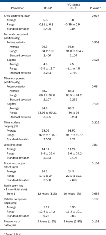
There were no significant differences between the groups in terms of the alignment of the knee (mean, 5.8° of valgus in both groups), the position of the femoral and tibial components in the coronal and sagittal planes, the patellar angle, the posterior slope of tibia (mean, 4.9° compared with 2.5°), the amount of the tibial surface area that was covered by the implants (tibial capping), the mean level of the joint line, the prevalence of radiolucent lines, or the posterior condylar offset (mean, 24.2 compared with 24.5 mm) (p > 0.05 for all comparisons; paired t test). The prevalence of radiolucent lines measuring <1 mm (on the tibial side only) was 11% (twelve knees) in the LCS RP group and 9% (ten knees) in the PFC Sigma PS-RP group. The prevalence of osteolysis was 1.9% (two knees) in the LCS RP group and 2.8% (three knees) in the PFC Sigma PS-RP group (Table I). In the LCS RP group, the estimated survival rate ac-cording to Kaplan-Meier analysis16
was 97.2% (95% confidence interval, 91% to 99%) at seven years, with an overall revision rate of 2.8% (three of 107 knees). In the PFC Sigma PS-RP group, the estimated survival rate was 98.1% (95% confidence interval, 92% to 99%) at seven years, with an overall revision rate of 1.9% (two of 107 knees).
Three knees (2.8%) in the LCS RP group and two knees (1.9%) in the PFC Sigma PS-RP group had a deep infection and required a two-stage revision. None of these five knees had had a recurrence of infection at the time of the latest follow-up. One knee (0.9%) in the LCS RP group required open reduction and internal fixation for the treatment of a supracondylar fracture of the femur. Two knees (1.9%) in the PFC Sigma PS-RP group had a patellar clunk syndrome and required arthroscopic de-bridement, with good results. Instability did not occur in any knee in either group.
Discussion
O
ur study investigated whether the PFC Sigma PS-RPprosthesis for total knee arthroplasty provides a greater benefit than the LCS RP prosthesis. Multiple studies2,5,17-26
have shown that, regardless of the criteria used to measure success or failure, the LCS RP prosthesis achieved essentially equal, or even better, results than the PFC Sigma PS-RP2,5,17-26
. We found that the intermediate-term clinical outcomes for both prostheses were similar in terms of the Knee Society score, radiographic results, and range of motion. Our survivorship data are agreement with those in other reports on these prosthetic designs17-26
. In previous studies, the mean flexion of the knee has ranged from 102° to 113° for knees with the LCS RP pros-thesis5,6,18-22
and from 101.7° to 130° for knees with the PFC Sigma PS-RP prosthesis22-24,26
. Our patients had comparable range of motion in comparison with the patients in those re-ports, and we found no clinical difference between the two designs (128° for the LCS RP, compared with 132° for the PFC Sigma PS-RP). Because all of our patients had full knee ex-tension, our findings suggest that the PFC Sigma PS-RP design provides no advantage in terms of knee motion.
TABLE I Radiographic Results at Time of Latest Follow-up
Parameter LCS RP
PFC Sigma
PS-RP P Value*
Knee alignment (deg) 0.937
Average 5.8 5.8 Range 0.81 to 6.8 –0.39 to 6.9 Standard deviation 2.496 2.84 Femoral component position (deg) Anteroposterior 0.951 Average 96.9 96.8 Range 84 to 103 91.8 to 104.2 Standard deviation 2.406 2.104 Sagittal 0.125 Average 4.9 2.5 Range –3.9 to 13.7 –1.1 to 4.5 Standard deviation 3.284 2.719 Tibial component position (deg) Anteroposterior 0.68 Average 88.2 88.3 Range 80.1 to 92.8 83.5 to 94.2 Standard deviation 2.107 2.235 Sagittal 0.103 Average 84.6 88.2 Range 73.96 to 89.21 86 to 93 Standard deviation 3.305 3.234 Tibial surface capping (%) 0.222 Average 98.06 98.52 Range 92.2 to 108.3 91.7 to 107.6 Standard deviation 3.558 3.22 Joint line (mm) 0.81 Average 14.31 14.24 Range 6.4 to 22.4 6.6 to 24.2 Standard deviation 3.333 3.196 Posterior condylar offset (mm) 0.102 Average 24.2 24.5 Range 17.2 to 34 20.1 to 32.1 Standard deviation 3.508 2.699 Radiolucent line <1 mm (tibial side)
Zone 1 12 knees (11%) 10 knees (9%) 0.653 Patellar component angle (deg) 0.105 Average 1.12 0.93 Range –12.4 to 14.2 –11.3 to 10.1 Standard deviation 9.25 9.86 Prevalence of osteolysis 2 knees (1.9%) 3 knees (2.8%) 0.138 *Paired t test.
In the current study, no knee had aseptic loosening or osteolysis that resulted in revision, similar to previous reports on these knee designs5,17
. Our findings of a low prevalence of osteolysis in both groups may be related to the inclusion of only female patients, the use of a polished cobalt-chromium tibial baseplate to reduce backside wear of the insert, the use of a polyethylene insert sterilized with gamma irradiation in a vacuum, and the short shelf life of the insert. It is possible that the duration of follow-up was not sufficiently long to reveal osteolysis.
It has been emphasized that an exacting surgical tech-nique, especially balancing of flexion and extension gaps, is mandatory during a mobile-bearing total knee arthroplasty in order to avoid bearing dislocation or instability of the knee27-29
. Many surgeons believe that the use of an unconstrained mobile-bearing total knee implant is contraindicated in cases of severe varus and valgus deformity27-29
. This idea was challenged by Beverland30
, who stated that a mobile-bearing total knee im-plant can be used for virtually every primary total knee ar-throplasty, irrespective of deformity. In our series, we were able to use a mobile-bearing total knee implant for every primary total knee arthroplasty selected by the process of randomiza-tion, irrespective of the range of deformity, with no postop-erative instability.
The present study had limitations. First, it is difficult for a patient who has undergone bilateral total knee ar-throplasty to distinguish the independent function of each knee. Although this was a problem when assessing function after the bilateral total knee arthroplasties, the patients
were able to grade which knee caused more functional limitation. Second, the duration of follow-up was seven years and long-term variability in outcome cannot be pre-dicted. Third, we performed no interobserver variability testing on the radiographic measurements. Finally, the range of knee motion was not determined under weight-bearing conditions.
The findings of the present intermediate-term, pro-spective, randomized clinical study suggest that these first and second-generation mobile-bearing knee designs perform well, with no significant differences between the two prostheses that were evaluated.
Appendix
A table summarizing the demographic data for the pa-tients is available with the online version of this article at jbjs.org.n
Young-Hoo Kim, MD Jun-Shik Kim, MD Jang-Won Park, MD Jong-Hwan Joo, MD
The Joint Replacement Center of Korea, Ewha Womans University MokDong Hospital, 911-1, Mokdong,
YangChun-Gu,
Seoul 158-710, South Korea.
E-mail address for Y.-H. Kim: [email protected]
References
1. Bartel DL, Bicknell VL, Wright TM. The effect of conformity, thickness, and ma-terial on stresses in ultra-high molecular weight components for total joint replace-ment. J Bone Joint Surg Am. 1986;68:1041-51.
2. Buechel FF, Pappas MJ. Long-term survivorship analysis of cruciate-sparing ver-sus cruciate-sacrificing knee prostheses using meniscal bearings. Clin Orthop Relat Res. 1990;260:162-9.
3. O’Connor JJ, Goodfellow JW. Theory and practice of meniscal knee replacement: designing against wear. Proc Inst Mech Eng H 1996;210:217-22.
4. Insall JN, Lachiewicz PF, Burstein AH. The posterior stabilized condylar prosthe-sis: a modification of the total condylar design. Two to four-year clinical experience. J Bone Joint Surg Am. 1982;64:1317-23.
5. Callaghan JJ, Squire MW, Goetz DD, Sullivan PM, Johnston RC. Cemented rotating-platform total knee replacement. A nine to twelve-year follow-up study. J Bone Joint Surg Am. 2000;82:705-11.
6. Callaghan JJ, O’Rourke MR, Iossi MF, Liu SS, Goetz DD, Vittetoe DA, Sullivan PM, Johnston RC. Cemented rotating-platform total knee replacement. A concise follow-up, at a minimum of fifteen years, of a previous report. J Bone Joint Surg Am. 2005;87:1995-8.
7. Kim YH, Yoon SH, Kim JS. The long-term results of simultaneous fixed-bearing and mobile-bearing total knee replacements performed in the same patient. J Bone Joint Surg Br. 2007;89:1317-23.
8. Dennis DA, Komistek RD, Hoff WA, Gabriel SM. In vivo knee kinematics derived using an inverse perspective technique. Clin Orthop Relat Res. 1996;331: 107-17.
9. Ahlb¨ack S. Osteoarthrosis of the knee. A radiographic investigation. Acta Radiol
Diagn (Stockh) 1968;Suppl 277:7-72.
10. Arima J, Whiteside LA, McCarthy DS, White SE. Femoral rotational alignment, based on the anteroposterior axis, in total knee arthroplasty in a valgus knee. A technical note. J Bone Joint Surg Am. 1995;77:1331-4.
11. Ewald FC. The Knee Society total knee arthroplasty roentgenographic evaluation and scoring system. Clin Orthop Relat Res. 1989;248:9-12.
12. Insall JN, Ranawat CS, Aglietti P, Shine J. A comparison of four models of total knee-replacement prostheses. J Bone Joint Surg Am. 1976;58: 754-65.
13. Landis JR, Koch GG. The measurement of observer agreement for categorical data. Biometrics 1977;33:159-74.
14. Bach CM, Biedermann R, Goebel G, Mayer E, Rachbauer F. Reproducible as-sessment of radiolucent lines in total knee arthroplasty. Clin Orthop Relat Res. 2005;434:183-8.
15. Tegner Y, Lysholm J. Rating systems in the evaluation of knee ligament injuries. Clin Orthop Relat Res. 1985;198:43-9.
16. Kaplan EL, Meier P. Nonparametric estimation from incomplete observations. J Am Stat Assoc. 1958;53:457-81.
17. Sorrells RB, Voorhorst PE, Murphy JA, Bauschka MP, Greenwald AS. Un-cemented rotating-platform total knee replacement: a five to twelve-year follow-up study. J Bone Joint Surg Am. 2004;86:2156-62.
18. Kim YH, Kim JS, Choi Y. Osteolysis after unidirectional and multidirectional mobile-bearing total knee arthroplasty in young patients. J Arthroplasty 2009; 24:586-93.
19. Hooper G, Rothwell A, Frampton C. The low contact stress mobile-bearing total knee replacement: a prospective study with a minimum follow-up of ten years. J Bone Joint Surg Br. 2009;91:58-63.
20. Kim YH, Kim JS. Prevalence of osteolysis after simultaneous bilateral fixed- and mobile-bearing total knee arthroplasties in young patients. J Arthroplasty 2009; 24:932-40.
21. Kim YH, Kook HK, Kim JS. Comparison of fixed-bearing and mobile-bearing total knee arthroplasties. Clin Orthop Relat Res. 2001;392:101-15.
22. Kim YH, Kim JS. Comparison of anterior-posterior-glide and rotating-platform low contact stress mobile-bearing total knee arthroplasties. J Bone Joint Surg Am. 2004;86:1239-47.
23. Ranawat AS, Rossi R, Loreti I, Rasquinha VJ, Rodriguez JA, Ranawat CS. Comparison of the PFC Sigma fixed-bearing and rotating-platform total knee
arthroplasty in the same patient: short-term results. J Arthroplasty 2004;19: 35-9.
24. Gioe TJ, Glynn J, Sembrano J, Suthers K, Santos ER, Singh J. Mobile and fixed-bearing (all-polyethylene tibial component) total knee arthroplasty designs. A prospective randomized trial. J Bone Joint Surg Am. 2009;91: 2104-12.
25. Luring C, Bathis H, Oczipka F, Trepte C, Lufen H, Perlick L, Grifka J. Two-year follow-up on joint stability and muscular function comparing rotating versus fixed bearing TKR. Knee Surg Sports Traumatol Arthrosc 2006;14:605-11. 26. Kim YH, Kim DY, Kim JS. Simultaneous mobile- and fixed-bearing total knee replacement in the same patients. A prospective comparison of mid-term outcomes using a similar design of prosthesis. J Bone Joint Surg Br. 2007;89:904-10.
27. Thompson NW, Wilson DS, Cran GW, Beverland DE, Stiehl JB. Dislocation of the rotating platform after low contact stress total knee arthroplasty. Clin Orthop Relat Res. 2004;425:207-11.
28. Bert JM. Dislocation/subluxation of meniscal bearing elements after New Jer-sey low-contact stress total knee arthroplasty. Clin Orthop Relat Res.
1990;254:211-5.
29. Weaver JK, Derkash RS, Greenwald AS. Difficulties with bearing dislocation and breakage using a movable bearing total knee replacement system. Clin Orthop Relat Res. 1993;290:244-52.
30. Beverland D. Management of the severe varus and valgus knee using the low contact stress rotating platform. Orthopedics 2006;29(9 Suppl): S60-3.