Effects of simultaneous combined exposure to CDMA and WCDMA
electromagnetic fields on serum hormone levels in rats
Yeung Bae JIN
1, Hyung-Do CHOI
2, Byung Chan KIM
2, Jeong-Ki PACK
3, Nam KIM
4and Yun-Sil LEE
5,*
1
Division of Radiation Effects, Korea Institute of Radiological and Medical Sciences, Seoul 139-706, Korea 2
Electromagnetic Engineering Team, Radio Technology Research Department, Electronics and Telecommunications Research Institute (ETRI), Daejon 305-350, Korea
3
Department of Radio Sciences and Engineering, College of Engineering, Chungnam National University, Daejon 305-764, Korea
4
School of Electrical and Computer Engineering, Chungbuk National University, Cheongju 361-763, Korea 5
College of Pharmacy and Division of Life and Pharmaceutical Sciences, Ewha Women’s University, Seoul 120-750, Korea
*Corresponding author. Tel: +82-2-3277-3022; Fax: +82-2-3277-2851; E-mail: [email protected] (Received 2 February 2012; revised 19 November 2012; accepted 20 November 2012)
Despite more than a decade of research on the endocrine system, there have been no published studies about the effects of concurrent exposure of radiofrequency electromagnetic fields (RF-EMF) on this system. The present study investigated the several parameters of the endocrine system including melatonin, thyroid stimulating hormone, stress hormone and sex hormone after code division multiple access (CDMA, 849 MHz) and wideband code division multiple access (WCDMA, 1.95 GHz) signals for simultaneous ex-posure in rats. Sprague-Dawley rats were exposed to RF-EMF signals for 45 min/day, 5 days/week for up to 8 weeks. The whole-body average specific absorption rate (SAR) of CDMA or WCDMA was 2.0 W/kg (total 4.0 W/kg). At 4 and 8 weeks after the experiment began, each experimental group’s 40 rats (male 20, female 20) were autopsied. Exposure for 8 weeks to simultaneous CDMA and WCDMA RF did not affect serum levels in rats of melatonin, thyroid stimulating hormone (TSH), triiodothyronine (T3) and thyroxin (T4), adrenocorticotropic hormone (ACTH) and sex hormones (testosterone and estrogen) as assessed by the ELISA method.
Keywords:CDMA; WCDMA; combined exposure; melatonin; hormone
INTRODUCTION
The steadily increasing use of radio frequency (RF) com-munication systems has raised public concerns about the safety of electromagnetic fields (EMF). Daily, over three billion people in more than 200 countries are exposed to EMF [1]. Most studies to date do not indicate a health risk of RF exposure. However, controversy has been stimulated by some epidemiologic studies reporting an association between magnetic field exposure and human diseases [2]. Moreover, a recent International Agency for Research on Cancer (IARC) report suggested that RF-EMF is regarded as a category 2B carcinogen [3]. Animal studies have shown that exposure to RF-EMF may alter the endocrine or
nervous systems [4–6]. Melatonin is a hormone, secreted by the pineal gland, which plays an important role in central nervous system. Melatonin levels vary in a circadian rhythm in both rats and humans, sunsetted to a 12 h light– dark (LD) cycle. The LD cycle causes blood and pineal body melatonin levels to increase during the dark period and decrease during the light period. Thyroid activity is regulated by the thyroid stimulating hormone (TSH) secreted by the pituitary gland. Elevated TSH levels induce the thyroid to elaborate triiodothyronine (T3) and thyroxin (T4), a hormone that functions in at least 20 enzyme systems, influencing the acceleration of protein synthesis. It has been suggested that exposure to RF-EMF may alter thyrotropin secretion [7].
doi: 10.1093/jrr/rrs120 Advance Access Publication 13 December 2012
© The Author 2012. Published by Oxford University Press on behalf of The Japan Radiation Research Society and Japanese Society for Therapeutic Radiology and Oncology.
This is an Open Access article distributed under the terms of the Creative Commons Attribution Non-Commercial License (http://creativecommons.org/licenses/ by-nc/3.0/), which permits unrestricted non-commercial use, distribution, and reproduction in any medium, provided the original work is properly cited.
Experimental data have shown that RF-EMF can act on the emotional state of people and on the anxiety-related be-havior of animals [8–10]. Both epidemiological and experi-mental studies indicate that ELF-EMF could increase anxiety in women [10] and enhance anxiety-like behavior in rats [8,9].
It is well known that activation of the central corticotrophin-releasing factor system and secretion of gluco-corticoids can evoke negative emotional states and can potentiate fear- and anxiety-related behaviors [11]. Therefore, it is reasonable to attribute elevation in anxiety level to the stimulating effect of RF-EMF on the hypothalamic– pituitary–adrenal system function. Adrenocorticotropic hormone (ACTH) triggers the secretion of glucocorticoids from the adrenal cortex, and overall functioning is con-trolled by several negative feedback loops [12]. ACTH, stress- induced hormone, is regarded as a major hallmark of the stress-activated hypothalamic–pituitary–adrenal axis. Despite these concerns, there have been few in vivo experi-ments on the effects of RF-EMF from personal telecommu-nication devices on the endocrine system. Moreover, the literature is contradictory and leaves open the question of whether RF-EMF may affect the endocrine system [7,13]. Human subjects use various types of telecommunication systems, raising concerns regarding the harmful effects of multi-signal RF-EMF exposure on human health. Despite more than a decade of research in this field, to the best of our knowledge there have been no published studies about the effects of concurrent exposure of RF-EMF on the endo-crine system, and no such studies have been identified in the endocrine experiment database. The present study investigated several parameters of the endocrine system such as melatonin, thyroid-related hormone, stress hormone and sex hormones. code division multiple access (CDMA, 849 MHz) and wideband code division multiple access (WCDMA) signals were selected for simultaneous expos-ure, because CDMA is used in classical cellular telephones in Korea and WCDMA is utilized in new types of mobile telecommunication systems.
MATERIALS AND METHODS Animals and animal husbandry
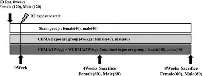
A total of 240 specific pathogen-free male (120) and female (120) Sprague-Dawley (SD) rats were obtained from the Japan SLC, Inc (Hamamatsu, Shizuoka, Japan) at an age of 8 weeks. The temperature and relative humidity in the exposure facility were maintained at 22 ± 2°C and 50 ± 10%, respectively, and were continuously monitored. Fluorescent lighting was provided for 12 h daily. The studies were performed according to the guidelines for use and care of laboratory animals and were approved by the
Institutional Animal Care and Use Committee of the Korea Institute Radiological and Medical Sciences (KIRAMS).
RF multi-exposure system
A reverberation chamber was designed as a whole-body ex-posure system for in vivo experiments to allow simultan-eous exposure to multiple mobile phone frequencies. The multiple-frequency whole-body exposure system generates CDMA signals and WCDMA signals simultaneously. To generate the signals, a microprocessor unit (MPU) chip with prewritten CDMA and WCDMA format codes was used. The MPU, in turn, controlled a central processing unit (CPU) to generate real CDMA and WCDMA test signals. For the cellular system, a real CDMA signal, at 848.5 MHz, was generated and subsequently amplified using a high power amplifier module (DCS60WHPA_CW; Kortcom, Anyang-si, Korea) after passing through a digital attenuator. For the WCDMA system, a real WCDMA signal at 1950 MHz was generated and subsequently ampli-fied using another high power amplifier module (PCS60WHPA_CW; Kortcom) after passing through a sep-arate digital attenuator. We used an 11-bit digital PIN diode attenuator (Model 349; General Microwave, Farmingdale, NY, USA) and the attenuator controlled the output power level. The maximum available input power was 60 W. The transmitting antennae used for the CDMA and the WCDMA frequencies are commercial products ( patch type; KCAN0800PA for CDMA, KCAN1900PA for WCDMA, Korea telecommunication component, Kyunggi-do, South Korea). A computer controlled the exposure level and the exposure time schedule. The external dimensions of the re-verberation chamber were 2295 mm × 2293 mm × 1470 mm, and the thickness of the walls was 2.3 mm. These dimensions satisfy the requirements for the minimum number of modes required at the lowest useable frequency (LUF) of interest. Additionally, the stainless wall of the chamber functioned as an electromagnetic shield. Eight cages were placed on the table (test area), which was located inside the exposed chamber and the field uniformity in the test area was confirmed by measuring the field strength for 1 min at 24 points. The field uniformity in the cage was also confirmed by measuring the field strength for 1 min at 27 points on the surface of the cage at a typical exposure location. The electric field distribution was measured inside the chamber using a three-axis isotropic probe (HI-6005; ETS-Lindgren, Cedar Park, TX, USA). The field distribution of both 848.5 and 1950 MHz was well within 3 dB in the region of interest. Assuming the field vectors were uniform in all directions, the SAR distri-bution for a caged rat was calculated using a finite differ-ence time domain (FDTD) tool (XFDTD version 6.5; Remcom, State College, PA, USA) in six orthogonal
directions. Two independent polarizations were used for each direction. The rat model (Chungnam National University, Daejon, Korea) used for simulation had 40 tissues and a 1-mm voxel size. The calculated SAR values were then averaged and multiplied by the measured root mean square electric field to estimate the real SAR values for a given input power. For 2.0 W/kg of whole-body aver-aged SAR, the power output was controlled at 30 W for CDMA and at18W for WCDMA [14,15].
Experimental design
The experimental design is shown in Fig. 1. RF exposure was performed by whole-body exposure in the exposure chamber for 45 min/day, 5 days per week for a total of 8 weeks from 9 a.m. to 2 p.m. CDMA exposed rats were exposed to CDMA (4.0 W/kg) signals. Combined RF exposed rats were simultaneously exposed to CDMA (2.0 W/kg) and WCDMA (2.0 W/kg) signals, 4 W/kg in total. All rats (five rats per cage) were housed in autoclaved polycarbonate cages (420 × 260 × 180 mm) on certified hardwood bedding. The position of the cages and turn of exposure were changed every day. Sham-exposed rats were placed in the exposure chamber without exposure to mag-netic field signals. Experimental animals were euthanized with CO2 gas. The data analysis was blinded to prevent treatment bias. Experimental animals were sacrificed in the morning, because of serum melatonin level.
Enzyme linked immunosorbent assay (ELISA) assay
Melatonin (Uscn Life Science Inc., Wuhan, P.R. China), TSH (Cusabio Biotech Co., Wuhan, P.R. China), T3, T4 (Calbiotech Inc., CA, USA), ACTH (Cusabio Biotech Co.), testosterone (Enzo Life Science, NY, USA) and estrogen
(Cusabio Biotech Co.) in the serum were measured using an ELISA Complete Kit according to the manufacturer’s in-struction sheet. Rat sera were collected from the rat abdom-inal aorta blood.
Briefly, standard solution or rat sera at various dilutions (testosterone; 1:20, estrogen; 1:100, T3, T4, TSH; 1:2, ACTH; 1:200, melatonin; 1:10) were added to each well and incubated at 37°C for 30–60 min. After washing three times with PBST (0.05%, Tween 20), Horseradish peroxid-ase (HRP)-labeled anti-rat testosterone, estrogen, T3, T4, TSH, ACTH and melatonin secondary antibody was added and incubated at 37°C for 1 h. After removing liquid from all wells, the wells were washed three times and the plates developed with 3,3′, 5,5′ tetra-methylbenzidine (TMB) color development solution. Sulfuric acid (2 mmol/10 ml) was added after the plates were allowed to react for 15 min at room temperature in the dark. Plates were read at 450 nm by ELISA reader (Multiskan MS photometer type 352, Labsystems, Helsinki, Finland). The final hormone con-centration was calculated based on a standard curve constructed for each assay using recombinant hormone standards.
Statistical analysis
Data are presented as mean ± SEM. The significance of the differences between group means of hormone levels was determined by one-way analysis of variance (ANOVA) fol-lowed by post hoc testing using the least significant differ-ence (LSD) method, performed with the SPSS 12.0 for Windows statistical package (SPSS, Inc., Chicago, IL, USA). The significance of the differences between the means of each age (4 weeks and 8 weeks) in the same gender was determined by Student’s t-test. Differences were considered statistically significant at P < 0.05.
RESULTS
Body weights and histopathological analysis
No deaths occurred during the 8-week experimental period. When compared to gender-matched controls after 4 and 8 weeks of exposure, there was no difference in body weight among the sham, CDMA alone and combined RF-exposed groups (Table 1). Histopathological analysis of all the organs including brain was performed and no significant alteration was found (data not shown).
Serum melatonin levels
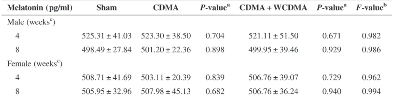
The effect of combined exposure on serum melatonin levels of both male and female SD rats was studied. Rats were exposed to RF-EMF during day time and autopsied at day time. The serum melatonin concentration was slightly lower at 8 weeks after exposure than 4 weeks in male rats, however, there were no significant differences. No alteration by exposure of CDMA alone or simultaneous exposure of CDMA and WCDMA was shown in serum melatonin levels (Table2).
Serum TSH, T3 and T4 levels
The effects of CDMA alone or combined exposure of CDMA and WCDMA on serum TSH and T3, T4 hormones levels of adult male and female SD rats were studied.
Table 3 showed TSH concentrations of sham-exposed, CDMA alone-exposed and combined RF exposed groups, after autopsy at 4 and 8 weeks post-exposure. Serum TSH concentration increased with age in both male and female rats, but only female TSH level in CDMA alone-exposed rats showed statistical significance. Moreover, there were no significant differences between sham and exposed groups (Table 3). Similarly, serum T3 and T4 hormone levels of CDMA alone or combined RF-exposed rats did not show any significant differences compared with sham exposed rats (Table3).
Serum ACTH level
Serum ACTH levels were assayed using the ELISA method. Serum ACTH levels significantly decreased with age in both male and female rats. However, CDMA alone or simultaneous combined exposure did not affect serum ACTH levels when compared with those of sham exposed rats (Table4).
Serum sex hormone levels
Sex hormones (testosterone in male rats and estrogen in female rats) were assayed. These sex hormone levels increased with age however, only estrogen level in the group of combined exposed rats showed statistical signifi-cance. CDMA alone or combined exposure of CDMA and Table 1. The body weights in RF-exposed rats
Body weight (g) Sham CDMA P-valuea CDMA + WCDMA P-valuea F-valueb
Male (weeksc) 4 414.73 ± 32.12 415.90 ± 27.59 0.902 425.25 ± 27.94 0.276 0.465 8 454.73 ± 70.11 465.55 ± 60.80 0.605 468.80 ± 52.29 0.476 0.751 Female (weeks3) 4 267.40 ± 20.95 263.33 ± 16.81 0.502 266.60 ± 18.11 0.898 0.767 8 288.10 ± 26.13 279.95 ± 26.64 0.335 286.03 ± 35.55 0.835 0.669
Values represent mean ± SD, aP-value: Student’s t-test, bF-value: one-way ANOVA test, cWeeks after exposure.
Table 2. The levels of melatonin in RF-exposed rat serum
Melatonin (pg/ml) Sham CDMA P-valuea CDMA + WCDMA P-valuea F-valueb
Male (weeksc) 4 525.31 ± 41.03 523.30 ± 38.50 0.704 521.11 ± 51.50 0.671 0.982 8 498.49 ± 27.84 501.20 ± 22.36 0.898 499.95 ± 39.46 0.929 0.986 Female (weeksc) 4 508.71 ± 41.69 503.11 ± 20.39 0.839 506.76 ± 39.07 0.729 0.962 8 505.95 ± 32.96 507.98 ± 45.13 0.682 506.76 ± 36.24 0.940 0.994
Values represent mean ± SD, aP-value: Student’s t-test, bF-value: one-way ANOVA test, cWeeks after exposure.
WCDMA, total SAR 4 W/kg, did not alter these serum hormone levels (Table5).
DISCUSSION
In the present study, we investigated the effects of simultan-eous combined exposure of CDMA and WCDMA RF-EMF,
especially focusing on serum hormone levels. We exposed rats to CDMA alone or to a combination of CDMA and WCDMA with total 4 W/kg for 8 weeks, a relatively long exposure in animal endocrine system experiments [7,16,17]. There is some research indicating that low frequency EMF decreases serum and pineal melatonin levels [18,19]. However, RF-EMF has not been often reported to affect Table 3. The levels of thyroid-stimulating hormone (TSH), thyroxine 3 (T3) and 4 (T4) in -F exposed rat serum
TSH (μU/ml) Sham CDMA P-valuea CDMA + WCDMA P-valuea F-valueb
Male (weeksc) 4 3.68 ± 0.99 3.43 ± 2.62 0.572 3.67 ± 1.47 0.810 0.956 8 4.91 ± 2.31 4.61 ± 2.31 0.374 4.99 ± 2.26 0.694 0.923 Female (weeks3) 4 2.85 ± 1.13 2.59 ± 1.01 0.716 2.76 ± 0.50 0.697 0.883 8 4.06 ± 1.93 4.28 ± 1.48* 0.616 4.11 ± 1.90 0.830 0.964 T3 (ng/ml) Male (weeksc) 4 19.33 ± 1.96 20.05 ± 0.49 0.472 19.42 ± 0.84 0.929 0.699 8 19.38 ± 1.41 19.92 ± 0.81 0.352 19.14 ± 1.17 0.712 0.350 Female (weeksc) 4 18.57 ± 1.93 18.86 ± 1.05 0.816 18.85 ± 0.39 0.897 0.907 8 16.68 ± 3.12 16.81 ± 2.91 0.716 16.20 ± 1.69 0.849 0.898 T4 (ng/dl) Male (weeksc 4 31.81 ± 1.69 31.35 ± 1.36 0.709 30.16 ± 0.51 0.768 0.731 8 31.55 ± 1.42 31.81 ± 3.53 0.618 31.47 ± 0.34 0.814 0.967 Female (weeksc) 4 33.46 ± 4.31 33.66 ± 1.69 0.897 34.09 ± 2.09 0.694 0.895 8 33.26 ± 4.04 33.87 ± 3.24 0.899 33.47 ± 2.24 0.765 0.923
Values represent mean ± SD,aP-value: Student’s t-test,bF-value: one-way ANOVA test,cWeeks after exposure. *Significant difference from the corresponding values of 4-week-old rats according to Student’s t-test.
Table 4. The levels of adrenocorticotropic hormone (ACTH) in RF-exposed rat serum.
ACTH (ng/ml) Sham CDMA P-valuea CDMA + WCDMA P-valuea F-valueb
Male (weeksc) 4 7.18 ± 0.26 7.21 ± 0.17 0.728 7.14 ± 0.38 0.802 0.853 8 6.28 ± 0.28* 6.42 ± 0.11* 0.884 6.39 ± 0.38* 0.423 0.643 Female (weeksc) 4 6.32 ± 0.23 6.40 ± 0.24 0.545 6.44 ± 0.20 0.883 0.614 8 5.07 ± 0.47* 5.10 ± 0.50* 0.713 5.04 ± 0.49* 0.880 0.957
Values represent mean ± SD, aP-value: Student’s t-test,bF-value: one-way ANOVA test,cWeeks after exposure.
melatonin level [20–22]. One explanation could be the rela-tively low SAR level and short-term exposure duration. However, our long-term exposure (8 weeks) and higher SAR level (4 W/kg) did not alter serum melatonin level either. One limitation of our study is the timing of exposure and the timing of sacrifice. We exposed RF-EMF to rats at day time and sacrificed at day time. Therefore, if we were to change the timing of exposure or sacrifice, the results may be different. While prior research shows that dark phase exposure does not affect serum and pineal melatonin level [16], exposure was limited to only 6 h of exposure with a SAR value of 2.0 W/kg, so more detailed experi-ments for combined exposure with longer exposure and higher SAR values were needed.
There is little prior work describing the relationship between RF-EMF and pituitary hormones such as TSH, T3 and T4 hormones in animals. GSM-EMF significantly lowered serum TSH and T3, T4 values [7] and increased thyroxine and triiodothyronine secretion when the thyroids of dogs were exposed to varying levels of 2.45 GHz RF-EMF at estimated very high SARs of 58–190 W/kg. Whole-body exposure to RF-EMF at 4 W/kg in rats caused rectal temperature rise to 40°C resulting in decreased circu-lating thyroxine and TSH levels [23]. However, 1.29 GHz RF-EMF at 3–4 W/kg did not change serum thyroxin [24]. Our data indicated that combined exposure to CDMA and WCDMA at 4 W/kg SAR for 8 weeks did not alter TSH, T3 and T3 hormone levels, and did not increase rectal tem-perature [25].
ACTH is a hormone induced by stress in the hypothal-amic–pituitary–adrenal axis (HPA). In a previous study 120 min of exposure of universal mobile telecomm-unications system (UMTS) to full brain-exposed rats with 10 W/kg SAR increased the plasma corticosterone level and ACTH level [26]. However, our 4 W/kg SAR with whole-body exposure for 8 weeks did not affect serum ACTH level, even with combination exposure of CDMA and WCDMA, suggesting higher SAR levels with focused exposure to the brain may be necessary to alter ACTH levels.
Serum testosterone levels were not changed by RF-EMF according to several papers [14, 27]. However, there are suggestions that damage to the testis may occur, resulting in reduced fertility potential of men [28–30] and experi-mental data have suggested that RF-EMF caused a signifi-cant increase in sperm cell death and abnormal clumping of sperm cells [31]. Others found no such adverse effects of RF-EMF on testicular function including testosterone level in animal experiments [25,32–34]. Consistently with these latter findings, our data show no alteration of serum testos-terone level from combined exposure of CDMA and WCDMA with 4 W/kg SAR and 8 weeks of exposure, a higher SAR level and longer exposure time. The literature on estrogen effects is sparse. Short-term exposure (4 h) of time division multiple access (TDMA) with higher SAR levels of 6.1 W/kg did not change estrogenic levels in rats [35]. Similarly, our combined exposure to CDMA and WCDMA with total 4 W/kg SAR for 8 weeks did not alter serum estrogen level.
In this study, we examined the various endocrine hormone levels in serum after 4 or 8 weeks’ exposure to RF-EMF. Even though age-related changes between 4- and 8-week-old rats were found in all the hormone levels we examined in this study, using the combination of CDMA and WCDMA as well as CDMA alone, a 4 W/kg SAR value did not affect hormone secretion in hormones such as melatonin, thyroid-related hormone, ACTH and sex hormone when they were detected in the serum of rats. However, our negative results are limited by the experimen-tal sensitivity for the detection of hormone levels and the absence of a positive control; therefore, more detailed experiments are needed in the future. In summary, our find-ings indicate that simultaneous combined exposure of CDMA and WCDMA with a total SAR dose of 4.0 W/kg for 45 min/day for 8 weeks, which is a relatively high SAR level and longer duration of exposure than the basic restric-tions recommended by ICNIRP for humans (frequency range of 100 kHz–10 GHz, 0.4 W/kg is the occupational exposure limit of whole-body average SAR and 0.08 W/kg is the general public exposure limit) has no significant Table 5. The levels of sexual hormones in RF-exposed rat serum
Testosterone (ng/ml) (weeksc) Sham CDMA P-valuea CDMA + WCDMA P-valuea F-valueb
4 12.39 ± 2.6 12.86 ± 2.4 0.558 12.30 ± 3.2 0.861 0.787
8 13.45 ± 2.6 13.84 ± 2.8 0.971 13.28 ± 1.5 0.332 0.751
Estrogen (ng/ml) (weeksc)
4 69.66 ± 4.06 68.51 ± 4.34 0.701 68.26 ± 3.71 0.543 0.576
8 71.40 ± 5.75 71.32 ± 8.73 0.474 72.99 ± 3.58* 0.522 0.805
Values represent mean ± SD,aP-value: Student’s t-test,bF-value: one-way ANOVA test,cWeeks after exposure. *Significant difference from the corresponding values of 4-week-old rats according to Student’s t-test.
effects on rat serum hormone levels in our limited experi-mental systems.
We also compared the effects between CDMA alone and a combination of CDMA and WCDMA with the same total SAR value (4 W/kg). Our results suggested that neither 4 W/kg of CDMA alone nor a combination of CDMA and WCDMA affected rat serum hormone levels.
ACKNOWLEDGEMENTS
A Grant from the Korea Communications Commission (2011) supported this work. Further support was provided by the Ewha Global Top 5 Grant 2011 of Ewha Womans University.
REFERENCES
1. Fragopoulou A, Grigoriev Y, Johansson O et al. Scientific panel on electromagnetic field health risks: consensus points, recommendations, and rationales. Rev Environ Health 2010;25:307–17.
2. Schüz J, Lagorio S, Bersani F. Electromagnetic fields and epidemiology: an overview inspired by the fourth course at the International School of Bioelectromagnetics. Bioelectromagnetics 2009;30:511–24.
3. IARC. IARC classifies radiofrequency electromagnetic fields as possibly carcinogenic to humans. International Agency for Research on Cancer 2011.
4. Lu ST, Lebda N, Pettit S et al. Microwave-induced tempera-ture, corticosterone, and thyrotropin inter relationships. J Appl Physiol 1981;50:399–405.
5. Lu ST, Lebda N, Michaelson SM et al. Serum-thyroxine levels in microwave-exposed rats. Radiat Res 1985;101:413–23. 6. Lu ST, Lebda NA, Lu SJ et al. Effects of microwaves on
three different strains of rats. Radiat Res 1987;110:173–91. 7. Koyu A, Cesur G, Ozguner F et al. Effects of 900 MHz
elec-tromagnetic field on TSH and thyroid hormones in rats. Toxicol Lett 2005;157:257–62.
8. Tamasidze AG. Influence of the chronic exposure to network frequency electromagnetic field on rats under interrupted and continuous action of EMF. [Article in Russian] Georgian Med News 2006;140:91–3.
9. Semenova TP, Medvinskaia NI, Bliskovka GI et al. Influence of electromagnetic fields on the emotional behaviour of rats. [Article in Russian] Radiats Biol Radioecol 2000;40:693–5. 10. Boscolo P, Di Giampaolo L, Di Donato A et al. The immune
response of women with prolonged exposure to electromag-netic fields produced by radiotelevision broadcasting stations. Int J Immunopathol Pharmacol 2006;19:43–8.
11. Szemerszky R, Zelena D, Barna I et al. Stress-related endo-crinological and psychopathological effects of short- and long-term 50Hz electromagnetic field exposure in rats. Brain Res Bull 2010;81:92–9.
12. Nussey S, Whitehead S. Endocrinology: An Integrated Approach. Oxford: BIOS Scientific Publishers. 2001, Chapter 4.
13. Black DR, Heynick LN. Radiofrequency (RF) effects on blood, cells, cardiac, endocrine and immunological functions. Bioelectromagnetics 2003;6:187–95.
14. Lee HJ, Lee JS, Pack JK et al. Lack of teratogenicity after combined exposure of pregnant mice to CDMA and WCDMA radiofrequency electromagnetic fields. Radiat Res 2009;172:648–52.
15. Jin YB, Lee HJ, Seon Lee J et al. One-year, simultaneous combined exposure of CDMA and WCDMA radiofrequency electromagnetic fields to rats. Int J Radiat Biol 2011; 87:416–23.
16. Hata K, Yamaguchi H, Tsurita G et al. Short term exposure to 1439 MHz pulsed TDMA field does not alter melatonin synthesis in rats. Bioelectromagnetics 2005;26:49–53. 17. Bakos J, Kubinyi G, Sinay H et al. GSM modulated
radiofre-quency radiation does not affect 6-sulfatoxymelatonin excre-tion of rats. Bioelectromagnetics 2003;24:531–4.
18. Grota LJ, Reiter RJ, Keng P et al. Electric field exposure alters serum melatonin but not pineal melatoninsynthesis in male rats. Bioelectromagnetics 1994;15:427–37.
19. Kato M, Honma K, Shigemitsu T et al. Circularly polarized 50-Hz magnetic field exposure reduces pineal gland and blood melatonin concentrations of Long-Evans rats. Neurosci Lett 1994;17:59–62.
20. Vollrath L, Spessert R, Kratzsch T et al. No short-term effects of high-frequency electromagnetic fields on the mammalian pineal gland. Bioelectromagnetics 1997;18: 376–87.
21. Radon K, Parera D, Rose DM et al. No effects of pulsed radio frequency electromagnetic fields on melatonin, cortisol, and selected markers of the immune system in man. Bioelectromagnetics 2001;22:280–7.
22. Bortkiewicz A, Pilacik B, Gadzicka E et al. The excretion of 6-Hydroxyl-melatonin sulfate in healthy young men exposed to electromagnetic fields emitted by cellular phone—an ex-perimental study. Neuro Endocrinol Lett 2002;23 (Suppl 1): 88–91.
23. Lu ST, Lebda N, Pettit S et al. Delineating acute neuroendo-crine responses in microwave-exposed rats. J Appl Physiol 1980;48:927–32.
24. Lotz WG, Podgorski RP. Temperature and adrenocortical responses in rhesus monkeys exposed to microwaves. J Appl Physiol 1982;53:1565–71.
25. Lee HJ, Jin YB, Kim TH et al. The effects of simultaneous combined exposure to CDMA and WCDMA electromagnetic fields on rat testicular function. Bioelectromagnetics 2012;33:356–64.
26. Prochnow N, Gebing T, Ladage K et al. Electromagnetic field effect or simply stress? Effects of UMTS exposure on hippocampal longterm plasticity in the context of procedure related hormone release. PLoS One 2011;5:6(5):e19437. 27. Djeridane Y, Touitou Y, de Seze R. Influence of
electromag-netic fields emitted by GSM-900 cellular telephones on the circadian patterns of gonadal, adrenal and pituitary hormones in men. Radiat Res 2008;169:337–43.
28. Fejes I, Zavaczki Z, Szollosi J et al. Is there a relationship between cell phone use and semen quality? Arch Androl 2005;51:385–93.
29. Agarwal A, Deepinder F, Sharma RK et al. Effect of cell phone usage on semen analysis in men attending infertility clinic: an observational study. Fertil Steril 2008;89:124–8. 30. Agarwal A, Desai NR, Makker K et al. Effects of
radiofre-quency electromagnetic waves (RF-EMW) from cellular phones on human ejaculated semen: an in vitro pilot study. Fertil Steril 2009;92:1318–25.
31. Yan JG, Agresti M, Bruce T et al. Effects of cellular phone emis-sions on sperm motility in rats. Fertil Steril 2007;88:957–64. 32. Dasdag S, Zulkuf Akdag M, Aksen F et al. Whole body
exposure of rats to microwaves emitted from a cell phone
does not affect the testes. Bioelectromagnetics 2003; 24:182–8.
33. Dasdag S, Akdag MZ, Ulukaya E et al. Mobile phone expos-ure does not induce apoptosis on spermatogenesis in rats. Arch Med Res 2008;39:40–4.
34. Wang XW, Ding GR, Shi CH et al. Effect of electromagnetic pulse exposure on permeability of blood–testicle barrier in mice. Biomed Environ Sci 2008;21:218–21.
35. Yamashita H, Hata K, Yamaguchi H et al. Short-term expos-ure to a 1439-MHz TDMA signal exerts no estrogenic effect in rats. Bioelectromagnetics 2010;31:573–5.