Risk Factors for Delayed post-EMR
Hemorrhage in Patients
with Gastric Neoplasms
Dong Hoon Park
The Graduate School
Yonsei University
Department of Medicine
Risk Factors for Delayed post-EMR
Hemorrhage in Patients
with Gastric Neoplasms
A Master’s Thesis
Submitted to the Department of Medicine
and the Graduate School of Yonsei University
in partial fulfillment of the
requirements for the degree of
Master of Medicine
Dong Hoon Park
This certifies that the master’s thesis of Dong Hoon Park is
approved.
___________________________
Hyun Soo Kim : Thesis Supervisor
___________________________
Dong Ki Lee : Committee Member
___________________________
Kap Jun Yoon : Committee Member
The Graduate School
Yonsei University
C
ONTENTS
List of figures ………... iv L i s t o f t a b l e s … … … . . . i v A b s t r a c t … … … . v I . I n t r o d u c t i o n … … … . 1 I I . P a t i e n t s a n d me t h o d s … … … . 3 1 . P a t i e n t s … … … . 3 2 . P r e p a r a t i o n s … … … . 3 3 . E M R m e t h o d s … … … . 4 4 . D e f i n i t i o n o f i m m e d i a t e a n d d e l a y e d P E H … … … . … 6 5 . Va r i a b l e s … … … . 7 6 . St a t i s t i c a l a n a l y s i s … … … . 8 I I I . R e s u l t s … … … . … … … … . 9 1 . C h a ra c t e r i s t i c s o f p a t i e n t s … … … . 92. Characteristics of gastric neoplasms ………….………. 10
3. Univariate analysis of post-EMR hemorrhage according to variables …… 12
4. Multivariate analyses of immediate delayed PEH according to variables ... 14 I V. D i s c u s s i o n … … … . 1 5 R e f e r e n c e s … … … . 2 1 A b s t r a c t i n K o r e a n … … … . 2 6
List of figure
Figure 1. Degree of immediate PEH ………
6List of Tables
Table 1. Characteristics of patients
……… 9Table 2. Characteristics of gastric neoplasms
……… 11Table 3. Univariate analysis of post-EMR hemorrhage according to variables
………
……….. 13Table 4. Multivariate analyses of immediate and delayed PEH according to
ABSTRACT
Risk Factors for Delayed post-EMR Hemorrhage
in Patients with Gastric Neoplasms
Background: Although endoscopic mucosal resection (EMR) has been recognized as the
standard treatment for mucosal gastric neoplasm, post-EMR hemorrhage (PEH) remains a
major complication of EMR, and this problem seems to be on the increase due to the
development of invasive techniques. The aims of this study were to determine the incidence and
grade of immediate or delayed PEH and to identify risk factors for delayed PEH in patients with
gastric neoplasm.
Methods: Data of EMRs performed by three endoscopists were retrospectively collected over 8
years and then analyzed. The clinical records, the EMR and 24 hr follow-up endoscopic
procedures were reviewed for 157 patients with gastric adenoma or early gastric cancer.
Immediate PEH was defined as the bleeding during the procedure. Delayed PEH was defined
or dizziness, 2) hemoglobin loss >2 g/dL, 3) blood pressure decrease >20 mmHg or pulse rate
increase >20 /min, and 4) Forrest I or IIa-IIb on follow-up endoscopy. The patient-related,
neoplasm-related and procedure-related variables were evaluated as potential risk factors for
PEH.
Results: A total of 157 patients (mean age: 64, M:F = 44:113) were reviewed. Twenty-nine
patients (18.5%) and thirteen patients (8.3%) presented with immediate and delayed PEH,
respectively. Multivariate logistic regression analysis revealed that the patient’s age (<65 years;
OR 0.18 95%, CI 0.03-0.96), the size of lesion (>15 mm; OR 5.45, 95% CI 1.03-28.79), and the
experience of the endoscopist (>5 years; OR 0.07, 95% CI 0.01-0.71) were significantly
predictive variables for the delayed PEH. However, immediate PEH was not related to any of
the studied variables.
Conclusions: Considering the higher risk of delayed PEH, careful preparation and close
monitoring are required for elderly patients (>65 years), large sized gastric neoplasms (>15
mm), or EMR performed by an inexperienced endoscopist (≤5 years).
Key words; Risk factor, Endoscopic mucosal resection, Hemorrhage, Gastric neoplasm
Risk Factors for Delayed post-EMR Hemorrhage
in Patients with Gastric Neoplasms
< Thesis Supervisor : Hyun Soo Kim >
Department of Medicine
The Graduate School of Yonsei University
Dong Hoon Park
I. INTRODUCTION
Endoscopic mucosal resection (EMR) has become a widely accepted therapeutic strategy for
gastric mucosal neoplasm because it is a minimally invasive and highly effective procedure. In
particular, EMR has come into the spotlight owing to improvements of the endoscopic devices
that allow for early detection of small lesions.1-5 Considering that it can be performed even in
outpatients, EMR is minimally invasive and more beneficial in the aspect of cost-effectiveness
when compared with surgical resection. EMR can provide long-term survival rates comparable
Even if EMR’s safety has been substantiated, unexpected complications can diminish the
effectiveness of EMR in some patients. Complications of EMR include pain, hemorrhage,
perforation and stricture formation. Of those, hemorrhage is the most common complication and
it can cause a fatal outcome. In addition, post-EMR hemorrhage (PEH) seems to be increasing
due to the generalization of invasive techniques such as endoscopic submucosal dissection.
Whereas a previous study reported on the frequency of colonoscopic post-polypectomy
bleeding,7 detailed studies concerning the risk factors for PEH in the stomach are scarce.8 In this
study, we investigated the incidences of immediate and delayed PEH, and we evaluated the
patient-related, gastric neoplasm-related and procedure-related factors that can predict the risk
II. PATIENTS AND METHODS
1. Patients
From January 1995 to June 2003, EMR was performed by three endoscopists on 157 patients
with gastric neoplasm at the Yonsei University Wonju Christian Hospital, Gangwondo, South
Korea. The 157 lesions consisted of 62 early gastric cancers and 95 gastric adenomas. The
indication for EMR in patients with EGC included intramucosal differentiated adenocarcinoma
without any ulceration or fibrosis being noted in the submucosal layer.
2. Preparations
EMR was performed using an endoscope (GIF-Q200, 240, 260, Olympus Optical Co., Ltd.,
Tokyo, Japan) under topical anesthesia of the pharynx and with intravenous injections of
midazolam (0.05 mg/kg, Roche Korea Ltd.) and meperidine (0.5 mg/kg, Korea). After careful
the lesion to be resected was marked by a needle-knife using coagulation current (40W).
Normal saline solution with or without epinephrine (1:10,000) was injected into the submucosa
to detach the lesion from the muscle layer and to prevent possible bleeding or perforation.
3. EMR methods
In the present study, we used two techniques of EMR, i.e. strip biopsy or ligation devices.
The strip biopsy method was performed in 58 patients and EMR with the ligation (EMRL)
method was performed in 99 patients. Strip biopsy was performed with a two-channel scope. A
snare and grasping forceps were inserted via the accessory channel. The snare loop was opened
in the stomach, and the grasping forceps was passed through the loop. The tissue adjoining the
lesion was next grasped and lifted with the grasping forceps. The snare was opened and the
lesion was then ensnared. Electrosurgical blended current (Valleylab generator, Boulder, Colo.,
USA), which was composed of coagulation (40W) and cutting (80W) current, was then applied
to resect the lesion. The resected specimens were removed and then examined
equipped with a ligation device (the O-ring from the Stiegman-Goff endoscopic ligator kit,
12-mm outer diameter, 10.5-12-mm inner diameter, 10-12-mm depth; Bard International Inc., Tewksbury,
MA, USA). The target lesion was aspirated into the O-type ligation device. The iatrogenic
pseudopolypoid lesion was resected by snare polypectomy with high-frequency
electrocauterization just below the ligation band. Because the maximal size of the specimen
resected by EMRL is 1.5 cm, the size of neoplasm as an independent variable was determined
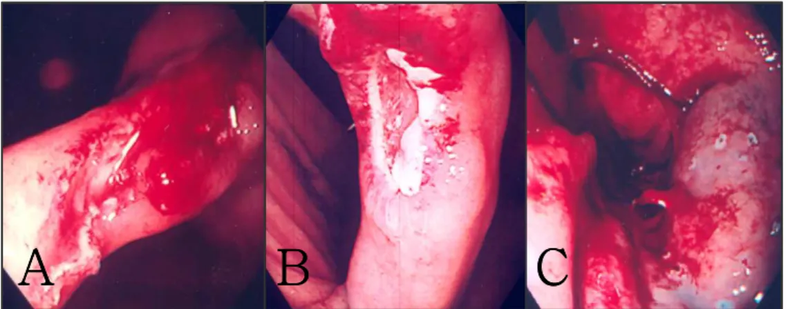
4. Definitions of immediate and delayed PEH
Immediate PEH was defined as the bleeding that occurred during the procedure, and the
grades of immediate PEH were classified as G1 (oozing, but not requiring endoscopic
intervention), G2 (continued oozing requiring endoscopic intervention), and G3 (active
spurting) (Figure 1). Delayed PEH was defined when 2 of 4 following parameters were satisfied
after the EMR procedure; 1) hematemesis, melena or dizziness, 2) hemoglobin loss >2 g/dL, 3)
a blood pressure decrease >20 mmHg or a pulse rate increase >20 /min, and 4) Forrest I, IIa-IIb
on follow-up endoscopy.
A
A
A
A
B
B
B
B
C
C
C
C
A
A
A
A
B
B
B
B
C
C
C
C
Figure 1. Degree of immediate PEH. A, Endoscopic view of grade I PEH; spontaneous hemostasis without endoscopic treatment. B, Endoscopic view of grade II PEH; oozing requiring endoscopic treatment. C, Endoscopic view of grade III PEH; active spurting
5. Variables
The patient-related variables (age, gender, presence of chronic disease, prior drugs such as
aspirin, NSAID or warfarin use, and the American Society of Anaesthesiologists (ASA)
physical status), the neoplasm-related variables (number, type, size, location, presence of en
bloc resection, gross and microscopic characteristics), and the procedure-related variables
(experience of the endoscopist, procedure time and type of electrosurgical current applied) were
evaluated as potential risk factors for PEH. Complete blood count (CBC), prothrombin time
(PT), and partial thromboplastin time (PTT) were measured before the procedure, and another
6. Statistical Analysis
All statistical tests were performed by two-sided tests and a p-value < 0.05 was considered
statistically significant. Statistical analysis was performed with the SPSS PC window program
(Statistical Package for the Social Science, SPSS Ins, Chicago, USA). Associations between the
categorical predictors and delayed PEH were assessed by the chi-square test. Multiple logistic
regression analysis for the risk factors of delayed PEH was performed to examine the effects of
the independent variables, and adjustments were made for the effects of each of the variables on
III. RESULTS
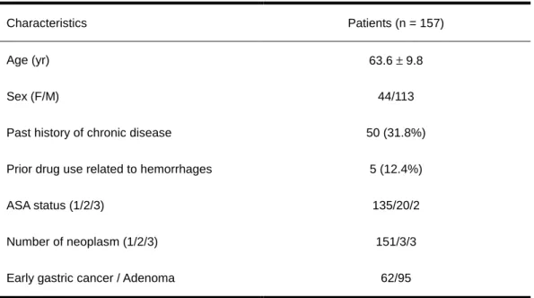
1. Characteristics of patients
A total of 157 patients (mean age: 64, M:F=44:113) were reviewed (Table 1). Fifty of the
patients had chronic concomitant diseases and five patients had been using drugs related to
bleeding tendencies. For the functional classification, 135 patients were ASA status 1, 20
patients were status 2, and 2 patients were status 3. Most of patients (151 of 157 patients,
96.2%) had one neoplasm at the time of the procedure. The removed lesions consisted of 62
EGCs and 95 adenomas.
Table 1. Characteristics of patients
Characteristics Patients (n = 157) Age (yr) 63.6 ± 9.8 Sex (F/M) 44/113 Past history of chronic disease 50 (31.8%) Prior drug use related to hemorrhages 5 (12.4%) ASA status (1/2/3) 135/20/2 Number of neoplasm (1/2/3) 151/3/3 Early gastric cancer / Adenoma 62/95
2. Characteristics of gastric neoplasms
The characteristics of the gastric neoplasms are shown in Table 2. The mean size of the
lesions was 11 mm. The overall en bloc resection rate was 59% (92/157). The most common
location was the antrum and greater curvature. Immediate PEH occurred in 29 of 157 patients
(18.5%; 25 were G1, 3 were G2 and 1 was G3). In the majority of the cases, endoscopic
intervention was not necessary due to spontaneous hemostasis. Three cases of G2 bleeding were
easily controlled by application of a hemoclip and 1 case of G3 bleeding was controlled by local
injection therapy. Delayed PEH occurred in 13 of 157 patients (8.4%). In all 17 cases, the
Table 2. Characteristics of gastric neoplasms
Characteristics Neoplasms Size of neoplasm (mm, range) 11.3 ± 5.7 (2-30) Location of neoplasm (body/antrum) 22/ 135 Location of neoplasm
(LC/GC/AW/PW) 41/ 65/ 26/ 25 Gross findings
EGC (I/ IIa/ IIb/ IIc/ IIa+c/ IIc+a) 9/ 15/ 10/ 15/ 11/ 2 Adenoma (flat/ Yamada-I/ LST) 36/ 57/ 2 Microscopic findings
EGC (W-D/ M-D) 38/ 24 Adenoma (LGD/ HGD/ Others) 62/ 12/ 21
En bloc / piecemeal 92/ 65
LC, lesser curvature; GC, greater curvature; AW, anterior wall; PW, posterior wall; LGD, low
grade dysplasia; HGD, high grade dysplasia, W-D, well differentiated; M-D, moderate differentiated; LST, laterally spreading neoplasm
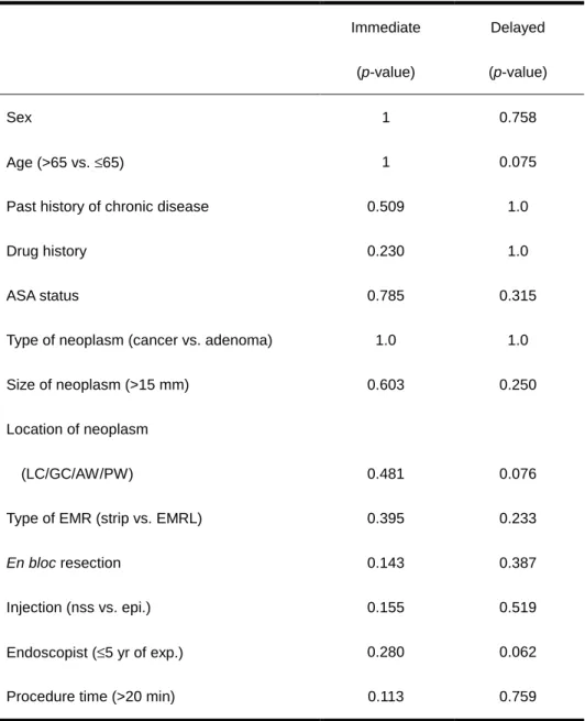
3. Univariate analysis of post-EMR hemorrhage according to
variables
On univariate analysis, we analyzed age, anticoagulant medication, type of neoplasm, size of
neoplasm, location of neoplasm, EMR method, En bloc resection, kind of injection solution,
procedure time and EMR experience of the endoscopist. No significant variable for immediate
or delayed PEH was found on the univariate analysis (Table 3). Among variables mentioned
above, the type of injection solution (normal saline solution or epinephrine-mix solution) was
not related to the immediate (p = 0.155) or delayed (p = 0.519) PEH. The EMR method
including strip biopsy or EMRL also did not have an influence on the frequency of immediate
(p = 0.395) or delayed (p = 0.233) PEH. However, age, location of neoplasm and experience of
Table 3. Univariate analysis of post-EMR hemorrhage according to variables Immediate (p-value) Delayed (p-value) Sex 1 0.758 Age (>65 vs. ≤65) 1 0.075 Past history of chronic disease 0.509 1.0 Drug history 0.230 1.0 ASA status 0.785 0.315 Type of neoplasm (cancer vs. adenoma) 1.0 1.0 Size of neoplasm (>15 mm) 0.603 0.250 Location of neoplasm
(LC/GC/AW/PW) 0.481 0.076 Type of EMR (strip vs. EMRL) 0.395 0.233
En bloc resection 0.143 0.387
Injection (nss vs. epi.) 0.155 0.519 Endoscopist (≤5 yr of exp.) 0.280 0.062 Procedure time (>20 min) 0.113 0.759
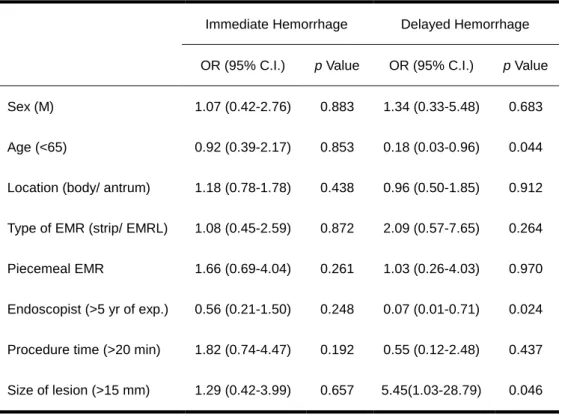
4. Multivariate analyses of immediate and delayed post-EMR
hemorrhage according to variables
On multivariate logistic regression analysis, the immediate PEH was not related to any of the
studied variables. On the other hand, the patient’s age (<65 years; OR 0.18, 95% C.I. 0.03-0.96),
size of the lesion (>15 mm; OR 5.45, 95% CI 1.03-28.79), and EMR experience of the
endoscopist (>5 years; OR 0.07, 95% CI 0.01-0.71) were significant predictive variables for the
delayed PEH (Table 4).
Table 4. Multivariate analyses of immediate and delayed PEH according to variables Immediate Hemorrhage Delayed Hemorrhage OR (95% C.I.) p Value OR (95% C.I.) p Value
Sex (M) 1.07 (0.42-2.76) 0.883 1.34 (0.33-5.48) 0.683 Age (<65) 0.92 (0.39-2.17) 0.853 0.18 (0.03-0.96) 0.044 Location (body/ antrum) 1.18 (0.78-1.78) 0.438 0.96 (0.50-1.85) 0.912 Type of EMR (strip/ EMRL) 1.08 (0.45-2.59) 0.872 2.09 (0.57-7.65) 0.264 Piecemeal EMR 1.66 (0.69-4.04) 0.261 1.03 (0.26-4.03) 0.970 Endoscopist (>5 yr of exp.) 0.56 (0.21-1.50) 0.248 0.07 (0.01-0.71) 0.024 Procedure time (>20 min) 1.82 (0.74-4.47) 0.192 0.55 (0.12-2.48) 0.437 Size of lesion (>15 mm) 1.29 (0.42-3.99) 0.657 5.45(1.03-28.79) 0.046
IV. DISCUSSION
On the grounds of its safety and effectiveness, EMR is acknowledged as an alternative
technique to surgery for the complete resection of gastric mucosal neoplasm.1-3 Nevertheless,
various complications can accompany EMR and these can cause unexpected and unwanted
results. In particular, post-EMR hemorrhage (PEH) is the most common complication and the
incidence of PEH reported in the previous literature ranges from 1.4% to 22%.5,9,12-18 In this
study, the frequency of overall (immediate and delayed) bleeding was 27% (42 of 157 patients),
which was a little higher than the previous results. 8 This discrepancy was probably related to
whether a mild degree of hemorrhage was included into the immediate PEH data. On the
contrary, the frequency of delayed PEH was 8.4% (13 of 157 patients), and this was not much
different from the previous studies. Delayed PEH was defined by relatively objective
parameters and these might be the preferable variables for comparing the results of different
studies. Additionally, in the view of the clinician, most of immediate PEH can be easily treated
by endoscopic hemostasis during the resection procedure, regardless of the severity. Ono et al. 3
endoscopic treatment, which would obviate the need for blood transfusion. Also in this study,
we made every effort to use hemostatic procedures whenever necessary during EMR because
we considered the presence of immediate PEH to be a warning sign of delayed PEH. For this
reason, all of the immediate PEHs in our study were completely controlled during the resection
procedure, and there was no occurrence of delayed PEH among these cases. Unlike immediate
PEH, delayed PEH can be easily overlooked due to the gradual onset of signs and symptoms,
and this can cause unpredictable patient deterioration. Furthermore, because delayed
hemorrhage can prolong the admission period,3,5,9-11 it is very important, in respect to clinical
practice, to find out the risk factors for delayed PEH. Even though there have been some reports
about the risk factors for overall (immediate and delayed) PEH, they failed to provide useful
predictors for delayed PEH. Therefore, we evaluated the patient-related, tumor-related and
procedure-related variables as the risk factors for delayed PEH.
There appears to be a recent increase in the rate of PEH for some reasons. The intention of
complete resection may seduce the endoscopist to attempt to do a more extensive resection. It
over 15 mm19, and there has also been a report that contradicts this finding.20 In cases of en bloc
resection, it’s been reported that the extensive EMR, which is a procedure prone to inadequate
coagulation around the resection margin, raised the possibility of PEH.22 Okano et al.8 proposed
that insufficient coagulation could be the cause of delayed bleeding.On the univariate anlaysis,
the size of the neoplasm and en bloc resection did not have significant influence on the rate of
overall (immediate and delayed) PEH. However, on multivariate analysis, the size of neoplasm
(>15 mm) was a risk factor for delayed PEH. Accordingly, for patients with large neoplasms,
every attention should be paid to the hemostasis during EMR, and a careful monitoring should
be done after EMR.
Some reports have argued that the recently adopted equipment or techniques were
responsible for the increase of PEH.15-18,22 As compared with the conventional methods (EMRL,
EMRC), the up-to-date techniques using the insulated tip (IT)-knife or the hook-knife make for
easy access to en bloc resection. Miyamoto et al.22 reported that the en bloc resection rate with
the IT-knife was high even for large lesions, including those lesions over 30 mm. On the
more time on resection. In addition, the methods of submucosal dissection, which can resect an
invasive lesion directly, are more likely to bring on a risk of vascular injury.4 Consequently,
while the endoscopists have achieved more favorable results with the new techniques in regard
to complete resection, the risk of PEH took a turn for the worse as a corollary. In the present
study, there was no difference in the rate of PEH between the conventional methods, strip
biopsy and the EMRL method. On multivariate analysis, the EMR performed by an
inexperienced endoscopist was a significant risk factor for delayed PEH. Therefore, the
endoscopist’s skill was a significant determinant for delayed PEH and it was a more important
factor than the EMR method.
Some authors have reported that the location of neoplasms could be implicated in PEH.15,23,24
There were some studies showing that bleeding occurred more frequently in the upper area of
the stomach compared with the middle and lower areas. Hirao et al.23 reported that the
submucosal arteries of the upper third were larger than the arteries in the other areas, and they
were stubby or thick upon histological examination. Also, there could be some difficulties for
location of the neoplasms. In the present study, there was a marginal statistical significance for
delayed PEH according to the location of the neoplasms. Probably, the reason is that the
majorities of lesions in our study (135 of 157 lesions, 85.9%) were disproportionately located in
the lower portion of the stomach. Also, this may be somewhat responsible to the difference of
accessibility among locations, and accessibility was a bit easier at the greater curvature and
anterior wall (91 of 157 patients, 57.9%).
Although it has recently become possible to perform a safe and effective procedure in the
elderly with the help of supplementary oxygen and monitoring devices, 25 it is rather common
knowledge that the general patient conditions such as age, the presence of comorbidities and
functional status could be the major determinants of wound healing and rebleeding for EMR.
Vogiagis et al.26 proposed that aging could alter the cyclooxygenase activity and prostaglandin
synthesis, and thereby compromise the healing of tissue. On multivariate analysis, an age over
65 was a significant risk factor for delayed PEH. So, we can recommend the endoscopist to pay
more attention to the elderly via careful monitoring and follow-up. Furthermore, a prospective
In conclusion, considering the higher risk of delayed PEH, careful preparation and close
monitoring are required with elderly patients (>65 years), with a large sized gastric neoplasm
(>15 mm), or when the the resection procedure is performed by an inexperienced endoscopist
REFERENCES
1. Ponchon T. Endoscopic mucosal resection. J Clin Gastroenterology 2001;32(1):6-10.
2. Ohyama T, Kobayashi Y, Mori K, Kano K, Sakurai Y, Sato Y. Factors affecting complete
resection of gastric neoplasms by the endoscopic mucosal resection procedure. J Gastroentorol
Hepatol 2002;17:844-8.
3. Ono H, Kondo H, Gotoda T, Shirao K, Yamaguchi H, Saito D, et al. Endoscopic mucosal
resection for treatment of early gastric cancer. Gut 2001;48:225-9.
4. Yahagi N, Fujishiro M, Iguchi M, Kakushima N, Omata M. Theoretical and technical
requirements to expand EMR indications. Dig Endosc 2003;15:S19-21.
5. Soetikno RM, Gotoda T, Nakanishi Y, Soehendra N. Endoscopic mucosal resection.
Gastrointest Endosc 2003;57:567-79.
6. Kim HS, Lee DK, Baik SK, Kim JM, Kwon SO, Kim DS, et al. Endoscopic mucosal
resection with a ligation device for early gastric cancer and precancerous lesions: comparison of
7. Parra-Blanco A, Kaminaga N, Kojima T, Endo Y, Uragami N, Okawa N, et al.
Hemoclipping for postpolypectomy and postbiopsy colonic bleeding. Gastrointest Endosc
2000;51:37-41.
8. Okano A, Hajiro K, Takakuwa H, Nishio A, Matsushita M. Predictors of bleeding after
endoscopic mucosal resection of gastric neoplasms. Gastrointest Endosc 2003;57:687-90.
9. Ahmad NA, Kochman ML, Long WB, Furth EE, Ginsberg GG. Efficacy, safety and clinical
outcomes of endoscopic mucosal resection: a study of 101 cases. Gastrointest Endosc
2002;55:390-6.
10. Adachi Y, Shiraishi N, Kitano S. Modern treatment of early gastric cancer: review of the
Japanese experience. Dig Surg 2002;19:333-9.
11. Tsunada S, Ogata S, Ohyama T, Ootani H, Oda K, Kikkawa A, et al. Endoscopic closure of
perforations caused by EMR in the stomach by application of metallic clips. Gastrointest
12. Kojima T, Parra-Blanco A, Takahashi H, Fujita R. Outcome of endoscopic mucosal
resection for early gastric cancer: review of the Japanese literature. Gastrointest Endosc
1998;48:550-5.
13. Lambert R. Treatment of esophagogastric neoplasms. Endoscopy 2003;35:118-26.
14. Tanabe S, Koizumi W, Kokutou M, Imaizumi H, Ishii K, Kida M, et al. Usefulness of
endoscopic aspiration mucosectomy as compared with strip biopsy for the treatment of gastric
mucosal cancer. Gastrointest Endosc 1999;50:819-22.
15. Ohkuwa M, Hosokawa K, Boku N, Ohtu A, Tajiri H, Yoshida S. New endoscopic treatment
for intramucosal gastric neoplasms using an insulated-tip diathermic knife. Endoscopy
2001;33:221-6.
16. Tanabe S, Koizumi W, Mitomi H, Nakai H, Murakami S, Nagaba S. Clinical outcome of
endoscopic aspiration mucosectomy for early stage gastric cancer. Gastrointest Endosc
2002;56:708-13.
17. Yamamoto H, Kawata H, Sunada K, Satoh K, Kaneko Y, Ido K, et al. Success rate of
submucosal injection of sodium hyaluronate. Gastrointest Endosc 2002;56:507-12.
18. Fujisaki J, Matsuda K, Tajiri H. Endoscopic mucosal resection for early gastric cancer:
aiming at safety, speed and reliability. Dig Endosc 2003;15:S8-11.
19. Akamatsu T, Miyata K, Hasebe O, et al. Complications in endoscopic polypectomy and
endoscopic mucosal resection [In Japanese with English abstract]. Endoscopia Digestiva
1994;6:487-94.
20. Muto M, Saito Y, Koike T, Ikeya S, Sasaki T, Hoshino E. Complications of endoscopic
polypectomy and endoscopic mucosal resection in the stomach [In Japanese with English
abstract]. Gastroenterol Endosc 1996;38:858-65.
21. Kida M, Tanabe S, Saigenji K. Endoscopic mucosal resection for gastric cancer: necessity
of ‘incision and stripping method’ and present status. Dig Endosc 2003;15(Suppl.):S15-S18.
22. Miyamoto S, Muto M, Hamamoto Y et al. A new technique for endoscopic mucosal
resection with an insulated-tip electrosurgical knife improves the completeness of resection of
23. Hirao M, Asanuma T, Masuda K, Miyazaki A. Endoscopic resction of early gastric cancer
following locally injecting hypertonic saline-epinephrine. [In Japanese with English abstract]
Stomach Intestine 1988;23;399-409.
24. Narimiya N, Sato H, Joki M, Odagiri M, Iwasaki M, Sugimoto I, et al. An experimental
study of submucosal vascular structure of the stomach after endoscopic mucosal resection. [In
Japanese with English abstract] Gastroenterol Endosc 1994;36:958-63.
25. Yamaguchi Y, Yamato T, Katsumi N et al. Endoscopic hemostasis: Safe treatment for peptic
ulcer patients aged 80 years or older? J Gastroenterol Hepatol 2003;18:521-25.
26. Vogiasis D, Glare EM, Misajon A et al. Cyclooxygenase-1 and an alternatively spliced
mRNA in the rat stomach: effects of aging and ulcers. Am J Physiol Gastrointest Liver Physiol
국문요약
국문요약
국문요약
국문요약
위
위
위
위 종양
종양
종양
종양 환자의
환자의
환자의
환자의 내시경적
내시경적 점막
내시경적
내시경적
점막
점막 절제술
점막
절제술
절제술
절제술 후
후
후
후
지연
지연
지연
지연 출혈
출혈
출혈
출혈 발생의
발생의
발생의 위험
발생의
위험
위험 인자
위험
인자
인자
인자
연세대학교
연세대학교
연세대학교
연세대학교 대학원
대학원
대학원
대학원
의학과
의학과
의학과
의학과
박동훈
박동훈
박동훈
박동훈
배경 배경배경배경: 내시경적 점막 절제술(Endoscopic mucosal resection, EMR)은 위 점막 종양의 주
된 치료로 인정받고 있지만, 시술 후 출혈(Post-EMR hemorrhage, PEH)이 주요 합병증
이며, 침습적인 시술방법이 개발되면서 발생 빈도가 증가하는 양상이다. 본 연구에 서는 시술후의 조기 또는 지연 출혈의 빈도와 정도를 알아보고, 위종양이 있는 환 자에서 지연 출혈의 발생에 관여하는 위험인자를 조사하였다. 방법 방법방법 방법: 1995년 1월부터 2003년 6월까지 세 명의 내시경 전문의에 의해 시행된 내시경 적 점막 절제술의 자료를 후향적으로 분석하였다. 위선종이나 조기 위암이 있었던 157명의 환자에서 임상 기록과 내시경적 점막 절제술 소견, 그리고, 시술 24시간 후 의 추적 내시경 검사 소견을 분석하였다. 시술 후 조기출혈은 시술중에 발생하는 출혈로 정의하였다. 지연 출혈은 시술 후에 다음의 네가지 조건 중 두가지 이상을
만족하는 경우로 정의하였다; 1) 토혈, 흑색변, 어지러움증이 있는 경우, 2) 혈색소 수치가 2 g/dL이상 감소한 경우, 3) 혈압이 20 mmHg이상 감소하거나, 맥박이 20/min 이상 증가한 경우, 4) 추적 내시경 소견상 Forrest I 이나 IIa-IIb 에 해당하는 경우. 시술 후 지연출혈의 위험인자를 알아보고자 환자, 종양, 그리고, 시술과 관련된 변 수들을 조사하였다. 결과 결과결과 결과: 대상 환자의 평균 연령은 64세 였으며, 남자가 44명, 여자가 113명이었다. 조 기 출혈은 29명(18.5%)의 환자에서, 지연 출혈은 13명(8.3%)의 환자에서 관찰되었다. 다변량 회귀 분석에서 환자의 나이(<65세; OR 0.18, 95% CI 0.03-0.96), 병변의 크기 (>15 mm; OR 5.45, 95% CI 1.03-28.79), 시술자의 경력(>5년; OR 0.07, 95% CI 0.01-0.71) 이 지연 출혈의 발생에 있어 의미 있는 위험인자였다. 그러나, 대상 변수들 중에서 조기출혈의 위험인자는 없었다. 결론 결론결론 결론: 지연출혈의 높은 위험성을 고려할 때, 환자의 나이가 65세 이상일 경우나, 병 변의 크기가 15 mm이상일 경우, 시술자의 경험이 5년 이하일 경우에는 보다 주의 깊은 사전준비와 철저한 환자감시가 요구된다. ___________________________________________________________________________