International Journal of COPD
Dove
press
2057
O r I g I n a l r e s e a r C h open access to scientific and medical research Open access Full Text article
relationship between airway obstruction and
incidence of metabolic syndrome in Korea:
a community-based cohort study
Chi Young Kim Youngmok Park ah Young leem Kyung soo Chung Ji Ye Jung
Moo suk Park Young sam Kim
Division of Pulmonology, Department of Internal Medicine, Institute of Chest Diseases, severance hospital, Yonsei University College of Medicine, seoul, south Korea
Background: Although studies have examined the relationship between metabolic syndrome
(MetS) and COPD, the incidence of MetS in individuals with COPD has not specifically been investigated. This study aimed to evaluate the incidence of MetS in subjects with airway obstruc-tion using data from a community-based cohort.
Patients and methods: Data representing 4 years of follow-up from the Ansung–Ansan cohort
were analyzed; a total of 6,184 adults, who were $40 years of age and underwent spirometry, were enrolled in this study. Airway obstruction was defined as forced expiratory volume in 1 s/forced vital capacity ratio ,70%, and MetS was determined according to the National Cholesterol Education Program Adult Treatment Panel III guidelines.
Results: A total of 419 patients were newly diagnosed with MetS, based on the National
Cholesterol Education Program Adult Treatment Panel III guidelines, during follow-up. MetS was more frequent in COPD subjects, relative to non-COPD subjects, in both sexes (14.7% vs 11.0% [men] and 14.7% vs 11.8% [women]). In men subjects, the risk for MetS was higher in subjects with airflow obstruction than in subjects without obstruction, after adjusting for age, body mass index, and smoking status.
Conclusion: The incidence of MetS was higher in men with airflow obstruction than in healthy
subjects.
Keywords: COPD, metabolic syndrome, incidence
Introduction
COPD is characterized by persistent air-flow limitation that is typically progressive1
and is a major cause of morbidity and mortality worldwide.2 Metabolic syndrome
(MetS) has been associated with several diseases, including cardiovascular disease, as well as with increased exacerbation of underlying disease and mortality risk.3–5
As COPD and MetS both constitute major public health problems, more precise and informative epidemiological data are needed regarding the relationship between COPD and MetS.
Previous studies have evaluated the prevalence of MetS in COPD patients and normal subjects.6,7 In addition, a Korean study reported a relatively higher prevalence of MetS
in patients with COPD, compared with healthy subjects.8,9 However, there have been no
community-based cohort studies investigating the incidence of MetS in COPD patients. Many of the previous studies regarding MetS in COPD were cross-sectional and primarily focused on the prevalence, rather than the incidence, of MetS in patients with COPD. Thus, the present study aimed to evaluate the incidence and characteristics of MetS in COPD patients using data from a large community-based cohort.
Correspondence: Young sam Kim Division of Pulmonology, Department of Internal Medicine, severance hospital, Institute of Chest Diseases, Yonsei University College of Medicine, 50-1, Yonsei-ro, seodaemun-gu, seoul 120-752, south Korea Tel +82 2 2228 1971 Fax +82 2 393 6884 email [email protected] Year: 2018 Volume: 13
Running head verso: Kim et al
Running head recto: Metabolic syndrome incidence in airflow obstruction DOI: 157453
This article was published in the following Dove Press journal: International Journal of COPD
Patients and methods
study population
Prospectively collected data from the Ansung–Ansan cohort were used for this analysis. The Ansung–Ansan cohort is a community-based sample that was evaluated in the Korean Genome and Epidemiology study (KoGES). The KoGES cohort included subjects from both the Ansan (urban) and Anseong (rural) areas of Korea for prospective investigation. Detailed information regarding the study design and protocols has been previously published.10 The baseline survey of the
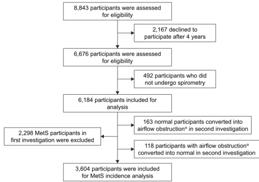
Ansung–Ansan cohort was conducted between May 2001 and February 2003; the present study included 8,843 eligible adults who were 40–69 years of age. Follow-up examinations were performed biennially. Of these subjects, 6,676 were reevaluated after 4 years, corresponding with a follow-up rate of 75.4% in the secondary survey (2005–2006). Subjects who did not undergo pulmonary function testing at baseline and follow-up after 4 years (n = 492) were excluded. Of the remaining 6,184 subjects, a further 2,298 were excluded because they exhibited MetS in the baseline survey. Although subjects were diagnosed with COPD at the time of the baseline survey, those exhibiting normal pulmonary function results in the secondary survey were excluded (n = 118), as were par-ticipants with normal lung function who exhibited COPD in the second investigation (n = 163). Ultimately, a total of 3,604 subjects were included in the final analysis (Figure 1).
Definition of MetS and airflow obstruction
MetS was defined clinically based on the presence of $3 of the following modified Adult Treatment Panel III revised guidelines:11 central obesity (with waist circumference cutoffpoints of .90 cm for men and .80 cm for women); an elevated triglyceride level ($1.7 mmol/L or undergoing drug treatment for elevated triglyceride levels); a reduced high-density lipoprotein cholesterol (HDL-C) level (,1.0 mmol/L [men], ,1.3 mmol/L [women], or drug treatment for reduced HDL-C); elevated blood pressure ($130 mmHg systolic, $85 mmHg diastolic, or antihypertensive drug treatment in patients with a history of hypertension); and an elevated fasting plasma glucose concentration ($5.6 mmol/L or drug treatment for diabetes).
Lung function tests were performed by a skilled tech-nician using a portable spirometer (Vmax-2130, Sensor Medics, Yorba Linda, CA, USA) in accordance with stan-dardized protocols from the American Thoracic Society.12
All participants underwent pulmonary function testing during each visit (at baseline as well as the first and second follow-up visits). Calibration and quality control of spiro-metric examinations were performed regularly, based on American Thoracic Society guidelines.12 Airflow
obstruc-tion was defined as a prebronchodilator forced expiratory volume in 1 second (FEV1)/forced vital capacity (FVC) ratio of ,0.7.
Figure 1 study population and design.
Note: *Airflow obstruction: forced expiratory volume in 1 second/forced vital capacity ratio , 0.7. Abbreviation: Mets, metabolic syndrome.
8,843 participants were assessed for eligibility
6,676 participants were assessed for eligibility
6,184 participants included for analysis
3,604 participants were included for MetS incidence analysis
163 normal participants converted into airflow obstruction* in second investigation
118 participants with airflow obstruction* converted into normal in second investigation 2,298 MetS participants in
first investigation were excluded
2,167 declined to participate after 4 years
492 participants who did not undergo spirometry
statistical analysis
All values are expressed as mean ± standard deviation. Continuous and categorical variables were compared using Student’s t-test and the chi-squared/Fisher’s exact test, respectively. Logistic regression analyses were performed to estimate odds ratios (ORs) with 95% confidence intervals (CIs) for MetS development, after adjusting for other con-founding variables; P , 0.05 was considered to be statisti-cally significant. Statistical analyses were performed using SAS 9.2 (SAS Institute Inc., Cary, NC, USA).
ethics approval
The Korean Centers for Disease Control and Prevention obtained written informed consent from all participants, and the Institutional Review Board of Severance Hospital (Seoul, South Korea) approved the study protocol (4-2016-0458). The data accessed from the KoGES cohort study is anonymous.
Results
Baseline patient characteristics
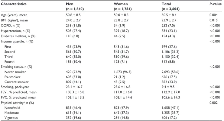
Table 1 describes the baseline characteristics of all patients who were included in the analysis. In total, 3,604 subjects (51.1% men, 45.9% women) were included in our analysis. The mean age of the patients was 50.5 ± 8.4 years. There was
an age difference between men and women (50.8 ± 8.5 vs 50.0 ± 8.3, P = 0.004). Among all included subjects, 23.1% and 4.3% exhibited hypertension and diabetes mellitus, respectively. The proportion of current smokers was 44.1% of male subjects and 2.5% of female subjects (P , 0.001).
The average body mass index (BMI) was 23.9 kg/m2, with
female subjects exhibiting a relatively lower mean BMI than male subjects (24.0 kg/m2 vs 23.8 kg/m2, P = 0.015). There
were no differences in lung function or physical activity between male and female subjects. At baseline, 252 of 3,604 (7.0%) subjects exhibited airflow obstruction. The propor-tion of male subjects with COPD was significantly higher than the proportion of female subjects with COPD (11.8% vs 1.9%, P , 0.001). The median follow-up duration was 4 years (interquartile range: 3.0–4.0 years).
Incidence of Mets
During the follow-up period, 11.6% (n = 419) of patients developed MetS based on the National Cholesterol Education Program Adult Treatment Panel III guidelines.11 The
inci-dence of MetS was higher in patients with COPD, relative to those without COPD, for both sexes (11.0% vs 14.7% [men] and 11.8% vs 14.7% [women]) (Figure 2). Table 2 shows the result of a multivariate logistic regression analysis of risk factors for MetS. After adjusting for confounding variables, Table 1 Baseline characteristics of men and women subjects included in the study
Characteristics Men (n = 1,840) Women (n = 1,764) Total (n = 3,604) P-value
age (years), mean 50.8 ± 8.5 50.0 ± 8.3 50.5 ± 8.4 0.004
BMI (kg/m2), mean 24.0 ± 2.7 23.8 ± 2.7 23.9 ± 2.7 0.015 COPD, n (%) 218 (11.8) 34 (1.9) 252 (7.0) ,0.001 hypertension, n (%) 505 (27.4) 329 (18.7) 834 (23.1) ,0.001 Diabetes mellitus, n (%) 110 (6.0) 44 (2.5) 154 (4.3) ,0.001 Income quartile, n (%) ,0.001 First 436 (23.9) 543 (31.6) 979 (27.6) second 561 (30.7) 545 (31.7) 1,106 (31.2) Third 640 (35.0) 510 (29.6) 1,150 (32.4) Fourth 189 (10.4) 123 (7.1) 312 (8.8) smoking status, n (%) ,0.001 never smoker 420 (22.9) 1,673 (96.3) 2,093 (58.6) ex-smoker 605 (33.0) 21 (1.2) 626 (17.5) Current smoker 809 (44.1) 43 (2.5) 852 (23.9) smoking, pack-year 23.1 ± 16.7 23.6 ± 16.8 9.4 ± 9.5 ,0.001
FeV1, % predicted, mean 108.3 ± 15.8 117.8 ± 16.8 112.9 ± 17.0 ,0.001
FVC, % predicted, mean 103.1 ± 13.5 108.1 ± 14.6 105.6 ± 14.3 ,0.001
Physical activity,a n (%) 0.002
None/mild 835 (46.4) 823 (47.9) 1,658 (47.1)
Moderate 613 (34.1) 642 (37.3) 1,255 (35.7)
Vigorous 352 (19.6) 254 (14.8) 606 (17.2)
Notes: Data are presented as mean ± standard deviation or n (%). aDefined as none/mild: 0–29 minutes/day; moderate 30–60 minutes/day; vigorous $60 minutes/day.
such as age, BMI, and smoking, male patients with airflow obstruction were 1.76-fold (95% CI 1.04–2.96, P = 0.028) more likely to develop MetS, relative to normal subjects. Thus, the risk for MetS in subjects with airway obstruction was higher in the subgroup of male subjects, following adjust-ment for age, BMI, smoking, and physical activity.
Discussion
This is the first study to confirm the incidence of Mets in COPD patients via data from a large community-based cohort. The incidence of MetS was higher in the airflow obstruction group, even after adjustment for other confound-ing factors, includconfound-ing age, BMI, smokconfound-ing, and physical activity, in male subjects. This finding is consistent with previous studies that have reported an increased prevalence of MetS in patients with COPD. However, clinical implica-tion of the present study is that it was the first to evaluate and compare the incidence of MetS between patients with and without airflow obstruction.
In the present study, incidence of MetS was higher in patients with impaired lung function. However, a strength of our study was that we defined the airflow obstruction group according to airflow limitation, rather than FEV1 quartile or changes in FVC. Previously, Kim et al investigated the incidence of MetS in patients with impaired lung function.13
In that study, changes in vital capacity over a 6-year period were associated with development of MetS, which itself was associated with obesity and abdominal obesity. However, the patients included in that study did not exhibit COPD, nor were they enrolled from a community-based sample. Another group attempted to characterize the incidence of MetS using 2-year follow-up data from Health Screening Centers in Taiwan.14 In contrast to the present study, the
follow-up duration in the Taiwan-based study was short and the target group was divided according to FEV1, rather than airflow limitation; furthermore, the target group was not a community-based cohort. There is significant disparity between the present study and previous studies regarding the accuracy of the definition of airflow obstruction.
Some studies have reported an association between COPD and MetS. Recent reviews revealed that the mean prevalence of MetS in patients with COPD was 32% (range 23%–58%), compared with 30% (range 17%–54%) in controls. Moreover, the average reported prevalence of MetS varies among different regions, with a lower prevalence in studies from Asia (28%), relative to European (41%) and American studies (53%).6 Furthermore, the difference in
MetS prevalence between COPD patients and controls was small, which may be related to the inclusion of subjects without COPD, but with other comorbidities, in the control group. In this study, we confirmed the prevalence of MetS in both populations (34.8%), relative to the control group (28.4%). The rates revealed in the present study are consistent with other recent reports. Additionally, the 4-year incidence of MetS was higher in the airflow obstruction group, rela-tive to the control group. Several groups have investigated the incidence of MetS using different diagnostic methods, 20% P = 0.116 14.7% 11.4% N = 382 N = 37 N = 178N = 32 N = 204 N = 5 11.0% 11.8% 14.7% 14.7% P = 0.106 P = 0.602 15% 10% 5% 0%
Total Men Women
COPD–
(N = 3,352) (N = 252)COPD+ (N = 1,622)COPD– (N = 218)COPD+ (N = 1,730)COPD– COPD+(N = 34) COPD (–) COPD (+)
Figure 2 Incidence of metabolic syndrome in patients with COPD at 4 years of follow-up.
Notes: Data are expressed as number and percentage. Metabolic syndrome devel-oped in 11.6% of the total subjects (419/3,604). Incidence of metabolic syndrome in patients with COPD was compared to incidence in patients without COPD, in both sexes.
Table 2 association between metabolic syndrome and lung function impairment at 4 years of follow-up
Characteristic Men OR (95% CI) P-value Women OR (95% CI) P-value Total OR (95% CI) P-value age 1.02 (1.00–1.05) 0.040 1.08 (1.05–1.10) ,0.001 1.05 (1.03–1.07) ,0.001 BMI 1.26 (1.18–1.34) ,0.001 1.31 (1.23–1.40) ,0.001 1.29 (1.23–1.35) ,0.001 smoking 1.73 (1.06–2.81) 0.035 1.30 (0.29–5.88) 0.730 1.33 (0.97–1.83) 0.082 COPD 1.76 (1.04–2.96) 0.028 0.35 (0.07–1.92) 0.229 1.23 (0.76–1.97) 0.401 Physical activity 0.89 (0.19–4.30) 0.875 1.08 (0.28–4.19) 0.914 1.08 (0.40–2.92) 0.877
Note: Data are presented as odds ratio (OR) (95% confidence interval [CI]), adjusted for age, body mass index (BMI), COPD, physical activity, and metabolic syndrome incidence in multivariable logistic regression analysis.
including spirometry and health care databases.15–17 A study
by Breyer et al revealed that the prevalence of MetS was higher in patients with COPD.18 Similarly, in a study
involving Asian subjects, the risk for MetS was higher in individuals with airflow obstruction, relative to individuals without obstruction, even after adjustment for potential confounders.19 Additionally, a Korean study that used data
from a nationwide survey reported that the risk for MetS was higher in sarcopenic males with COPD;8 notably, the
prevalence of MetS was also higher.9 Previous studies,
however, have sought to evaluate the relationship between COPD and MetS primarily through measurement of the prevalence of MetS. The findings from our study are unique in that we are the first to report the incidence of MetS in patients with COPD using data from a large community-based cohort. In contrast to other studies, the present study involved repeated measurements of pulmonary function and participants were more accurately categorized according to airflow obstruction, which differs from the methods used in previous investigations.
Many studies have investigated the relationship between COPD and the presence of comorbidities. In patients with COPD and MetS, cardiovascular comorbidities are more prevalent and are associated with an increased mortality, largely related to the aggravation of underlying disease.20,21
A possible explanation for higher incidence of MetS among COPD patients may be that COPD patients with MetS are physically less active and exhibit increased levels of systemic inflammation relative to healthy patients.22,23 However in this
study, these particular differences were not clearly observed, perhaps because the number of patients with MetS was insufficient; moreover, patients with severe and very severe airflow obstruction were not included in the analysis.
Although pathophysiological consequences of MetS remain controversial, insulin resistance may play a crucial role.24 Findings of the present study regarding airflow
obstruction and occurrence of insulin resistance, which was defined as increased fasting plasma glucose levels, are generally consistent with previous studies.14 For example,
Lawlor et al25 observed an inverse association between
insulin resistance and lung function, following adjustment for important confounding factors. In addition, Lazarus et al26 reported that decreased lung function at baseline
predicted insulin resistance over 20 years of follow-up, independent of age, BMI, waist-to-hip ratio, or cigarette smoking status. Additional studies have demonstrated that patients with impaired lung function carry an increased risk for developing insulin resistance and type 2 diabetes
mellitus.27–29 Consistent with these findings, we found that
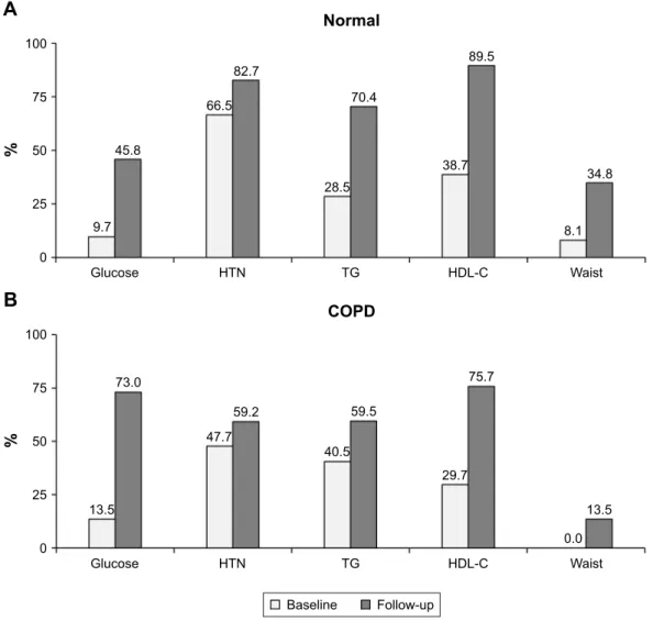
the percentage change in glucose level was higher in patients with COPD, suggesting that insulin resistance may be an independent risk factor for the incidence of MetS among COPD patients (Figure 3).
According to the International Diabetes Federation, the prevalence of MetS is largely affected by abdominal obesity. Indeed, patients with obstructive lung disease exhibit more visceral fat mass, relative to healthy subjects.30 In the present
study, these findings are indirectly confirmed by the observa-tion that the incidence of MetS was higher in individuals with a higher BMI (25.5 kg/m2 vs 23.7 kg/m2). It remains unclear
why abdominal obesity is more prevalent in COPD patients than in healthy subjects; however, various factors, including poor nutrition and inactive lifestyle, may play an important role.30 Future longitudinal studies with larger sample sizes
are required to further investigate the causal relationship between COPD and MetS.
Our study has several strengths. To our knowledge, this was the first study to evaluate the incidence of MetS in patients with airflow obstruction. Second, we evaluated the risk for MetS using a relatively large community-based popu-lation with a relatively long duration of follow-up. Third, we accurately classified patients as COPD or non-COPD through repeated pulmonary function tests; moreover, we obtained MetS data via laboratory tests and accurate medical history, rather than through patient interviews.
Our study also included multiple limitations. First, we could not analyze other important comorbidities, such as cardiovascular disease and malignancies, because of the absence of objective data regarding both factors. Second, most patients with airway obstruction were classified as exhibiting mild or moderate disease; the exclusion of patients with severe disease, therefore, may have influenced our results. Third, the incidence of MetS was higher in only male subjects, which might be influenced by smoking status and low event number in female subjects (n = 5). Further, large prospective studies are needed to more clearly elucidate the relationship between airflow obstruction and the incidence of MetS in the general population.
Conclusion
The incidence of MetS was higher in COPD patients, relative to controls, and was most influenced by fasting blood glucose level and high blood pressure. The incidence of MetS was higher in older, male COPD subjects, who exhibited higher BMI and more severe smoking status. Future longitudinal and interventional studies are needed to elucidate the relationship
between airflow obstruction and the incidence of MetS in the general population.
Data availability
The data are not available for public access because of patient privacy concerns.
Author contributions
All authors contributed toward data analysis, drafted and critically revised the paper, gave final approval of the ver-sion to be published, and agreed to be accountable for all aspects of the work.
Disclosure
The authors report no conflicts of interest in this work.
References
1. Vogelmeier CF, Criner GJ, Martinez FJ, et al. Global Strategy for the Diagnosis, Management, and Prevention of Chronic Obstructive Lung Disease 2017 Report. GOLD executive summary. Am J Respir Crit Care
Med. 2017;195(5):557–582.
2. Mannino DM, Buist AS. Global burden of COPD: risk factors, preva-lence, and future trends. Lancet. 2007;370(9589):765–773.
3. Alberti KG, Zimmet P, Shaw J. Metabolic syndrome – a new world-wide definition. A consensus statement from the International Diabetes Federation. Diabet Med. 2006;23(5):469–480.
4. Isomaa B, Almgren P, Tuomi T, et al. Cardiovascular morbidity and mor-tality associated with the metabolic syndrome. Diabetes Care. 2001; 24(4):683–689.
5. Lakka HM, Laaksonen DE, Lakka TA, et al. The metabolic syndrome and total and cardiovascular disease mortality in middle-aged men. J Am
Med Assoc. 2002;288(21):2709–2716.
6. Cebron Lipovec N, Beijers RJ, van den Borst B, Doehner W, Lainscak M, Schols AM. The prevalence of metabolic syndrome in chronic obstructive pulmonary disease: a systematic review. COPD. 2016;13(3):399–406.
A
Normal 45.8 9.7 Glucose HTN TG HDL-C Waist 66.5 82.7 100 % % 75 50 25 0 70.4 28.5 38.7 89.5 8.1 34.8 Baseline Follow-up COPDB
73.0 13.5 Glucose HTN TG HDL-C Waist 47.7 59.2 100 75 50 25 0 59.5 40.5 29.7 75.7 0.0 13.5Figure 3 Individual components of metabolic syndrome in normal and COPD patients. (A) Individual component changes (expressed as percentage) in metabolic syndrome in normal subjects. (B) Individual component changes (expressed as percentage) in metabolic syndrome in subjects with COPD.
Notes: Individual component is defined as central obesity (with waist circumference cutoff points of .90 cm for men and .80 cm for women), an elevated triglyceride (Tg) level ($1.7 mmol/L or undergoing drug treatment for elevated triglyceride levels), reduced high-density lipoprotein cholesterol (HDL-C) level (,1.0 mmol/L [men] and ,1.3 mmol/L [women], or drug treatment for reduced HDL-C), elevated blood pressure ($130 mmhg systolic, $85 mmhg diastolic, or antihypertensive drug treatment in patients with a history of hypertension [HTN]), and an elevated fasting plasma glucose concentration ($5.6 mmol/L or drug treatment for diabetes).
International Journal of COPD
Publish your work in this journal
Submit your manuscript here: http://www.dovepress.com/international-journal-of-chronic-obstructive-pulmonary-disease-journal
The International Journal of COPD is an international, peer-reviewed journal of therapeutics and pharmacology focusing on concise rapid reporting of clinical studies and reviews in COPD. Special focus is given to the pathophysiological processes underlying the disease, intervention programs, patient focused education, and self management protocols.
This journal is indexed on PubMed Central, MedLine and CAS. The manuscript management system is completely online and includes a very quick and fair peer-review system, which is all easy to use. Visit http://www.dovepress.com/testimonials.php to read real quotes from published authors.
Dove
press
7. Baffi CW, Wood L, Winnica D, et al. Metabolic syndrome and the lung.
Chest. 2016;149(6):1525–1534.
8. Chung JH, Hwang HJ, Han CH, Son BS, Kim DH, Park MS. Association between sarcopenia and metabolic syndrome in chronic obstructive pul-monary disease: the Korea National Health and Nutrition Examination Survey (KNHANES) from 2008 to 2011. COPD. 2015;12(1):82–89. 9. Park BH, Park MS, Chang J, et al. Chronic obstructive pulmonary
disease and metabolic syndrome: a nationwide survey in Korea. Int J
Tuberc Lung Dis. 2012;16(5):694–700.
10. Shin C, Abbott RD, Lee H, Kim J, Kimm K. Prevalence and correlates of orthostatic hypotension in middle-aged men and women in Korea: the Korean Health and Genome Study. J Hum Hypertens. 2004;18(10): 717–723.
11. Grundy SM, Cleeman JI, Daniels SR, et al. Diagnosis and management of the metabolic syndrome: an American Heart Association/National Heart, Lung, and Blood Institute Scientific Statement. Circulation. 2005; 112(17):2735–2752.
12. Standardization of spirometry, 1994 update. American Thoracic Society. Am J Respir Crit Care Med. 1995;152(3):1107–1136. 13. Kim SK, Bae JC, Baek JH, et al. Decline in lung function rather than
baseline lung function is associated with the development of meta-bolic syndrome: a six-year longitudinal study. PLoS One. 2017;12(3): e0174228.
14. Hsiao FC, Wu CZ, Su SC, et al. Baseline forced expiratory volume in the first second as an independent predictor of development of the metabolic syndrome. Metabolism. 2010;59(6):848–853.
15. Ford ES, Cunningham TJ, Mercado CI. Lung function and metabolic syndrome: findings of National Health and Nutrition Examination Survey 2007–2010. J Diabetes. 2014;6(6):603–613.
16. Diez-Manglano J, Barquero-Romero J, Almagro P, et al. COPD patients with and without metabolic syndrome: clinical and functional differ-ences. Intern Emerg Med. 2014;9(4):419–425.
17. Choi JH, Park S, Shin YH, Kim MY, Lee YJ. Sex differences in the relationship between metabolic syndrome and pulmonary function: the 2007 Korean National Health and Nutrition Examination Survey.
Endocr J. 2011;58(6):459–465.
18. Breyer MK, Spruit MA, Hanson CK, et al. Prevalence of metabolic syndrome in COPD patients and its consequences. PLoS One. 2014; 9(6):e98013.
19. Lam KB, Jordan RE, Jiang CQ, et al. Airflow obstruction and meta-bolic syndrome: the Guangzhou Biobank Cohort Study. Eur Respir J. 2010;35(2):317–323.
20. Mannino DM, Thorn D, Swensen A, Holguin F. Prevalence and out-comes of diabetes, hypertension and cardiovascular disease in COPD.
Eur Respir J. 2008;32(4):962–969.
21. Divo M, Cote C, de Torres JP, et al. Comorbidities and risk of mortality in patients with chronic obstructive pulmonary disease. Am J Respir
Crit Care Med. 2012;186(2):155–161.
22. Waschki B, Kirsten A, Holz O, et al. Physical activity is the strongest predictor of all-cause mortality in patients with COPD: a prospective cohort study. Chest. 2011;140(2):331–342.
23. Watz H, Waschki B, Kirsten A, et al. The metabolic syndrome in patients with chronic bronchitis and COPD: frequency and associated consequences for systemic inflammation and physical inactivity. Chest. 2009;136(4):1039–1046.
24. Schmidt AM. Insulin resistance and metabolic syndrome: mechanisms and consequences. Arterioscler Thromb Vasc Biol. 2012;32(8):1753. 25. Lawlor DA, Ebrahim S, Smith GD. Associations of measures of lung
function with insulin resistance and type 2 diabetes: findings from the British Women’s Heart and Health Study. Diabetologia. 2004;47(2): 195–203.
26. Lazarus R, Sparrow D, Weiss ST. Baseline ventilatory function predicts the development of higher levels of fasting insulin and fasting insulin resistance index: the Normative Aging Study. Eur Respir J. 1998;12(3): 641–645.
27. Lin WY, Yao CA, Wang HC, Huang KC. Impaired lung function is associated with obesity and metabolic syndrome in adults. Obesity
(Silver Spring). 2006;14(9):1654–1661.
28. Engstrom G, Hedblad B, Nilsson P, Wollmer P, Berglund G, Janzon L. Lung function, insulin resistance and incidence of cardiovascular disease: a longitudinal cohort study. J Intern Med. 2003;253(5):574–581. 29. Ford ES, Mannino DM. Prospective association between lung
func-tion and the incidence of diabetes: findings from the Nafunc-tional Health and Nutrition Examination Survey Epidemiologic Follow-up Study.
Diabetes Care. 2004;27(12):2966–2970.
30. van den Borst B, Gosker HR, Koster A, et al. The influence of abdominal visceral fat on inflammatory pathways and mortality risk in obstructive lung disease. Am J Clin Nutr. 2012;96(3):516–526.