EUKARYOTICCELL, Nov. 2008, p. 2008–2011 Vol. 7, No. 11 1535-9778/08/$08.00⫹0 doi:10.1128/EC.00105-08
Copyright © 2008, American Society for Microbiology. All Rights Reserved.
Hydrogen Peroxide Induces Hyphal Differentiation in Candida albicans
䌤
Olviyani Nasution,
1Kavitha Srinivasa,
1Minsun Kim,
1Yeo-Jung Kim,
1Wankee Kim,
3Woojin Jeong,
1and Wonja Choi
1,2*
Division of Life and Pharmaceutical Sciences,1and Microbial Resources Research Center,2Ewha Womans University, Seoul 120-750,
South Korea, and Institute for Medical Sciences, School of Medicine, Ajou University, Suwon 442-749, South Korea3
Received 24 March 2008/Accepted 3 September 2008
In this study, we demonstrate that hyphal differentiation is induced by the subtoxic concentration of exogenous H2O2in Candida albicans. This finding is confirmed by the changing intracellular concentration of
H2O2. In order to induce the same level of differentiation, low concentrations of exogenous H2O2are required
for the null mutants of the thiol-specific antioxidant and catalase, while higher concentrations are needed for cells treated with ascorbic acid, an antioxidant chemical.
Hydrogen peroxide (H2O2) directly affects various redox sys-tems to regulate cell differentiation, proliferation, death, signal transduction, and ion transport (3, 12, 13, 19, 20) at subtoxic concentrations (23, 27–29). Therefore, the homeostatic mainte-nance of H2O2at low levels should be tightly regulated (1, 9, 28).
The yeast Candida albicans is a pleomorphic human patho-gen. An important virulence factor is the morphological tran-sition involving hyphae formation (6, 16, 24), which is regu-lated by signaling pathways, including the cyclic AMP/protein kinase A and mitogen-activated protein kinase pathways (4, 7,
* Corresponding author. Mailing address: Science Building A, Room 211, Ewha Womans University, Seoul 120-750, South Korea. Phone: 82-2-3277-2892. Fax: 82-2-3277-2385. E-mail: wjchoi@ewha .ac.kr.
䌤Published ahead of print on 12 September 2008.
FIG. 1. Hyphal induction by exogenous H2O2. (A) Microscopic images of H2O2-induced hyphae. Wt cells were grown on YPD solid plates
supplemented with the indicated concentrations of H2O2at 30°C for 6 days. Representative colonies were photographed with a stereomicroscope
(top). Cells in the mid-log phase were cultured in YPD liquid medium containing H2O2for 6 h at 30°C and observed with a light microscope
(bottom). (B) Cytotoxicity of H2O2. Standardized cell suspensions were challenged with the indicated concentrations of H2O2for 30 min, plated
onto YPD solid medium, and incubated at 30°C for 2 days. The survival rate was expressed as a percentage of the number of colonies in the presence of H2O2divided by the number of colonies in the absence of H2O2. (C) Efficiency of hyphal differentiation. Cells were grown on YPD
solid medium containing the indicated concentrations of H2O2and incubated at 30°C for 6 days. The percentage of hyphal differentiation was
expressed as the number of hyphal colonies divided by the total number of colonies.
2008
on March 16, 2017 by Ewha Womans Univ
http://ec.asm.org/
18, 21, 22). Pathway triggers are varied (8) and include specific carbohydrates or amino acids (5, 26), serum (11), temperature (17), pH (10),N-acetylglucosamine (2), and starvation (7).
Following infection, C. albicans encounters macrophages but survives ingestion by rapidly adopting a hyphal morphology (25). Since the intracellular concentration of H2O2in a mac-rophage is intrinsically high, it was presently germane to ex-amine whether H2O2can induce hyphal differentiation.
Hyphal differentiation by H2O2.When wild-type (wt) SC5314
cells were grown on YPD solid or liquid medium containing 0, 0.4, 1, 4, or 10 mM H2O2, the extent of differentiation was augmented in a dose-dependent manner (Fig. 1A). At the 10 mM concentration, however, the cells were severely swollen due to the cytotoxic effects of H2O2, which was inferred by the survival rate (35%) in contrast to the survival rate at 0.4 mM and 1 mM (90%) (Fig. 1B). Interestingly, undifferentiated col-onies also appeared at all concentrations, enabling the evalu-ation of induction efficiency expressed as a percentage of the number of differentiated colonies in the total number of col-onies. The induction efficiency was dose dependent as ex-pected, but 100% differentiation did not occur even at 10 mM (Fig. 1C).
Next, we increased or decreased the endogenous intracellu-lar H2O2. The increase was achieved by nullifying two H2O2 -scavenging genes, the thiol-specific antioxidant C. albicans
TSA1 (30, 31) and the catalase C. albicans CAT1, individually
(tsa1⌬ or cat1⌬) or simultaneously (tsa1⌬ cat1⌬) (Fig. 2). The
growth of null mutants was impeded, and mutants were more H2O2 sensitive than the wt over the concentration range (data not shown). The decrease was achieved by the addi-tion of ascorbic acid, an antioxidant chemical (14, 15) (see Fig. 4 and 5).
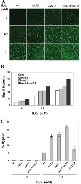
The enhanced sensitivity of the mutants to exogenous H2O2 was presumably caused by an increase in the concentration of intracellular H2O2. The relative amount of intracellular H2O2 was measured by visualizing fluorescent dichlorodihydrofluo-rescein (DCF) produced by esterase and H2O2from 5-chlo-romethyl-2⬘,7⬘-DCF diacetate (CM-H2DCFDA) (Invitrogen, Carlsbad, CA). At exogenous H2O2concentrations of 0.2 mM and 1 mM, fluorescent intensity was enhanced to some degree in the tsa1⌬ and cat1 mutants and in the tsa1⌬ cat1⌬ mutant (Fig. 3A). When the intensities were converted to arbitrary units for quantitative comparison, the intracellular H2O2 con-centration increased about 1.5-fold in the tsa1⌬ and cat1⌬ mutants and about twofold in the tsa1⌬ cat1⌬ mutant com-pared with that of the wt (Fig. 3B).
The hyphal differentiation efficiencies of the wt and null mutants were compared using 0.2 mM exogenous H2O2. As shown in Fig. 3C, efficiency was considerably enhanced from 5% in the wt to about 25% in the tsa1⌬ and cat1⌬ mutants and to about 35% in the tsa1⌬ cat1⌬ mutant. This efficiency was obtained when the wt cells were treated with 1 mM H2O2(Fig. 1C). The effective promotion of hyphal differentiation at a low concentration of exogenous H2O2in mutants in which intra-cellular H2O2increased indicated that H2O2is a genuine in-ducer of C. albicans hyphal differentiation. When the func-tional CAT1 gene was reintroduced, the percentage of hyphae reduced to the level between the wt and the cat1⌬ mutant.
The effects of a decreased intracellular H2O2concentration on hyphal differentiation were examined in the presence of ascorbic acid, which reduces the number of intracellular reac-tive oxygen species in some organisms. When wt cells were cultured under the full differentiation conditions (YPD plus 10% fetal bovine serum [FBS], 37°C), the level of intracellular H2O2increased about sevenfold, from 8 to 65 arbitrary units (Fig. 4B and C). However, the addition of 50 mM or 100 mM ascorbic acid to the medium reduced the amount of intracel-lular H2O2 to the same or a lower level of serum depletion (Fig. 4B and C). Microscopic examination revealed that hyphal differentiation was markedly inhibited by ascorbic acid (Fig. 4A). Although the mechanisms of H2O2-induced hyphal tran-sition are unclear, it is highly possible that increased intracel-lular H2O2might be partly or completely involved. We further confirmed the above effects in the tsa1⌬, cat1⌬, and tsa1⌬
cat1⌬ mutants. When 50 mM ascorbic acid, an antioxidant
chemical, was added to the medium 30 min after the treatment of mutant cells with different concentrations of exogenous H2O2, hyphal differentiation was induced even at otherwise toxic concentrations: 10 mM for the wt, 4 mM for the tsa1⌬ and
cat1⌬ mutants, and 1 mM for the tsa1⌬ cat1⌬ mutant (Fig. 5).
Thus, ascorbic acid lowered the intracellular concentration of H2O2 and inhibited hyphal differentiation. Also, efficient hy-phal differentiation in the presence of ascorbic acid required exogenous H2O2.
The above results suggest that the mere increase of intra-cellular H2O2 is insufficient for complete hyphal differentia-tion. The intracellular H2O2concentration of cells cultured in
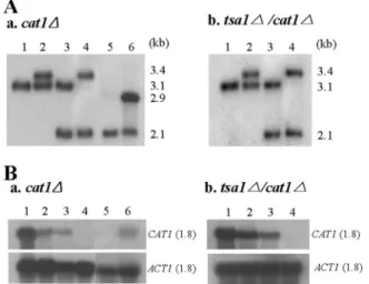
FIG. 2. Construction of CAT1 null mutants and a revertant. The CAT1 genes of the wt and the tsa1⌬ mutant (30, 31) were disrupted using URA3-dpl200 (32), yielding the cat1⌬ and tsa1⌬ cat1⌬ mutants, respectively. The sense and antisense primers were nucleotide posi-tions 754 to 823 and 2312 to 2381, respectively, of the CAT1 open reading frame (ORF). To construct a revertant, the DNA fragment containing its own promoter, ORF, and terminator was cloned into pLUX, linearized with NheI, and transformed into the cat1⌬ mutant. Southern (A) and Northern (B) analyses were performed to confirm the authenticity of the constructed strains, using the32P-labeled probe
prepared from the MfeI fragment of the CAT1 ORF. For the Southern analyses, genomic DNA was digested with NsiI and NcoI. Lanes 1, parental strains (CAI4 and the tsa1⌬ mutant in panels A and B, respectively); lanes 2, strains with one allele disrupted; lanes 3, strains with URA3 popped out from the lane 2 strains; lanes 4, null mutants (the cat1⌬ and tsa1⌬ cat1⌬ mutants in panels A and B, respectively); lanes 5, strains with URA3 popped out from the cat1⌬ mutant; lanes 6, CAT1-reintroduced strains of the cat1⌬ mutant.
VOL. 7, 2008 NOTES 2009
on March 16, 2017 by Ewha Womans Univ
http://ec.asm.org/
FBS-supplemented YPD was identical to cells grown in YPD in the presence of exogenous 4 mM H2O2(Fig. 4C and 5B), although differentiation was 100% and 60%, respectively (Fig. 1C). This indicates that some factors present in the serum are required for full hyphal differentiation in addition to increased intracellular H2O2. Based on these observations, we propose that hyphal differentiation in C. albicans occurs through two separate, but not mutually exclusive, steps: (i) initiation by
FIG. 3. Effects of increased intrinsic H2O2on hyphal
differenti-ation. Cells were grown in YPD medium containing 0.2 and 1 mM H2O2 for 6 h, washed, and resuspended in Hank’s balanced salt
solution. After the addition of CM-H2DCFDA (10M final), the cells were further incubated at RT for 10 min. (A) Images of DCF fluorescence were taken by using a confocal microscope with exci-tation and emission wavelengths at 488 nm and 520 nm, respec-tively. (B) Relative concentrations of intracellular H2O2 were
derived from the confocal microscope-aided integration of fluores-cence signal intensity within a scope. (C) Efficiency of hyphal dif-ferentiation at 0.2 mM H2O2was determined as described in the
legend to Fig. 1C. CAT1-R represents the strain into which the functional CAT1 gene was introduced.
FIG. 4. Effects of ascorbic acid on hyphal differentiation in FBS-treated cells. Wt cells were grown in YPD in the absence (⫺) or presence (⫹) of 10% FBS for 30 min, followed by supplementation with 50 mM or 100 mM ascorbic acid. A portion of the cells was removed to take light microscopic images (A). For the rest of cells, fluorescence images (B) were taken, and the relative concentrations of intracellular H2O2(C) were determined as described in the legend to
Fig. 3.
FIG. 5. Effects of ascorbic acid on hyphal differentiation. After the wt and tsa1⌬, cat1⌬, and tsa1⌬ cat1⌬ mutant cells were grown in YPD medium supplemented with 4 concentrations of exogenous H2O2for
30 min at 30°C, 100 mM ascorbic acid was added, and the cells were further grown for 6 h at 30°C, the cultures were observed with a light microscope (magnification,⫻400).
2010 NOTES EUKARYOT. CELL
on March 16, 2017 by Ewha Womans Univ
http://ec.asm.org/
intracellular H2O2above a certain concentration and (ii) pro-motion by currently unknown additional factors in serum.
The PCR product-directed disruption cassette URA3-dpl200 and pLUX were kindly provided by D. Davis and W. Fonzi, respectively.
This work was supported by a Korea Research Foundation grant (KRF-2006-005-JO4003) and a Korea Science and Engineering Foun-dation (KOSEF) grant funded by the Korea government (MOST) (no. 2006-0063-2). O.N. and K.S. were recipients of the Brain Korea 21 project and the Ewha Global Partnership Program 2006.
REFERENCES
1. Aguirre, J., M. Rios-Momberg, D. Hewitt, and W. Hansberg. 2005. Reactive oxygen species and development in microbial eukaryotes. Trends Microbiol.
13:111–118.
2. Alvarez, F. J., and J. B. Konopka. 2007. Identification of an N-acetylglu-cosamine transporter that mediates hyphal induction in Candida albicans. Mol. Biol. Cell 18:965–975.
3. Bienert, G. P., J. K. Schjoerring, and T. P. Jahn. 2006. Membrane transport of hydrogen peroxide. Biochim. Biophys. Acta 1758:994–1003.
4. Biswas, S., P. Van Dijck, and A. Datta. 2007. Environmental sensing and signal transduction pathways regulating morphopathogenic determinants of
Candida albicans. Microbiol. Mol. Biol. Rev. 71:348–376.
5. Brega, E., R. Zufferey, and C. B. Mamoun. 2004. Candida albicans Csy1p is a nutrient sensor important for activation of amino acid uptake and hyphal morphogenesis. Eukaryot. Cell 3:135–143.
6. Calderone, R. A., and W. A. Fonzi. 2001. Virulence factors of Candida albicans. Trends Microbiol. 9:327–335.
7. Csank, C., K. Schroppel, E. Leberer, D. Harcus, O. Mohamed, S. Meloche,
D. Y. Thomas, and M. Whiteway. 1998. Roles of the Candida albicans mitogen-activated protein kinase homolog, Cek1p, in hyphal development and systemic candidiasis. Infect. Immun. 66:2713–2721.
8. Dhillon, N. K., S. Sharma, and G. K. Khuller. 2003. Signaling through protein kinases and transcriptional regulators in Candida albicans. Crit. Rev. Microbiol. 29:259–275.
9. Droge, W. 2002. Free radicals in the physiological control of cell function. Physiol. Rev. 82:47–95.
10. El Barkani, A., O. Kurzai, W. A. Fonzi, A. Ramon, A. Porta, M. Frosch, and
F. A. Muhlschlegel.2000. Dominant active alleles of RIM101 (PRR2) bypass the pH restriction on filamentation of Candida albicans. Mol. Cell. Biol.
20:4635–4647.
11. Feng, Q., E. Summers, B. Guo, and G. Fink. 1999. Ras signaling is required for serum-induced hyphal differentiation in Candida albicans. J. Bacteriol.
181:6339–6346.
12. Finkel, T. 2003. Oxidant signals and oxidative stress. Curr. Opin. Cell Biol.
15:247–254.
13. Foreman, J., V. Demidchik, J. H. Bothwell, P. Mylona, H. Miedema, M. A.
Torres, P. Linstead, S. Costa, C. Brownlee, J. D. Jones, J. M. Davies, and L. Dolan.2003. Reactive oxygen species produced by NADPH oxidase regulate plant cell growth. Nature 422:442–446.
14. Frei, B., L. England, and B. N. Ames. 1989. Ascorbate is an outstanding antioxidant in human blood plasma. Proc. Natl. Acad. Sci. USA 86:6377– 6381.
15. Frei, B., R. Stocker, L. England, and B. N. Ames. 1990. Ascorbate: the most effective antioxidant in human blood plasma. Adv. Exp. Med. Biol.
264:155–163.
16. Haynes, K. 2001. Virulence in Candida species. Trends Microbiol. 9:591–596. 17. Kadosh, D., and A. D. Johnson. 2005. Induction of the Candida albicans
fila-mentous growth program by relief of transcriptional repression: a genome-wide analysis. Mol. Biol. Cell 16:2903–2912.
18. Kohler, J. R., and G. R. Fink. 1996. Candida albicans strains heterozygous and homozygous for mutations in mitogen-activated protein kinase signaling components have defects in hyphal development. Proc. Natl. Acad. Sci. USA
93:13223–13228.
19. Kwak, J. M., I. C. Mori, Z. M. Pei, N. Leonhardt, M. A. Torres, J. L. Dangl,
R. E. Bloom, S. Bodde, J. D. Jones, and J. I. Schroeder.2003. NADPH oxidase AtrbohD and AtrbohF genes function in ROS-dependent ABA signaling in Arabidopsis. EMBO J. 22:2623–2633.
20. Lambeth, J. D. 2004. NOX enzymes and the biology of reactive oxygen. Nat. Rev. Immunol. 4:181–189.
21. Leberer, E., D. Harcus, I. D. Broadbent, K. L. Clark, D. Dignard, K.
Ziegelbauer, A. Schmidt, N. A. Gow, A. J. Brown, and D. Y. Thomas.1996. Signal transduction through homologs of the Ste20p and Ste7p protein kinases can trigger hyphal formation in the pathogenic fungus Candida albicans. Proc. Natl. Acad. Sci. USA 93:13217–13222.
22. Liu, H. 2001. Transcriptional control of dimorphism in Candida albicans. Curr. Opin. Microbiol. 4:728–735.
23. Livolsi, A., V. Busuttil, V. Imbert, R. T. Abraham, and J. F. Peyron. 2001. Tyrosine phosphorylation-dependent activation of NF-kappa B. Require-ment for p56 LCK and ZAP-70 protein tyrosine kinases. Eur. J. Biochem.
268:1508–1515.
24. Lo, H. J., J. R. Kohler, B. DiDomenico, D. Loebenberg, A. Cacciapuoti, and
G. R. Fink.1997. Nonfilamentous C. albicans mutants are avirulent. Cell
90:939–949.
25. Lorenz, M. C., J. A. Bender, and G. R. Fink. 2004. Transcriptional response of Candida albicans upon internalization by macrophages. Eukaryot. Cell
3:1076–1087.
26. Maidan, M. M., J. M. Thevelein, and P. Van Dijck. 2005. Carbon source induced yeast-to-hypha transition in Candida albicans is dependent on the presence of amino acids and on the G-protein-coupled receptor Gpr1. Bio-chem. Soc. Trans. 33:291–293.
27. Reth, M. 2002. Hydrogen peroxide as second messenger in lymphocyte activation. Nat. Immunol. 3:1129–1134.
28. Rhee, S. G., T. S. Chang, Y. S. Bae, S. R. Lee, and S. W. Kang. 2003. Cellular regulation by hydrogen peroxide. J. Am. Soc. Nephrol. 14:S211–S215. 29. Rhee, S. G., S. W. Kang, W. Jeong, T. S. Chang, K. S. Yang, and H. A. Woo.
2005. Intracellular messenger function of hydrogen peroxide and its regula-tion by peroxiredoxins. Curr. Opin. Cell Biol. 17:183–189.
30. Shin, D. H., S. Jung, S. J. Park, Y. J. Kim, J. M. Ahn, W. Kim, and W. Choi. 2005. Characterization of thiol-specific antioxidant 1 (TSA1) of Candida albicans. Yeast 22:907–918.
31. Urban, C., X. Xiong, K. Sohn, K. Schroppel, H. Brunner, and S. Rupp. 2005. The moonlighting protein Tsa1p is implicated in oxidative stress response and in cell wall biogenesis in Candida albicans. Mol. Microbiol. 57:1318– 1341.
32. Wilson, R. B., D. Davis, B. M. Enloe, and A. P. Mitchell. 2000. A recyclable Candida albicans URA3 cassette for PCR product-directed gene disruptions. Yeast 16:65–70.
VOL. 7, 2008 NOTES 2011