Effects of Five-month Training of Playing Harmonica on Pulmonary Function in Patients With Neuromuscular Disease: A Pilot Study
Bit-na-rae Kim1,2,3, BHSc, PT, Heon-seock Cynn2,3, PhD, PT
1Department of Rehabilitation Medicine and Pulmonary Rehabilitation Center, Gangnam Severance Hospital, Yonsei University College of Medicine, Seoul, Korea
2Applied Kinesiology and Ergonomic Technology Laboratory, Dept. of Physical Therapy, The Graduate School, Yonsei University
3Dept. of Physical Therapy, College of Health Science, Yonsei University
Abstract
1)Background: Progressive muscle weakness is aggravated not only in the skeletal muscles but also in the respiratory muscles in many patients with neuromuscular diseases (NMD). Inspiratory muscle training (IMT) has been reported as therapy for pulmonary rehabilitation to improve respiratory strength, endurance, exercise capacity, and quality of life, and to reduce dyspnea.
Objects: The purpose of this study was to determine the effect of playing harmonica for 5 months on pulmonary function by assessing the force vital capacity (FVC), peak cough flow (PCF), maximal inspiratory pressure (MIP), maximal expiratory pressure (MEP), and maximal voluntary ventilation (MVV) in patients with NMD.
Methods: Six subjects with NMD participated in this study. The subjects played harmonica once a week for 2 hours at a harmonica academy and twice a week for 1 hour at home. Thus, training was performed thrice a week for 23 weeks. The examiner assessed pulmonary function by measuring FVC in the sitting and supine positions and PCF, MIP, MEP, and MVV in the sitting position at the beginning of training and once a month for 5 months.
Results: Both sitting and supine FVC significantly increased after playing harmonica (p=.042), as did MIP (p=.043) and MEP (p=.042).
Conclusion: Playing harmonica can be used as an effective method to improve pulmonary function in patients with NMD.
Keywords: Harmonica; Neuromuscular disease; Pulmonary function test.
Introduction
Neuromuscular diseases (NMD) are characterized by progressive weakness of respiratory, skeletal, and bul- bar muscles, as well as myopathies and cardiac muscle weakness (Griggs et al, 1981). Pulmonary complications lead to increase in mortality and morbidity in patients with NMD (McCool and Tzelepis, 1995). Patients with NMD are noted to have an advance restrictive pulmo- nary disease pattern due to the progressive weakening of the respiratory muscles. Patients with NMD have severe expiratory and inspiratory muscle insufficiency
that decreases tidal volumes, sighing, and coughing those results in reduction in lung insufflation and thoracic chest wall (McCool et al, 1986). That symp- tom leads to ineffective coughing, decreased respiratory muscle power, and decreased vital capacity during oth- erwise benign chest infections (Bach et al, 1998;
Gibson et al, 1977).
A previous study has reported on the benefits of moderate-resistance training in progressive NMD for respiratory muscle power and especially reported that training for 12 weeks or longer would be needed to increase maximal expiratory pressure (MEP) or max-
Corresponding author: Heon-seock Cynn [email protected]
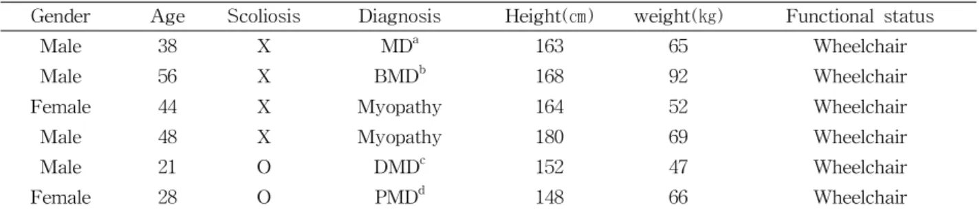
Gender Age Scoliosis Diagnosis Height(㎝) weight(㎏) Functional status
Male 38 X MDa 163 65 Wheelchair
Male 56 X BMDb 168 92 Wheelchair
Female 44 X Myopathy 164 52 Wheelchair
Male 48 X Myopathy 180 69 Wheelchair
Male 21 O DMDc 152 47 Wheelchair
Female 28 O PMDd 148 66 Wheelchair
amuscular dystrophy, bBecker muscular dystrophy, cDuchenne muscular dystrophy, dprogressive muscular dystrophy.
Table 1. Characteristics of the six subjects with neuromuscular disorders
imal inspiratory pressure (MIP) (Aitkens et al, 1993).
In many patients with NMD patients, progressive muscle weakness is aggravated not only in the skel- etal muscles, but also in the respiratory muscles.
Inspiratory muscle training (IMT) has been reported as therapy for pulmonary rehabilitation to improve respiratory strength, endurance (Dimarco et al, 1985), exercise capacity, and quality of life, and to reduce dyspnea (Lötters and Kwakkel et al, 2002; Weiner and McConnell, 2005). In the early stages of Duchenne muscular dystrophy (DMD), IMT with pressure threshold device was more effective than other IMT devices when the forced vital capacity (FVC) is preserved (Kang et al, 1998). Other studies have also proven that expiratory and inspiratory muscle endurance can be improved by specific ex- ercises involving resistant inspiratory breathing in patients with various types of nonmyotonic muscular dystrophies (Dimarco et al, 1985; Kang et al, 1998;
McCool and Tzelepis, 1995).
Playing harmonica has an effect similar to IMT based on the similarity of performing inspiration and expiration through a device that provides resistance with nose clip (due to the size of the holes in the harmonica), and provides an advantage similar to IMT (Alexander and Wagner, 2012). In addition, playing harmonica may be enjoyable, user-friendly, and useful for improving their breathing dynamics (Jeffery et al, 2012). A previous study has reported on the benefits of However, in the present, no stud- ies have reported a therapeutic effect of playing har- monica in patients with NMD.
Therefore, the purpose of this study was to de- termine whether 5-month training of playing har- monica can affect pulmonary function by assessing FVC, peak cough flow (PCF), MIP, MEP and max- imal voluntary ventilation (MVV) in patients with NMD. We hypothesized that FVC, PCF, MIP, MEP, and MVV would increase after 5 months of harmon- ica playing in patients with NMD.
Methods
Subjects
G*power software (G*power software 3.1.2; Franz Faul, University of Kiel, Kiel, Germany) was used for power analysis. From the data of a pilot study of eight subjects, the necessary sample size was three subjects, with a significance level of .05, power of .8, and effect size of 5.5. The subjects from the Korean Muscular Dystrophy Association volunteered to par- ticipate in the study. Finally, six subjects with NMD were selected from eight subjects of the pilot study.
Two subjects dropped out from the study because of deteriorating general medical condition, such as pneumonia and facial muscle weakness. The charac- teristics of the six subjects with NMDs are pre- sented in Table 1. NMD diagnosis was established by standard criteria (Emery et al, 1997). We eval- uated six subjects who were diagnosed with NMD based on case history, clinical electromyogram, and muscle biopsy. Exclusion criteria include those who refused to undergo the test for maneuvers, those
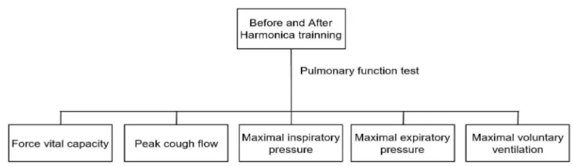
Figure 1. Flow chart of Measurement procedure.
with acute pulmonary disease conditions (e.g. pneu- monia, pneumothorax, etc.), and those with an in- dwelling tracheostomy tube. The subjects with in- ability to cooperate due to mental retardation were also excluded for this study. Before the measure- ments, the subjects were informed of the ex- perimental protocol. The subjects read and signed an informed consent form before participation. This study was approved by the Gang-Nam Severance Hospital Institutional Review Board (approval num- ber: 3-2017-0137).
Harmonica playing
All subjects used the same 22-hole harmonica (Weissenburg, 2202F, Taichung, Taiwan). The cover plates of the harmonica were made of vacuum titanium. The subjects used a harmonica holder as an adjunctive tool. All subjects played the harmonica in the harmonica academy of the Korean Muscular Dystrophy Association with and without an in- structor at home. The subjects played harmonica once a week for 2 hours at the harmonica academy and twice a week for 1 hour at home. Thus, training was performed thrice a week for 23 weeks.
Measurement procedure
The subjects were assessed for pulmonary func- tion using routine pulmonary function test and con- tinuous routine check of FVC, PCF, MIP, MEP, and MVV at the beginning of training, after the end of the 5-month training. Pulmonary function test was performed in sitting position. The FVC tests were
performed in both sitting and supine positions. In the supine position, the abdominal contents exert an up- ward pressure on the diaphragm, which pushes the thoracic cavity, thereby decreasing the FVC. All pul- monary function data were collected in each subject’s hospital room. The subjects performed the tests at least thrice, and resting time was given. The highest record was selected (Won et al, 2015).
Forced vital capacity
FVC was measured in both the sitting and supine positions by using a spirometer (CareFushion, Micro
™ Spirometer, Kent, UK). Each subject sat in a wheelchair with back support and lay on the bed for the supine position. The subjects inhaled as deeply as possible, and then maximally exhaled into the spirometer. We calculated the predicted FVC values (FVC pre) based on age, height, and weight (Wilson et al, 1984). The relative FVC values were reported as FVC/FVC pre (%).
Peak cough flow
PCF was measured by a peak flow-meter (Health Scan Products Inc., Assess Peck Flowmeter, NJ, USA). The subjects were instructed to take the deepest breath and then to cough as strongly as pos- sible through a peak flow-meter (Park et al, 2010).
Maximal inspiratory pressure and maximal expiratory pressure
Maximal respiratory pressure and reflecting pul- monary muscle strength were measured using
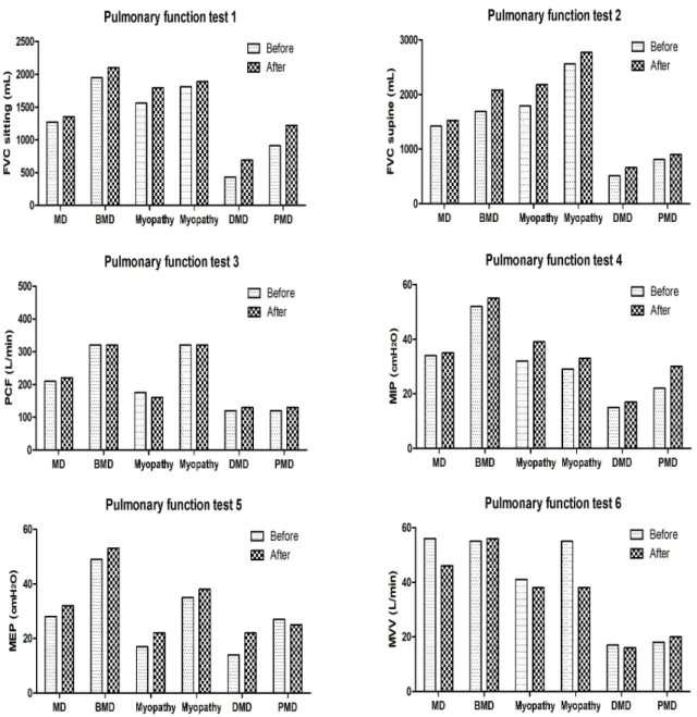
Figure 2. Before and After of pulmonary function measurement (BMD: Becker muscular dystrophy, DMD: Duchenne muscular dystrophy, MD: muscular dystrophy, PMD: progressive muscular dystrophy FVC: forced vital capacity, PCF: peak cough flow, MIP: maximal inspiratory pressure, MEP: maximal expiratory pressure, MVV: maximal voluntary ventilation).
mouth pressure (CareFushion, Micro RPM™, Hoechberg, Germany) in a sitting position. To meas- ure MEP, the subjects performed maximal expiratory effort after maximal inspiration. MIP was measured by exerting maximal inspiratory effort after maximal expiration. A conventional nose clip and mouthpiece were used to prevent air leakage. If the subjects
have difficulty in sealing their mouths, the examiner helped sealing the subject’s mouth firmly around the mouthpiece. To measure these pressures, effort was maintained for at least 1 second. We calculated the predicted MEP (MEP pre) and MIP (MIP pre) values based on age, height, and weight (Wilson et al., 1984; Won et al., 2015). The relative MEP and MIP
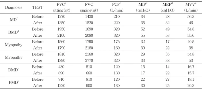
Diagnosis TEST FVCa sitting(㎖)
FVC supine(㎖)
PCFb (L/min)
MIPc (㎝H2O)
MEPd (㎝H2O)
MVVe (L/min)
MDf Before 1270 1420 210 34 28 56.3
After 1350 1520 220 35 32 46
BMDg Before 1950 1690 320 52 49 54.8
After 2100 2080 320 55 53 55.6
Myopathy Before 1560 1790 175 32 17 40.5
After 1790 2180 160 39 22 38
Myopathy Before 1810 2560 320 29 35 54.8
After 1890 2770 320 33 38 53
DMDh Before 430 510 120 15 14 16.7
After 690 660 130 17 22 15.7
PMDi Before 910 810 120 22 27 18.1
After 1220 900 130 30 25 20.3
aforced vital capacity, bpeak cough flow, cmaximal inspiratory pressure, dmaximal expiratory pressure, emaximal voluntary ventilation, fmuscular dystrophy, gBecker muscular dystrophy, hDuchenne muscular dystrophy, iprogressive muscular dystrophy.
Table 2. Evaluation of pulmonary function measurement values were presented as MEP/MEP pre (%) and MIP/MIP pre (%).
Maximal voluntary ventilation
MVV is an index of pulmonary function variable used to determine respiratory muscle endurance (Kor et al, 2004). The traditional intervention of measuring MVV was to let the subject breathe the largest ven- tilation volume that could be breathed into and out of the lungs during a 12-second interval with maximal voluntary effort. The subjects inhaled deeply (with a volume greater than the tidal volume but lower than the FVC) and rapidly for a 12-second interval, with ventilation flow measured using a pony FX (COSMED, Pony FX, Rome, Italy).
Statistical analysis
Because the sample size was small, the Wilcoxon signed-rank test was used to compare pulmonary func- tions (FVC, PCF, MIP, MEP and MVV) between the beginning of training and after the end of the 5-month training. Statistical significance was set at .05. All data were analyzed using statistical package for the SPSS ver. 23.0 (IBM corp., Armonk, NY, USA).
Results
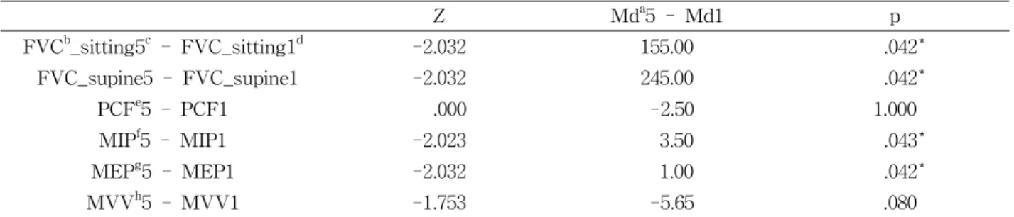
Table 2 and Figure 2 shows the collected data of each patient during pulmonary function test. Table 3 shows the pulmonary function measurement results, comparisons of before and after playing harmonica.
Both sitting and supine FVC significantly increase after playing harmonica (p= .042), as well as MIP (p= .043) and MEP (p= .042). Both PCF and MVV did not significantly increase after playing harmonica (p=1.000, p=.080, respectively).
Discussion
The purpose of this study was to determine the effect of 5-month training of playing harmonica on pulmonary function by assessing FVC, PCF, MIP, MEP, and MVV in patients with NMD. This was the first study to objectively assess the effect of playing harmonica on pulmonary function in patients with NMD. Our result partially supported the re- search hypothesis. Playing harmonica was effective in increasing FVC, MIP, and MEP in patients with
Z Mda5 - Md1 p
FVCb_sitting5c - FVC_sitting1d -2.032 155.00 .042*
FVC_supine5 - FVC_supine1 -2.032 245.00 .042*
PCFe5 - PCF1 .000 -2.50 1.000
MIPf5 - MIP1 -2.023 3.50 .043*
MEPg5 - MEP1 -2.032 1.00 .042*
MVVh5 - MVV1 -1.753 -5.65 .080
amedian, bforced vital capacity, cbefore 5-month training, dafter 5-month training-5, epeak cough flow, fmaximal inspiratory pressure, gmaximal expiratory pressure, hmaximal voluntary ventilation, *p<.05.
Table 3. Pulmonary function test result
NMD. The reason this study set the experiment pe- riod as 5 months was that no study has been con- ducted on harmonica training for patients with re- strictive lung disease, while previous research find- ings suggested that training for ≥12 weeks would be needed to improve the pulmonary function of pa- tients with neuromuscular disease (Aitkens et al., 1993). The training period was determined as the longer period of 5 months.
After a 5-month training, FVC was significantly greater in both the sitting and supine positions.
Sitting FVC increased by 12%, and supine FVC in- creased by 18%. These findings support our research hypothesis. Both deep breathing exercises and play- ing harmonica produced comparable results although both exercises are different (Jeffery et al, 2012). A previous study indicated that deep breathing exercise increased FVC in patients with NMD (Adams and Chandler, 1974). Playing harmonica might be similar to deep breathing exercises in increasing VC (Jeffery et al, 2012). Previous studies have reported that FVC was greater in the sitting position than in the supine position because the diaphragm maintains as a pri- mary inspiratory muscle in patients with NMD (Mcdonald et al, 1995; Park et al, 2010). However, in this study, interestingly FVC was greater in the su- pine position than in the sitting position in two subjects. This result could be because the two sub- jects had scoliosis, and the scoliotic curve and rib cage malposition might reduce FVC in the sitting position. Decreased FVC in the sitting position can
also occur in patients with NMD (Park et al, 2010).
Thus, playing harmonica might be useful to improve both sitting and supine FVC in patients with NMD.
Our results showed that MIP and MEP sig- nificantly increased after training. MIP and MEP in- creased by 10% and 11%, respectively. These find- ings support our research hypothesis. Our result is consistent with the finding of previous studies that investigated the effects of IMT on MIP and MEP (Ansved, 2001; Jeffery et al., 2012; Kang et al, 2006a). The only difference between previous studies and our study was that we used playing harmonica as an independent variable to increase MIP and MEP. In previous studies, MIP increased after the training when patients with NMD used IMT with pressure threshold device for respiratory muscle training (Kang et al, 2006a; McCool and Tzelepis, 1995). Moreover, playing harmonica was an effective therapy to improve breathing, similar to IMT (Jeffery et al, 2012). Most of the muscle endurance studies have reported increased MIP instead of MEP (Kang et al, 2006a; McCool and Tzelepis, 1995), but playing harmonica improved not only MIP, but also MEP in our study. The possible reason for improvement in MIP and MEP may be because playing harmonica played a crucial role similar to IMT and expiratory muscle training. Respiratory endurance training leads to an improvement in pulmonary muscle function through its effect on relatively conserved respiratory muscle fibers by increasing capillary and mitochondrial density and overall oxidative enzyme capacity
(Dimarco et al, 1985; Park et al, 2010). Therefore, all subjects who participated in playing harmonica showed improvement caused by respiratory muscle pressure.
After 5-month training, no significant difference was found in PCF. These findings did not support our research hypothesis. Contrary to the findings of our study, a previous study reported that IMT was effective in increasing muscle power and coughing ability (Kang, 2006b). PCF accompanies increases in abdominal and intrathoracic pressures and requires the appropriate strength of the core muscles, and di- aphragm and intercostal muscles, which are indis- pensable when inducing a reflexive cough (Torres et al, 2014). In this regard, a possible reason behind these findings could be that the subjects in this study had a disease that made it difficult to improve such muscles, and harmonica training did not involve similar motions that occur during a cough. Thus, manually assisted techniques were more useful in patients with NMD with PCF level <160 L/min (Torres et al, 2014).
Contrary to our hypothesis, no significant MVV was observed between periods. The possible explanation could be that the subjects who participated in our study used motorized wheelchair for ambulation. Kang et al. (1998) reported that subjects with NMD who use wheelchairs showed significantly lower MVV during IMT compared with the NMD subjects capable of walking. As MVV is a dynamic evaluation indicator, the NMD subjects capable of walking preserved respi- ratory muscle better than patients with NMD who uses wheelchair. Furthermore, the results were attrib- utable to the characteristics of the neuromuscular dis- ease, in which complete and accurate performance of MVV is difficult, requiring repeated MIP and MEP for 12 seconds as an indicator of the ability to respond when MVV is needed.
This study has several limitations. First, the sub- jects had different ages and diagnoses. This is be- cause of the difficulty in finding and recruiting NMD patients who could move independently while playing the harmonica as subjects. Subjects with the same
diagnosis should be recruited in future studies.
Second, this study included a relatively small number of subjects, and training duration and frequency were not individually customized. Therefore, further stud- ies, including large sample size, are necessary to confirm our results. Future studies should determine the long-term effects of playing harmonica on differ- ent pulmonary function parameters.
Conclusion
The purpose of this study was to determine whether 5-month training of harmonica can affect pulmonary function by assessing FVC, PCF, MIP, MEP and MVV in patients with NMD. Both supine and sitting positions of FVC, MIP, and MEP sig- nificantly increased after the training. Therefore, playing harmonica can be an effective method for improving pulmonary function in subjects with NMD.
References
Adams MA, Chandler LS. Effects of physical therapy program on vital capacity of patients with mus- cular dystrophy. Physical Ther. 1974;54(5):494-496.
Aitkens SG, McCrory MA, Kilmer DD, et al.
Moderate resistance exercise program: its effect in slowly progressive neuromuscular disease.
Arch Phys Med Rehabil. 1993;74(7):711-715.
Alexander JL, Wagner CL. Is harmonica playing an effective adjunct therapy to pulmonary re- habilitation? Rehabil Nurs. 2012;37(4):207-212.
https://doi.org/10.1002/rnj.33
Ansved T. Muscle training in muscular dystrophies.
Acta Physiol scand. 2001;171(3):359-366.
Bach JR, Rajaraman R, Ballanger F, et al.
Neuromuscular ventilatory insufficiency: Effect of home mechanical ventilator use vs oxygen ther- apy on pneumonia and hospitalization rate. Am J Phys Med Rehabil. 1998;77(1):8-19.
This article was received July 7, 2018, was re- viewed July 7, 2018, and was accepted September 10, 2018.
DiMarco AF, Kelling JS, DiMarco MS, et al. The ef- fects of inspiratory resistive training on respira- tory muscle function in patients with muscular dystrophy. Muscle Nerve. 1985;8(4):284-290.
Emery AE. Diagnostic criteria for neuromuscular disorders. Electroencephalogr Clin Neurophysiol.
1994;103(5):578. https://doi.org/10.1016/0960-8966 (94)90038-8
Gibson GJ, Pride NB, Davis JN, et al. Pulmonary me- chanics in patients with respiratory muscle weakness. Am Rev Respi Dis. 1977;115(3):389-395.
Griggs RC, Donohoe KM, Utell MJ, et al. Evaluation of pulmonary function in neuromuscular disease.
Arch Neurol. 1981;38(1):9-12.
Kang SW, Na YM, Baek SK, et al. Clinical im- plications of inspiratory muscle training in pa- tients with Duchenne muscular dystrophy. J Kor Acad Rehabil Med. 1998;22(2):361-368.
Kang, SW, Kang YS, Shon HS, et al. Respiratory muscle strength and cough capacity in patients with Duchenne muscular dystrophy. Yonsei Med J. 2006a;47(2):184-190.
Kang SW. Pulmonary rehabilitation in patients with neuromuscular disease. Yonsei Med J. 2006b;
47(3):307-314.
Kor AC, Ong KC, Earnest A, et al. Prediction of the maximal voluntary ventilation in healthy adult Chinese subjects. Respirology. 2004;9(1):76-80.
McCool FD, Mayewski RF, Shayne DS, et al.
Intermittent positive pressure breathing in pa-
tients with respiratory muscle weakness. Chest.
1986;90(4):546-552.
McCool FD, Tzelepis GE. Inspiratory muscle training in the patient with neuromuscular disease. Phys Ther. 1995;75(11):1006-1014.
McDonald CM, Abresch RT, Carter GT, et al.
Profiles of neuromuscular diseases: Duchenne muscular dystrophy. Am J Phys Med rehabil.
1995;74(5):S70-92.
Park JH, Kang SW, Lee SC, et al. How respiratory muscle strength correlates with cough capacity in patients with respiratory muscle weakness.
Yonsei Med J. 2010;51(3):392-397. https://do- i.org/10.3349/ymj.2010.51.3.39
Weiner P, McConnell A. Respiratory muscle training in chronic obstructive pulmonary disease: in- spiratory, expiratory, or both? Curr Opin Pulm Med. 2005;11(2):140-144.
Wilson SH, Cooke NT, Edwards RH, et al. Predicted normal values for maximal respiratory pressures in Caucasian adults and children. Thorax.
1984;39(7):535-538.
Won YH, Choi WA, Kim DH, et al. Postural vital ca- pacity difference with aging in Duchenne muscu- lar dystrophy. Muscle Nerve. 2015;52(5):722-727.
https://doi.org/10.1002/mus.24623