A Novel PHKA1 Mutation in a Patient with Glycogen Storage Disease Type IXD
Hye Jin Kim
1, Soo Hyun Nam
2,3, Sang Beom Kim
4, Ki Wha Chung
5* and Byung-Ok Choi
1,2,3*
1Department of Health Sciences and Technology, Samsung Advanced Institute for Health Science & Tech., Sungkyunkwan University, 81 Irwon-ro, Gangnam-gu, Seoul 06351, Korea
2Stem Cell & Regenerative Medicine Institute, Samsung Medical Center, 81 Irwon-ro, Gangnam-gu, Seoul 06351, Korea
3Department of Neurology, Samsung Medical Center, Sungkyunkwan University School of Medicine, 81 Irwon-ro, Gangnam-gu, Seoul 06351, Korea
4Department of Neurology, Kyung Hee University Hospital at Gangdong, Kyung Hee University School of Medicine, Seoul 05278, Korea
5Department of Biological Science, Kongju National University, Gongju 32588, Korea Received April 22, 2020 /Revised June 29, 2020 /Accepted August 4, 2020
Distal myopathy is a clinically and genetically heterogeneous group of degenerative diseases of the distal muscle. Glycogen storage disease type IXD (GSD9D) is a metabolic distal myopathy charac- terized by muscle deficiency of phosphorylase kinase, a key regulatory enzyme in glycogen metabo- lism. Affected individuals may develop muscle weakness, degeneration, and cramps, as well as abnor- mal muscle pain and stiffness after exercise. It has been reported that mutations in the PHKA1 gene which encodes the alpha subunit of muscle phosphorylase kinase cause GSD9D. In this study, we ex- amined a Korean GSD9D family with a c.3314T>C (p.I1105T) mutation in the PHKA1 gene. This muta- tion has not been previously reported in any mutation database nor was it found in 500 healthy controls. The mutation region is well conserved in various other species, and in silico analysis predicts that it is likely to be pathogenic. To date, only seven mutations in the PHKA1 gene have been docu- mented, and this is the first report of Korean GSD9D patients. This study also describes and compares the clinical symptoms and pathological conditions of previously reported cases and these Korean patients. We believe that our findings will be useful for the molecular diagnosis of GSD9D.
Key words : Distal myopathy, Glycogen storage disease type 9D, MRI, mutation, PHKA1
*Corresponding authors
Tel : +82-2-2148-9496, Fax : 82-2-3410-0052
*E-mail : [email protected] (Byung-Ok Choi) [email protected] (Ki Wha Chung)
This is an Open-Access article distributed under the terms of the Creative Commons Attribution Non-Commercial License (http://creativecommons.org/licenses/by-nc/3.0) which permits unrestricted non-commercial use, distribution, and reproduction in any medium, provided the original work is properly cited.
Introduction
Distal myopathy is a degenerative disease of the distal muscle and is a clinically and genetically heterogeneous group [7]. Distal myopathy is usually classified as Nonaka, Miyoshi, Laing, Welander, Udd and Markesbery-Griggs dis- tal myopathy [7]. Metabolic myopathy is a myopathy that causes distal weakness [14]. Glycogen storage disease type 9D (GSD9D, OMIM 300599) is one of the metabolic myo- pathies caused by the lack of decomposition of glycogen due to the deficiency of Phosphorylase kinase (PhK), so that sufficient energy for muscle contraction cannot be obtained [18, 20]. GSD9D belongs to a mild metabolic disorder, and is characterized by progressive muscle weakness, exercise
intolerance and cramps, and extensive death in muscle tis- sue [1, 17, 20, 21]. Most patients develop symptoms as adults and mainly show weakness in the distal muscles and muscle atrophy [1, 17, 20, 21].
Phosphorylase kinase is composed of four homotetr- amers: α (PHKA1, PHKA2), β (PHKB), γ (PHKG1, PHKG2), and δ (CALM1) [2, 4, 10, 11-13, 16, 19]. The γ subunit acts as a catalyst and is regulated by the α and β subunits, and δ is calmodulin, which gives the enzyme Ca
2+sensitivity [2].
There are two types of α subunits, liver-specific PHKA2 and muscle-specific PHKA1, and both genes exist on the X chro- mosome [11, 20]. To date, over 100 PHKA2 mutations in HGMD (http://www.hgmd.cf.ac.uk/ac/index.php) have been reported to cause GSD9A. It has also been reported in Korea [5]. However, only 7 mutations in PHKA1 have been reported to cause GSD9D and have not been reported in Korea [1, 3, 8, 10, 15, 18, 20, 21].
We investigated patients with GSD9D who visited the
hospital suspected of distal hereditary motor neuronopathies
(dHMN) in another hospital. dHMN does not show any
damage to the sensory nerves, and only the motor nerves
have abnormalities, so it shows symptoms of weakness in
A B
C D
Fig. 1. X-linked recessive metabolic myopathy family with novel hemizygous mutation in PHKA1. (A) Pedigrees of FC975 family.
Arrows indicate probands whose DNA were used for the WES (□, ○ : unaffected; ■ : affected). (B) Chromatograms of the mutation sites by capillary sequencing method. The mutation of c.3314T>C (p.I1105T) in PHKA1 is clearly shown in the mutant alleles (arrows). (C) PHKA1 protein structure and causative mutations. The present five mutations as well as previously reported mutations are indicated below the diagram. (D) Conservation of amino acid sequences in the mutation site. Multiple protein sequence alignment revealed strong conservation of amino acid sequences at the p.I1105T mutation site among different vertebrate species (Human: NP_002628.2, Mouse: NP_032858.2, Cow: XP_002700055.1, Frog: NP_001121 510.1, Zebrafish: XP_005166576.1).
the distal muscles [9]. Distal myopathy also indicates weak- ness in the distal muscles and muscle atrophy, making dHMN difficult to distinguish clinically from distal myo- pathy. Therefore, it is very necessary to distinguish dHMN from distal myopathy when the patient visits with distal muscular weakness and gait disorder. Because dHMN and distal myopathy are caused by mutations in different genes, genetic testing is a good way to distinguish these two dis- eases.
Therefore, we performed genetic analysis in Korean GSD9D patients and found PHKA1 mutation. In addition, the patient's clinical symptoms, pathological features, and MRI results were described. Accordingly, we attempted to compare the mutations and clinical patterns of patients en- rolled in this study and several previously reported GSD9D patients.
Materials and Methods Patients
This study enrolled a X-linked recessive Korean family with GSD9D (FC975, Fig. 1A). This study also included 500 healthy controls who had no clinical features or family his- tory of distal myopathy, which was confirmed after careful clinical and electrophysiological examinations. Written in- formed consent was obtained from all participants according to the protocol approved by the institutional review board for Sungkyunkwan University, Samsung Medical Center.
Exome sequencing and filtering of variants Exome sequencing was performed with the Human SeqCap EZ Human Exome Library v3.0 (Roche/NimbleGen, Madison, WI), and the HiSeq2500 Genome Analyzer (Illumina, San Diego, CA) for 1 sample from the proband of FC975.
The University of California, Santa Cruz assembly hg19 was
the reference sequence. We selected functionally significant
variants (missense, nonsense, exonic indel, and splicing site
variants) from the whole exome sequencing data, and then
variants registered as novel or uncommon variants (minor
allele frequencies ≤ 0.01) in dbSNP150 (http://www.ncbi.
nlm.nih.gov), the 1,000 Genomes project database (http://
www.1000genomes.org/) were further filtered.
In silico analysis
The Sanger sequencing method confirmed the variant us- ing the genetic analyzer ABI3130XL (Life Technologies, Foster City, CA). The genomic evolutionary rate profiling (GERP) scores were determined by the GERP program (http://mendel.
stanford.edu/SidowLab/downloads/gerp/index.html). We performed conservation analysis of the protein sequences us- ing MEGA5, version 6.06 (http://www.megasoftware.net/).
In silico analyses were done with the prediction algorithms SIFT (http://sift.jcvi.org), MUpro (http://www.ics.uci.edu/~
baldig/mutation), and PolyPhen-2 (http://genetics.bwh.
harvard.edu/pph2/). The SMART program predicted do- mains for the PHKA1 protein (http://smart.embl.de/).
Clinical and electrophysiological examinations Clinical information included assessments of age at onset, muscle impairments, sensory loss, deep tendon reflexes, and muscle atrophy. The muscle strength of the flexor and ex- tensor muscles was assessed manually with the standard Medical Research Council scale. The age at onset was de- termined by asking patients for their ages when symptoms, including distal muscle weakness, first appeared. Neuro- physiological studies were done on proband. Motor and sen- sory conduction studies of the median, ulnar, peroneal, ti- bial, and sural nerves were tested, and needle electromyog- raphy was performed in the bilateral upper and lower limb muscles. In a patient, serum CK levels were measured.
Muscle biopsy and histological examination Histopathological analyses including immunohistochem- istry of the left vastus lateralis muscles were done in the proband (Ⅲ-2). Frozen 10 mm sections were examined after staining with hematoxylin and eosin (H&E), modified Gomori trichrome (GT), nicotinamide adenine dinucleotide- tetrazolium reductase (NADH-tr), and myosin adenosine tri- phosphatase (ATPase) preincubated at pH4.3, 4.6, and 9.4.
Staining with Congo red, acid phosphatase, and periodic acid-Schiff (PAS) was performed on a frozen muscle speci- men of Patient.
Lower limb MRI
The patient was examined by lower limb magnetic reso- nance imaging (MRI) of the hip, thigh, and calf muscles at
32 years. MRI was undertaken by a 1.5T system (Siemens Vision, Erlan-gen, Germany). The imaging was done in the axial (field of view [FOV]524–32cm, slice thickness510mm, slice gap50.5–1.0mm) and coronal planes (FOV538–40cm, slice thickness54–5mm, slice gap50.5–1.0mm). The follow- ing protocol was used for all patients: T1-weighted spin-echo (SE) (repetition time [TR]5570–650 milliseconds, echo time [TE]514–20 milliseconds, 512 matrixes), T2-weighted SE (TR52,800–4,000 milliseconds, TE596–99 milliseconds, 512 matrixes), and fat-suppressed T2-weighted SE (TR53,090–
4,900 milliseconds, TE585–99 milliseconds, 512 matrixes)
Results
Identification of novel mutation in PHKA1
Whole exome sequencing (WES) was performed to find the causative mutation of the patient. The patient was screened for mutations in the distal myopathy and metabolic myopathy genes and peripheral neuropathy genes. As a re- sult of the screening, we did not find any causative candi- dates within the distal myopathy and peripheral neuropathy genes. However, within the metabolic myopathy gene, a c.3314T> C (p.I1105T) missense mutation was found in exon 31 of the PHKA1 gene (Table 3). Other mutations were fre- quently found in several public mutation databases (dbSNP 153, 1000 Genomes project and Exome Variant Server) and 500 healthy controls. The PHKA1 p.I1105T mutation was pre- viously not reported in any mutation database and was not found in 500 healty controls. This mutation was located be- low the glycosidase family 15 domain (Fig. 1C) and this re- gion was well conserved within various other species (Fig.
1D). The GERP score of the mutation was significantly high at 5.12, and three in silico analyzes predicted that the muta- tion is likely to be pathogenic. (SIFT: 0.02, Damaging; Poly Phen-2: 0.975, Probably damaging; MUpro: -0.997, Decrease the stability of protein structure).
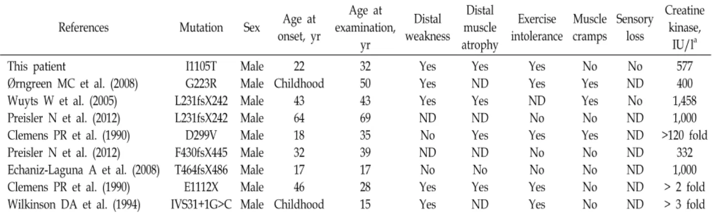
Clinical manifestations and electrophysiologic feature Table 1 shows the clinical symptoms of the patients in- cluded in this study and the 7 previously reported cases.
The patient found no abnormalities in the developmental
process and found it difficult to run at age 22. Along with
this, he noticed that it was difficult to walk long. At age
27, he noticed that both toes weren't moving, and he noticed
that his leg muscles were weakening. When he was 32, he
visited an outside hospital with symptoms of both his toes
Table 1. Clinical features in patients with mutation in the PHKA1 gene
References Mutation Sex Age at
onset, yr
Age at examination,
yr
Distal weakness
Distal muscle atrophy
Exercise intolerance
Muscle cramps
Sensory loss
Creatine kinase,
IU/la This patient
Ørngreen MC et al. (2008) Wuyts W et al. (2005) Preisler N et al. (2012) Clemens PR et al. (1990) Preisler N et al. (2012) Echaniz-Laguna A et al. (2008) Clemens PR et al. (1990) Wilkinson DA et al. (1994)
I1105T G223R L231fsX242 L231fsX242 D299V F430fsX445 T464fsX486 E1112X IVS31+1G>C
Male Male Male Male Male Male Male Male Male
22 Childhood
43 64 18 32 17 46 Childhood
32 50 43 69 35 39 17 28 15
Yes Yes Yes ND No ND No Yes Yes
Yes ND Yes ND Yes ND No Yes ND
Yes Yes ND No Yes No No Yes Yes
No Yes Yes No Yes No No No No
No ND No ND ND ND ND ND ND
577 400 1,458 1,000
>120 fold 332 1,000
> 2 fold
> 3 fold ND Not done
aCreatine kinase: normal range in our laboratory < 185IU/l
Table 2. Electrophysiological features of the patient with PHKA1 mutation
Patient Normal value
Side Motor nerve studies Median motor nerve TL (ms)
CMAP (mV) MNCV (m/s) Ulnar motor nerve TL (ms) CMAP (mV) MNCV (m/s) Peroneal nerve TL (ms) CMAP (mV) MNCV (m/s) Tibial nerve TL (ms) CMAP (mV) MNCV (m/s) Sensory nerve studies Median sensory nerve SNAP (μV) SNCV (m/s) Ulnar sensory nerve SNAP (μV) SNCV (m/s) Sural nerve SNAP (μV) SNCV (m/s)
Right
3.6 27.8 52.2 2.8 18.7 52.9 5.0 2.6 43.8 A A A
51.1 42.7 33.0 42.9 19.1 40.0
Left
3.6 29.2 54.5 2.9 13.4 50.2 5.5 2.1 43.0 A A A
47.0 41.3 33.0 39.3 26.1 41.2
<3.9
>6.0
>50.5
<3.0
>8.0
>51.1
<5.3
>1.6
>41.2
<5.4
>6.0
>41.1
>8.8
>39.3
>7.9
>37.5
>6.0
>32.1 Abbreviations: NP: no potential, CMAP: compound muscle ac- tion potential, MNCV: motor nerve conduction Velocity, SNAP:
sensory nerve action potential, SNCV: sensory nerve con- duction velocity.
not moving. At the time, the neuroscientist's opinion was suspected of dHMN, and he came to the hospital.
However, when examined in the neurology department of our hospital, there was no loss of sensory nerves and only impaired motor function was observed. As a result of the nerve conduction test, sensory nerves showed normal SMAP and NCS in both upper and lower extremities. Normal CMAP and NCS were found in the median, ulnar nerve, and fibula nerves of the motor nerve. However, CMAP was not measured in the left and right tibial nerves of the motor nerve (Table 2). Electromyography showed reasonable re- sults for myopathy. The CK concentration in the blood was 577 IU / l, showing a nearly three-fold increase compared to normal subjects.
The patient's type did not show any neurological abnor- malities and neither father nor mother (Fig. 1A). In addition, the parents and siblings of the patient's mother showed no abnormality based on the history examination.
Histopathological findings
Muscle histological examination revealed that angulated muscle fibers and subsarcolemmal vacuoles were observed in H&E staining, and atrophy and abnormal hypertrophy of muscle fibers were also observed (Fig. 2A). In addition, PAS positive muscle fibers were observed. These patho- logical features were similar to those of previously reported GSD9D patients [20]. Electron microscopy revealed glycogen granules of muscle fibers and thickened Z-band and mi- tochondrial swelling and degradation were also observed (Fig. 2B).
Fatty infiltration of the calf muscle
A patient's lower extremity MRI study was performed at
Fig. 2. Histopathologic observations of vastus lateralis muscle biopsies. (A) H&E showing angulated small size of myofibers with atrophy (arrow) and myofibers (arrowhead). (B) Electron micrograph showing a lot of glycogen (arrow) and and Z-band thickening (arrowhead).
Fig. 3. T1-weighted magnetic resonance imaging (MRI) of patient.
Coronal images of thigh (A) and calf (B). Axial image of hip (C) and thigh (D), calf (E).
age 33 (Fig. 3A - Fig. 3E). In the patient, no damage was observed in the hip and thigh muscles, and only moderate damage was observed in the calf. Severe gastrocnemius mus- cle damage was found in the right calf muscle, and soleus and gastrocnemius muscles were equally damaged in the left muscle. In the calf, the rear compartments soleus and gastrocnemius showed severe muscle atrophy and fat in- filtration, while the anterior and lateral compartment mus- cles were not damaged.
Discussion
This study describes the genetic and MRI data and clinical
characteristics of Korean GSD9D patients with PHKA1 muta- tions compared to previously reported cases. A total of seven
PHKA1 mutations have been reported to cause GSD9D todate [1, 3, 8, 10, 15, 18, 20, 21]. Table 1 compares the clinical symptoms of these patients. Patients enrolled in this study had similar symptoms, such as late onset age, distal weak- ness, and intolerance to exercise, with previously reported GSD9D patients. However, no muscle cramp was found in our patients, which was also not found in some of the pre- viously reported patients. The onset age of GSD9D was re- ported to be 17-64 years (average age of 34.6 years), exclud- ing 2 children onset, and this patient was also 22 years old.
The PHKA1 mutation was inherited from the patient's mother. Mothers showed little clinical symptoms because they had mutations as heterozygotes. The mutation was not found in the patient's older brother and the father's and mother's siblings. It was also not found in 500 healthy con- trols and was also not reported in the dbSNP153 and 1,000 genome databases. Therefore, this PHKA1 mutation is con- sidered a novel mutation. Previously reported mutations were mainly located in the glycoside hydrolase family 15 domain, but our patient mutations were located under the domain with p.E1112X and IVS31+1G>C. There were no symptom differences between the mutations in the domain and the mutations below.
In addition, this study performed MRI examination of the
lower limbs for the first time in patients with GSD9D. As
a result of MRI, fat infiltration was not observed in the hip
and thigh, and fat infiltration was observed only in the calf,
distal to the lower extremity. In the calf muscle, no damage
was observed in the anterior and lateral compartments,
whereas atrophy and severe fat infiltration were observed
Table 3. Functionally significant variants found in 44 metabolic myopathy genes
Chr Gene Nt changea AA
change dbSNP153 1,000 G Inheritanceb In-house
Freq (n=500) Description
chrX PHKA1 c.3314T>C (hem) I1105T ․ ․ XR 0 Pathogenic
chr01 AGL splicing site mutation rs2307130 0.43
AR 0.83 Polymorphic
c.1109G>A (het) R370Q rs17121464 0.06 0.4 Polymorphic
chr03 GBE1 c.1000A>G (hom) I334V rs2172397 0.99
AR 1 Polymorphic
c.568A>G (hom) R190G rs2229519 0.33 0.71 Polymorphic
chr04 HADH c.257T>C (hom) L86P rs4956145 0.89 AR 1 Polymorphic
chr04 ETFDH c.92C>T (hom) T31I rs11559290 0.72 AR 1 Polymorphic
chr12 PFKM c.5A>T (het) H2L rs11609399 0.35 AR 0.8 Polymorphic
chr12 ISCU
c.19T>G (hom) F7V rs10778647 0.88
AR
0.38 Polymorphic
c.20T>G (hom) F7C rs10778648 0.88 0.37 Polymorphic
c.35C>T (hom) A12V rs2287555 0.53 0.1 Polymorphic
chr16 PHKB c.2653G>A (het) G885R rs149983469 . AR 0 Polymorphic
chr17 ENO3 c.212A>G (het) N71S rs238238 0.6
AR 0.66 Polymorphic
c.254T>C (het) V85A rs238239 0.35 0.2 Polymorphic
chr17 GAA
c.596A>G (hom) H199R rs1042393 0.62
AR
0.84 Polymorphic
c.668G>A (hom) R223H rs1042395 0.61 0.85 Polymorphic
c.1726G>A (het) G576S rs1800307 0.05 0.36 Polymorphic
c.2065G>A (het) E689K rs1800309 0.09 0.53 Polymorphic
c.2338G>A (hom) V780I rs1126690 0.69 0.92 Polymorphic
chr19 GYS1 c.1246A>G (het) M416V rs5447 0.03 AR 0.25 Polymorphic
chr19 ETFB c.461C>T (het) T154M rs1130426 0.46 AR 0.54 Polymorphic
ahet: heterozygous, hom: homozygous and hem: hemizygous.
bAD: autosomal dominant, AR: autosomal recessive and XR: X-linked recessive
in the anterior compartment soleus and gastrocnemius muscles. This phenomenon is thought to be a singularity, so the patient showed disagreement with distal myopathy.
In summary, we wanted to present clinical features to pa- tients with new PHKA1 mutations. We report clinically typi- cal GSD9D symptoms and detect and report distal muscle damage by MRI. Since there are no reported mutations in Korea among the 7 cases, this study is the first to report a case of 9D type Glycogen patients in Korea. This study will expand the clinical spectrum of GSD9D with the PHKA1 mutation and suggest that it will be useful for molecular diagnosis of heterogeneous metabolic myopathy.
Acknowledgements
This work was supported by grants from the National Research Foundation (2016R1A5A2007009, 2018R1A4A1024 506 and 2019R1A2C1087547) and the Korean Health Tech- nology R&D Project, Ministry of Health & Welfare (HI14C 3484 and HI16C0426), Republic of Korea.
The Conflict of Interest Statement
The authors declare that they have no conflicts of interest with the contents of this article.
References
1. Bruno, C., Manfredi, G., Andreu, A. L., Shanske, S., Krishna, S., Ilse, W. K. and DiMauro, S. 1998. A splice junction muta- tion in the alpha(M) gene of phosphorylase kinase in a pa- tient with myopathy. Biochem. Biophys. Res. Commun. 249, 648-651.
2. Brushia, R. J. and Walsh, D. A. 1999. Phosphorylase kinase:
the complexity of its regulation is reflected in the complex- ity of its structure. Front. Biosci. 4, D618-641.
3. Burwinkel, B, Hu, B., Schroers, A., Clemens, P. R., Moses, S. W., Shin, Y. S., Pongratz, D., Vorgerd, M. and Kilimann, M. W. 2003. Muscle glycogenosis with low phosphorylase kinase activity: mutations in PHKA1, PHKG1 or six other candidate genes explain only a minority of cases. Eur. J.
Hum. Genet. 11, 516-526.
4. Burwinkel, B., Maichele, A. J, Aagenaes, O., Bakker, H. D., Lerner, A., Shin, Y. S., Strachan, J. A. and Kilimann, M.
W. 1997. Autosomal glycogenosis of liver and muscle due
to phosphorylase kinase deficiency is caused by mutations in the phosphorylase kinase beta subunit (PHKB). Hum.
Mol. Genet. 6, 1109-1115.
5. Choi, R., Park, H. D., Kang, B., Choi, S. Y., Ki, C. S., Lee, S. Y., Kim, J. W., Song, J. and Choe, Y. H. 2016. PHKA2 mutation spectrum in Korean patients with glycogen stor- age disease type IX: prevalence of deletion mutations. BMC.
Med. Genet. 17, 33.
6. Clemens, P. R., Yamamoto, M. and Engel, A. G. 1990. Adult phosphorylase b kinase deficiency. Ann. Neurol. 28, 529-538.
7. Dimachkie, M. M. and Barohn, R. J. 2014. Distal myopathies.
Neurol. Clin. 32, 817-842.
8. Echaniz-Laguna, A., Akman, H. O., Mohr, M., Tranchant, C., Talmant-Verbist, V., Rolland, M. O. and Dimauro, S.
2010. Muscle phosphorylase B kinase deficiency revisited.
Neuromuscul. Disord. 20, 125-127.
9. Harding, A. E. and Thomas, P. K. 1980. The clinical features of hereditary motor and sensory neuropathy types I and II. Brain 103, 259-280.
10. Harmann, B., Zander, N. F. and Kilimann, M. W. 1991.
Isoform diversity of phosphorylase kinase alpha and beta subunits generated by alternative RNA splicing. J. Biol.
Chem. 266, 15631-15637.
11. Hendrickx, J., Coucke, P., Dams, E., Lee, P., Odièvre, M., Corbeel, L., Fernandes, J. F. and Willems, P. J. 1995. Muta- tions in the phosphorylase kinase gene PHKA2 are respon- sible for X-linked liver glycogen storage disease. Hum. Mol.
Genet. 4, 77-83.
12. Koller, M., Schnyder, B. and Strehler, E. E. 1990. Structural organization of the human CaMIII calmodulin gene. Bio- chim. Biophys. Acta. 1087, 180-189.
13. Maichele, A. J., Burwinkel, B., Maire, I., Søvik, O. and Kilimann, M. W. 1996. Mutations in the testis/liver isoform of the phosphorylase kinase γ subunit (PHKG2) cause auto-
somal liver glycogenosis in the gsd rat and in humans.
Nat. Genet. 14, 337-340.
14. Mastaglia, F. L. and Laing, N. G. 1999. Distal myopathies:
clinical and molecular diagnosis and classification. J. Neurol.
Neurosurg. Psychiatry 67, 703-707.
15. Ørngreen, M. C., Schelhaas, H. J., Jeppesen, T. D., Akman, H. O., Wevers, R. A., Andersen, S. T., ter Laak, H. J., van Diggelen, O. P., DiMauro, S. and Vissing, J. 2008. Is muscle glycogenolysis impaired in X-linked phosphorylase B kinase deficiency? Neurology 70, 1876-1882.
16. Pegues, J. C. and Friedberg, F. 1990. Multiple mRNAs en- coding human calmodulin. Biochem. Biophys. Res. Commun.
172, 1145-1149.
17. Preisler, N., Orngreen, M. C., Echaniz-Laguna, A., Laforet, P., Lonsdorfer-Wolf, E., Doutreleau, S., Geny, B., Akman, H. O., Dimauro, S. and Vissing, J. 2012. Muscle phosphor- ylase kinase deficiency: a neutral metabolic variant or a disease? Neurology 78, 265-268.
18. Schneider, A., Davidson, J. J., Wüllrich, A. and Kilimann, M. W. 1993. Phosphorylase kinase deficiency in I-strain mice is associated with a frameshift mutation in the alpha subunit muscle isoform. Nat. Genet. 5, 381-385.
19. Wehner, M. and Kilimann, M. W. 1995. Human cDNA en- coding the muscle isoform of the phosphorylase kinase γ subunit (PHKG1). Hum. Genet. 96, 616-618.
20. Wehner, M., Clemens, P. R., Engel, A. G. and Kilimann, M. W. 1994. Human muscle glycogenosis due to phosphor- ylase kinase deficiency associated with a nonsense mutation in the muscle isoform of the alpha subunit. Hum. Mol.
Genet. 3, 1983-1987.
21. Wuyts, W., Reyniers, E., Ceuterick, C., Storm, K., de Barsy, T. and Martin, J. J. 2005. Myopathy and phosphorylase kin- ase deficiency caused by a mutation in the PHKA1 gene.
Am. J. Med. Genet. A. 133A, 82-84.
초록:당원 축적병 9D (GSD9D) 환자의 신규 PHKA1 돌연변이
김혜진
1․남수현
2,3․김상범
4․정기화
5*․최병옥
1,2,3*
(1성균관대학교 삼성융합의과학원 융합의과학과, 2삼성서울병원 줄기세포 재생의학 연구소, 3성균관대학교 의과대학
신경과학교실, 4강동경희대학교 의과대학 신경과학교실, 5공주대학교 생명과학과)