• 검색 결과가 없습니다.

Evaluation of Stability Following Two-Jaw Surgery

N/A
N/A
Protected

Academic year: 2021

Share "Evaluation of Stability Following Two-Jaw Surgery"

Copied!
7
0
0

로드 중.... (전체 텍스트 보기)

전체 글

(1)

Original Article

원고 접수일 2010년 10월 20일, 게재 확정일 2011년 1월 28일 책임저자 김수관

(501-759) 광주시 동구 서석동 375번지, 조선대학교 치의학전문대학원 구강악안면 외과학교실

Tel: 062-220-3819, Fax: 062-228-7316, E-mail: [email protected]

RECEIVED October 20, 2010, ACCEPTED January 28, 2011 Correspondence to Su-Gwan Kim

Department of Oral and Maxillofacial Surgery, School of Dentistry, Chosun University

375, Seosuk-dong, Dong-gu, Gwangju 501-759, Korea

Tel: 82-62-220-3819, Fax: 82-62-228-7316, E-mail: [email protected]

CC This is an open access article distributed under the terms of the Creative Commons Attribution Non-Commercial License (http://creativecommons.org/licenses/

by-nc/3.0) which permits unrestricted non-commercial use, distribution, and reproduction in any medium, provided the original work is properly cited.

양악 수술 후 안정성 평가

이승용ㆍ김수관ㆍ김서윤ㆍ오지수ㆍ문경남ㆍ윤대웅ㆍ김 훈1ㆍ김정선2

조선대학교 치의학전문대학원 구강악안면외과학교실, 1을지대학병원 치과, 2광주보건대학교 치위생과

Abstract

Evaluation of Stability Following Two-Jaw Surgery

Sung-Yong Lee, Su-Gwan Kim, Seo-Yoon Kim, Ji-Su Oh, Kyung-Nam Moon, Dae-Woong Yoon, Hoon Kim

1

, Jeong-Sun Kim

2

Department of Oral and Maxillofacial Surgery, School of Dentistry, Chosun University,

1

Department of Dentistry, Eulji University Hospital,

2

Department of Dental Hygieve, Gwangju Health College University

Purpose: Orthognathic surgery is required in patients with severe skeletal disharmony and facial asymmetry, which results in functional and esthetic improvement. Recently, bimaxillary surgery has become generalized. Establishment of the occlusal plane among several other factors included in the surgery plan is a major consideration for the diagnosis and treatment plan and it is also an important factor for postoperative stability.

Methods: In this study, we assessed postoperative stability of occlusal plane, B-point, and pogonion point on 20 patients who underwent two-jaw surgery in the Chosun Dental Hospital from 2000 to 2007. Preoperative and postoperative states and at least a one year postoperative follow-up were compared.

Results: The postsurgical relapse volume of the occlusal plane to the SN plane and the FH plane was −0.26±2.8o and

−0.44±3.29o, respectively and after two-jaw surgery, the stability of occlusal plane was maintained. The horizontal relapse degree was 0.85±0.46 mm and 0.76±0.48 mm, respectively, and the vertical relapse degree was 1.16±0.36 mm and 1.13±0.71 mm of the B point and the Pogonion point at the time after minimal 1 year.

Conclusion: The vertical relapse amount was shown to be slightly larger than the horizontal relapse amount.

Key words: Bilateral sagittal split ramus osteotomy, Le Fort I osteotomy, Orthognathic surgery, Relapse, Stability

Introduction

Dentofacial deformity patients have functional and es-

thetical impairment due to skeletal disharmony, and the

patients could not solve the dentofacial skeleton problem

itself by growth control or orthodontic therapy alone. In

(2)

dontic treatments, realign the jaw by accompanying orthog- nathic surgery, or relocate the segments of alveolar bone, and thus appropriate function and esthetics could be ob- tained[1-3].

Initially, orthognathic surgery was performed only on the mandible, however, the skeletal Class III malocclusion does not necessarily imply mandibular prognathism.

Particularly, for cases with the substantial antero-posterior, vertical, and horizontal skeletal disharmony, orthognathic surgery is required for the maxilla and mandible. Such Le-Fort I osteotomy on the maxilla allows the three-dimen- sional move of the maxilla and permits to obtain more satisfactory results for dentofacial deformity patients.

Therefore, two-jaw surgery has been improved afterward, and it became one of methods used widely for the treat- ment of dentofacial deformity patients[4].

In such orthognathic, Le Fort I osteotomy and diverse mandibular ramus surgeries became traditional surgeries to be used for the skeletal disharmony and the recovery of malocclusion in dentofacial deformity patients, nonethe- less, consequent relapse, TMJ dysfunction, dysesthesia, in- fection, bleeding, scar, etc. have been also reported abun- dantly[5,6]. Among them, stability and the relapse tendency after orthognathic surgery are important factors determin- ing the success or failure of surgery itself. Bothur et al.

have reported that the movement within 5 mm range is stable regardless of the fixation method or with or without bone graft[7].

Proffit et al.[8] have reported that in two-jaw surgery, the relapse trait is comparable to one-jaw surgery, how- ever, in the class III malocclusion cases, two-jaw surgery was more stable than one-jaw surgery, and in the class III malocclusion patients, two-jaw surgery was more stable than in the class II malocclusion patients. Donasky et al.[9]

have reported that after 1 year, two-jaw surgery accom- panied Le Fort I osteotomy and mandibular advancement surgery was more stable than two-jaw surgery accom- panied mandibular set-back surgery. However, in Franco et al. cases, two-jaw surgery showed 53.4% relapse rate, and thus it showed a higher relapse rate than 43.7% relapse rate shown in surgery performed only on the mandible[10].

In two-jaw surgery, the change of the occlusion plane occurs unavoidably, and the rotation of such change of

factor for the stability of bone fragment after surgery.

Wolford et al.[11] and Reyneke and Erans[12] reported the effects obtained by changing the occlusion plane during surgery, and Sinobad examined the association of the oc- clusion plane with the palatal surface.

Therefore, in this study, the stability after two-jaw sur- gery was evaluated by measuring the standard plane pres- ent in the craniofacial plane, the B point in the mandible (Pogonion point), and the relation to the occlusion plane of which location is inevitably changed during two-jaw surgery.

Materials and Methods

1. Patients

This study was conducted on patients with the skeletal class III malocclusion, visited department of oral and max- illofacial surgery of Chosun Dental Hospital, from 2000 to 2007, and among patients underwent two-jaw surgery in the department of oral maxillofacial surgery at our hospi- tal (Le Fort I osteotomy in the maxilla and bilateral saggital split osteotomy in the mandible), the difference of the man- dibular set-back degree was less than 3 mm and thus severe facial asymmetry was not shown, and 20 patients able to followed up for minimum 1 year. All patients underwent orthodontic therapy prior to surgery, the fixation using min- iplates was performed on the maxilla and the mandible, and after surgery, intermaxillary fixation was performed by placing splints approximately for 2 weeks. As the gender of the subject patients, the male was 14 patients and the female was 6 patients, their age distribution was form 18.1 years to 32.8 years, and the mean age was 21.9 years.

2. Methods

In this study, the time immediately prior to surgery was defined as T1, immediately after surgery was T2 (average 21.7 days), and more than one year after surgery was T3 (average 13.4 months).

The lateral cephalometric radiographs of all patients

were taken by the Planmeca Proline XC (PLANMECA,

Finland, Helsinki), in the department of diagnostic radiol-

ogy, dental clinic, Chosun University, at the time points

(3)

Fig. 1. Reference points. ANS, anterior nasal spine; Me, menton;

S, sella; N, nasion; PNS, posterior nasal spine; A, subspinale; B, supramentae; Pg, pogonion; Or, orbitale; P, porion.

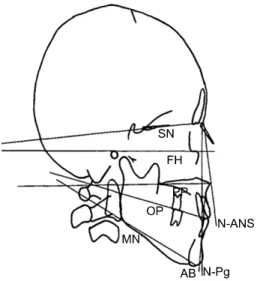
Fig. 2. Reference planes for measurement of occlusal plane angle.

SN, sella-nasion; FH, frankfort horizontal; PP, palatal plane; MN, mandibular plane; OP, Occlusal plane; N-ANS, nasion-anterior na- sal spine; AB, subspinale-supramentae; N-Pg, nasion-pogonion.

T1, T2, and T3 under identical conditions.

On each lateral cephalometric radiographs, projection images were prepared, the standard point (Fig. 1) and the standard plane (Fig. 2), and the measurement catego- ries were determined, and the distance as well as the angle were measured up to 0.1 mm and 0.5

o

, respectively.

1) Landmarks

(1) ANS (Anterior nasal spine): anterior nasal spine. The most anterior point of nasal floor, or the tip of premaxilla on the median plane.

(2) Me (Menton): the lowest point of mandibular median area.

(3) S (Sella): The center of sella turcica.

(4) N (Nasion): The most anterior point of fronto-nasal suture on the median plane.

(5) PNS (Posterior nasal spine): The most posterior area of nasal floor

(6) A (Subspinale): The deepest point between the ANS and the Prosthion on the median plane.

(7) B (Supramentae): The deepest point between the Infradentale and the Pogonion on the median plane.

(8) Pg (Pogonion): The most anterior point of chin con- tour on the median plane.

(9) Or (Orbitale): The lowest point of bony orbit contour.

(10) P (Porion): The highest point of external auditory canal.

2) Standard lines <Horizontal planes>

(1) SN plane :The line connecting the nasion in the center of sella turcica

(2) HP (Horizontal plane): The horizontal standard line on the SN plane with 7°ascending slope.

(3) FH plane (Frankfort horizontal plane): The line con- necting the orbitale on the anatomical porion.

(4) Palatal plane: The line connecting the ANS to the PNS.

(5) Mandibular plane : The line connecting the lowest point of mandibular ramus and the menton.

<Vertical planes>

(1) VP (Vertical plane): The vertical standard line vertical to the HP on the N point.

(2) N-ANS plane: The line connecting the nasion and the ANS.

(3) N-Pg plane: The line connecting the nasion and the pogonion.

(4) AB plane: The line connecting the point A and the point B.

3) Measurement categories <The slope of occlusal plane>

(1) SN plane - Occlusal plane

(2) FH plane - Occlusal plane

(3) Palatal plane - Occlusal plane

(4)

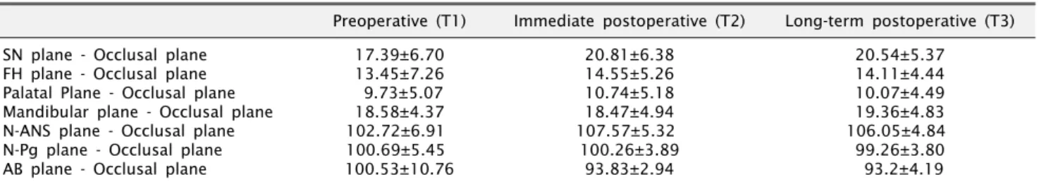
Preoperative (T1) Immediate postoperative (T2) Long-term postoperative (T3) SN plane - Occlusal plane

FH plane - Occlusal plane Palatal Plane - Occlusal plane Mandibular plane - Occlusal plane N-ANS plane - Occlusal plane N-Pg plane - Occlusal plane AB plane - Occlusal plane

17.39±6.70 13.45±7.26 9.73±5.07 18.58±4.37 102.72±6.91 100.69±5.45 100.53±10.76

20.81±6.38 14.55±5.26 10.74±5.18 18.47±4.94 107.57±5.32 100.26±3.89

93.83±2.94

20.54±5.37 14.11±4.44 10.07±4.49 19.36±4.83 106.05±4.84 99.26±3.80 93.2±4.19

SN plane, sella-nasion plane; FH plane, frankfort horizontal plane; N-ANS, nasion-anterior nasal spine; N-Pg, nasion-pogonion; AB, subspinale-supramentae.

Table 2. Average immediate surgical (T2-T1), follow-up (T3-T2), and total changes (T3-T1) of occlusal plane angle Immediate surgical

change (T2-T1) Follow-up change (T3-T2) Total change (T3-T1) SN plane - Occlusal plane

FH plane - Occlusal plane Palatal Plane - Occlusal plane Mandibular plane - Occlusal plane N-ANS plane - Occlusal plane N-Pg plane - Occlusal plane AB plane - Occlusal plane

3.42±3.64 5.1±5.65 1.0±3.64

−6.11±3.72 4.86±5.26

−1.43±3.74

−6.7±9.44

−0.26±2.8

−0.44±3.29

−0.17±2.37 0.39±3.21

−1.52±2.29

−0.3±2.2

−0.63±2.64

3.15±4.25 4.65±5.14 0.97±3.76

−6.28±3.42 3.34±5.27

−1.53±3.6

−7.33±8.46

SN plane, sella-nasion plane; FH plane, frankfort horizontal plane; N-ANS, nasion-anterior nasal spine; N-Pg, nasion-pogonion; AB, subspinale-supramentae.

Table 3. Distance of between B/Pogonion point

Preoperative (T1) Immediate postoperative (T2) Long-term postoperative (T3) Horizontal distance of B point

Horizontal distance of Pogonion point Vertical distance of B point

Vertical distance of Pogonion point

70.88±7.07 72.76±7.49 99.06±6.83 111.03±10.68

65.98±6.83 68.15±7.42 96.01±5.98 108.39±9.65

66.85±7.54 69.01±8.36 98.23±7.54 109.19±9.45

Table 4. Horizontal and vertical movement of B/Pogonion point

Immediate surgical change (T2-T1) Follow-up change (T3-T2) Horizontal distance of B point

Horizontal distance of Pogonion point Vertical distance of B point

Vertical distance of Pogonion point

−6.23±5.65

−6.54±5.46

−4.34±1.49

−3.98±1.76

0.85±0.46 0.76±0.48 1.16±0.36 1.13±0.71

(4) Mandibular plane - Occlusal plane (5) N. ANS plane - Occlusal plane (6) N. Pg plane - Occlusal plane (7) AB plane - Occlusal plane

<The horizontal as well as vertical distance of the B point and the Pogonion point>

(1) Horizontal distance of B point: VP - B

(2) Horizontal distance of Pogonion point: VP - Pg (3) Vertical distance of B point: HP - B

(4) Vertical distance of Pogonion point: HP - Pg

Results

The mean of the occlusal plane angle to the horizontal

as well as vertical measurement plane measured immedi-

ately prior to surgery (T1), immediately after surgery (T2),

and one year after surgery (T3) is shown in Table 1. The

angle of the SN plane and the occlusal plane immediately

prior to surgery was found to be 17.39±6.70

o

, immediately

after surgery was 20.81±6.38

o

, and one year after surgery

was 20.54±5.37

o

, and the angle of the FH plane and the

(5)

occlusal plane immediately prior to surgery was shown to be 13.45±7.26

o

, immediately after surgery was 14.55±5.26

o

, and one year after surgery was 14.11±4.44

o

. The mean change amount according to the occlusal plane to the horizontal and vertical measurement plane calculated by the immediate surgical change (T2-T1), the long-term follow-up change (T3-T2), and the total change (T3-T1) was shown in Table 2. The change of the angle of the SN plane and occlusal plane of the immediate surgi- cal change was 3.42±3.64

o

, and the follow-up change was shown to be −0.26±2.8

o

, and regarding the change of the angle of the FH plane and the occlusal plane, the imme- diate surgical change was 5.1±5.65

o

, and the follow-up change was shown to be −0.44±3.29

o

.

The mean of the location the B point and the Pogonion point measured immediately prior to surgery (T1), immedi- ately after surgery (T2), and one year after surgery (T3) is shown in Table 3, and similarly, the mean value of the movement amount caused by surgery and during the follow up period is shown in Table 4. The horizontal and vertical relapse movement of the B point was 0.85±0.46 mm and 1.16±0.36 mm, and the horizontal and vertical relapse movement of the Pogonion point were shown to be 0.76±0.48 mm and 1.13±0.71 mm.

Discussion

For dentofacial deformity patients, by performing ortho- dontic therapy and orthognathic surgery, not only the mal- occlusion, but also the severe disharmony of the tempor- omandibular bone could be improved. Recently, simulta- neous two-jaw surgery that performs surgery not only on the mandible but also on the maxilla has been performed widely. As orthognathic surgery is performed frequently, the accurate prediction of the change of soft tissues caused by the amount of movement of hard tissues is required, and much attentions have been paid on the changes oc- curred after long term postsurgical follow up.

The definition of the relapse trait varies depending on the point of view of investigators. Pepersack and Chausse[13]

defined relapse as cases that anatomical structures are moved to the anterior by more than 1.5 mm based on the maxillar tooth in cases treated for mandibular prognathism. MacIntosh[14] defined clinical relapse as cas-

es that the chin contour was changed by more than 1.0 mm in regard to skeletal changes. Perez et al.[15] consid- ered the relapse less than 2 mm after surgery as stable.

Martis[16] considered relapse as one of postsurgical compli- cations, and the postsurgical change less than 1 mm was not included in relapse, and it was not clinical problems.

The occlusal plane is the functional plane formed by the complex effects of the growth and development of the tooth and the alveolar bone[17]. All intrinsic and ex- trinsic factors and environmental factors acting on this area exert effects on the formation of the occlusal plane by the remodeling of the maxilla and the mandible, mastica- tory muscles, and teeth[18]. In dentofacial deformity pa- tients, the diagnosis of the occlusal plane associated with the craniofacial structure, the establishment of treatment protocols, and the evaluation of the stability after surgery could be considered to be greatly meaningful.

The occlusal plane is formed by the occlusal plane of each tooth and the incisal edge, and depending on in- dividuals and depending on the pattern of malocclusion, it presents as diverse patterns.

The occlusal plane could be changed artificially by two-jaw surgery, and thus it is desirable to diagnose dento- facial deformity based on the understanding of the occlusal plane and to treat. The establishment of the new occlusal plane could be classified to the clockwise rotation of the maxillo-mandibular complex that is the direction expand- ing the occlusion plane during two-jaw surgery and the counterclock wise rotation treatment of the maxillo-man- dibular complex that reduces the occlusal plane. Generally, in the orthognathic surgery for open bite, the procedure of the clockwise rotation of the maxillo-mandibular com- plex that increases the occlusal plane angle to the skull base reference plane has been recognize as a stable surgery.

Reyneke and Evans[12] have reported that the clockwise

rotation of the occlusal plane by raising the posterior area

of the maxilla to the upper part that increases the occlusal

plane angle against the cranial standard plane is advanta-

geous for stability, and additionally, the posterior mandible

is moved and thus more esthetic. However, Wolford et

al. have reported that if it is used appropriately to each

case, both the clockwise and counterclock wise rotation

do not create problems in maintaining stability after sur-

(6)

the occlusal plane artificially without understanding the mutual functional relationship of cranial facial area.

In this study, the change of the occlusal plane angle by two-jaw surgery on the SN plane as well as the FH plane was 3.42±3.64

o

and 5.1±5.65

o

, respectively, and it was smaller than 5.6±2.8

o

of the change of the occlusal plane angle in clockwise rotation reported by Wolford et al.[11] At the follow up performed 1 year after surgery, the change was −0.26±2.8

o

and −0.44±3.29

o

, respec- tively. The change of the occlusal plane angle was values comparable to −0.6±1.5

o

(clock-wise rotation) and 0.2±1.3

o

(counterclock-wise rotation) that are the changed amount of the surgery on the occlusal plane angle during long term follow ups reported by Wolford et al.[11], and it was found that the occlusal plane was maintained stably even after surgery.

Moss[20] has reported in a study performed on 31 pa- tients that during the long term observation, the maxillary incisors became protruded, the mandibular incisors be- came upright, and the SNB was decreased, however, the SNA was hardly changed, and the relapse trait in the Pog area was shown to be greatest.

In this study, the horizontal as well as vertical relapse volume of the B point and the Pogonion point were examined. After longer than 1 year, the horizontal and vertical relapse of the B point was 0.85±0.46 mm and 1.16±0.36 mm, respectively, the horizontal and vertical relapse volume of the Pogonion point was 0.76±0.48 mm and 1.13±0.71 mm, respectively. The relapse occurred in the antero-posterior direction, and considering the pre- vious studies reported by Pepersack and Chausse[13] and MacIntosh[14], it is evaluated that in both directions, post- surgical stability was maintained.

Conclusions

On the skeletal class III malocclusion patients performed two-jaw surgery using Le-Fort I osteotomy and bilateral mandibular sagittal split osteotomy, the lateral cephalo- metric radiographs of the time points immediately prior to surgery, immediately after surgery, and follow up per- formed after more than 1 year were analyzed and the fol- lowing conclusion was obtained.

to the SN plane and the FH plane was −0.26±2.8

o

and

−0.44±3.29

o

, respectively, and after two-jaw surgery, the stability of occlusal plane was maintained. Regarding the angle formed by the N. ANS plane and the occlusal plane, a significant difference between the time point T2 and T3 was shown, and other than that, in the slope of occlusal plane, a significant difference was not shown.

2. In regard to the relapse of the B point and the Pogonion point at the time after minimal 1 year, it advanced to forward by 0.85±0.46 mm and 0.76±0.48 mm, re- spectively, and moved to upward by 1.16±0.36 mm and 1.13±0.71 mm, respectively, and the vertical relapse amount was shown to be slightly larger than the horizontal relapse amount.

Acknowledgements

This study was supported by research fund from Chosun University, 2010.

References

1. Björk N, Eliasson S, Wictorin L. Changes in facial profile af- ter surgical treatment of the mandibular protrusion. A ceph- alometric study, 1 and 11 years after treatment. Scand J Plast Reconst Surg 1971;5:41-6.

2. Connor AM, Moshiri F. Orthognathic surgery norms for American black patients. Am J Orthod 1985;87:119-34.

3. Kajikawa Y. Changes in soft tissue profile after surgical cor- rection of skeletal class III malocclusion. J Oral Surg 1979;

37:167-74.

4. Steinhäuser EW. Historical development of orthognathic surgery. J Cranio Maxillofac Surg 1996;24:195-204.

5. Ive J, McNeil RW, West RA. Mandibular advancement: skel- etal and dental changes during fixation. J Oral Surg 1977;35:

881-6.

6. Schendel SA, Epker BN Results after mandibular advance- ment surgery: an analysis of 87 cases. J Oral Surg 1980;38:

265-82.

7. Bothur S, Blomqvist JE, Isaksson S. Stability of Le Fort I os- teotomy with advancement: a comparison of single maxillary surgery and a two-jaw procedure. J Oral Maxillofac Surg 1998;56:1029-33; discussion 1033-4.

8. Proffit WR, Philips C, Turvey TA. Stability after surgi- cal-orthodontic corrective of skeletal Class III malocclusion.

3. Combined maxillary and mandibular procedures. Int J Adult Orthod Orthognath Surg 1991;6:211-25.

9. Donatsky O, BjØrn-JØrgensen J, Holmqvist-Larsen M, Hille-

rup S. Computerized cephalometric evaluation of orthog-

nathic surgical precision and stability in relation to maxillary

superior repositioning combined mandibular advancement or

(7)

set back. J Oral Maxillofac Surg 1997;55:1071-9; discussion 1079-80.

10. Franco JE, Van Sicles JE, Thrash WJ. Factors contributing to relapse on rigidly fixed mandibular setbacks. J Oral Maxillofac Surg 1989;47:451-6.

11. Wolford LM, Chemello PD, Hilliard FW. Occlusal plane al- teration in orthognathic surgery. J Oral Maxillfoac Surg 1993;

51:730-40; discussion 740-1.

12. Reyneke JP, Evans WG. Surgical manipulation of the oc- clusal plane. Int J Adult Orthod Orthogn Surg 1990;5:99-110.

13. Pepersack WJ, Chausse JM. Long term follow up of the sag- ittal splitting technique for correction of mandibular prognatism. J Maxillofac Surg 1978;6:117-40.

14. MacIntosh RB. Experience with the sagittal osteotomy of the mandibular ramus: a 13-year review. J Maxillofac Surg 1981;9:151-65.

15. Perez MM, Sameshima GT, Sinclair PM. The long-term sta-

bility of LeFort I maxillary downgrafts with rigid fixation to correct vertical maxillary deficiency. Am J Orthod Dentofac Orthop 1997;112:104-8.

16. Martis CS. Complications after mandibular sagittal split os- teotomy. J Oral Maxillofac Surg 1984;42:101-7.

17. Enlow DH, Kuroda T, Lewis AB. Intrinsic cranioacial compensations. Angle Orthod 1971;41:271-85.

18. Harvold EP, Hierici G, Vargervic K. Experiments on the de- velopment of dental malocclusions. Am J Orthod 1972;61:

38-44.

19. Wolford LM, Chemello PD, Hilliard FW. Occlusal plane al- teration in orthognathic surgery. J Oral Maxillofac Surg 1993;

51:730-40.

20. Moss JP. A cephalometris and electromyographic investiga-

tion of patients treated for the correction of mandibular

prognathism by mandibular surgery only. Int J Orthod

1990;28:13-20

수치

Fig. 1. Reference points. ANS, anterior nasal spine; Me, menton;
Table 2. Average immediate surgical (T2-T1), follow-up (T3-T2), and total changes (T3-T1) of occlusal plane angle Immediate surgical

참조

관련 문서

The purpose of this study was to examine the soft tissue changes in skeletal class II patients after mandibular advancement by bilateral sagittal split ramus osteotomy (BSSRO)..

Objectives: Buccal fracture of the mandibular proximal bone segment during bilateral sagittal split ramus osteotomy (SSRO) reduces the postopera- tive stability.. The primary aim

Bilateral sagittal split ramus osteotomy(BSSRO) of the mandible is an essential and commonly used procedure to correct dentofacial deformities and malocclusion.

Orthognathic Treatment Using Combination Surgery (Unilateral Sagittal Ramus Osteotomy and Unilateral Intraoral Vertical Ramus Osteotomy) for Skeletal Class III

Objective: To investigate demographic and skeletodental characteristics of one-jaw (1J-OGS) and two-jaw orthognathic surgery (2J-OGS) in patients with skeletal Class

Bilateral sagittal split ramus osteotomy (BSSRO) is the most widely used mandibular surgical technique in orthognathic surgery and is easy to relocate the distal

Fang B, Shen GF, Yang C et al (2009) Changes in condylar and joint disc positions after bilateral sagittal split ramus osteotomy for correction of mandibular prognathism..

Stability of Le Fort I osteotomy with maxillary advancement: a comparison of combined wire fixation and rigid fixation. The stability of double jaw surgery: