R E V I E W Open Access
Orthognathic surgery and temporomandibular joint symptoms
Hwi-Dong Jung 1 , Sang Yoon Kim 2 , Hyung-Sik Park 1 and Young-Soo Jung 1*
Abstract
The aim of this article is to review temporomandibular joint symptoms as well as the effects of orthognathic surgery(OGS) on temporomandibular joint(TMJ). The causes of temporomandibular joint disease(TMD) are multifactorial, and the symptoms of TMD manifest as a limited range of motion of mandible, pain in masticatory muscles and TMJ, Joint noise (clicking, popping, or crepitus), myofascial pain, and other functional limitations.
Treatment must be started based on the proper diagnosis, and almost symptoms could be subsided by reversible options. Minimally invasive options and open arthroplasty are also available following reversible treatment when indicated.
TMD manifesting in a variety of symptoms, also can apply abnormal stress to mandibular condyles and affect its growth pattern of mandible. Thus, adaptive developmental changes on mandibular condyles and post-developmental degenerative changes of mandibular condyles can create alteration on facial skeleton and occlusion. The changes of facial skeleton in DFD patients following OGS have an impact on TMJ, masticatory musculature, and surrounding soft tissues, and the changes of TMJ symptoms. Maxillofacial surgeons must remind that any surgical procedures involving mandibular osteotomy can directly affect TMJ symptoms, thus pre-existing TMJ symptoms and diagnoses should be considered prior to treatment planning and OGS.
Introduction
Dentofacial Deformity (DFD) is derived from many fac- tors including genetic predisposition, environmental ex- posure, childhood facial trauma or infection, cyst or tumor, parafunctional habit causing developmental mal- occlusion, unilateral condylar hyperplasia, mandibular hypoplasia, prior surgical procedures, or temporoman- dibular joint disorder(TMD) [1]. Patients with dentofa- cial deformity (DFD) require an orthognathic surgery (OGS) for an improved facial profile and a correction of skeletal malocclusion and asymmetry. The motivating factors for patients undergoing OGS are to improve mastication, speech, and swallowing functions as well as facial esthetic and psychosocial factors [2]. The man- dibular condyle is one of the anatomic structures that consist of TMJ, and the position of condyles in relation to temporal bone can be altered via various movement during OGS. Thus, OGS can affect both functional and
esthetic components including mastication, promouncia- tion, and TMJ functions.
Tempromandibular joint disorders (TMDs) include any clinical conditions associated with masticatory musculature, temporomandibular joint (TMJ), surrounding bony and soft tissue components, and any combinations of these struc- tures. The symptoms of TMD manifest as a limited range of motion of mandible, pain in masticatory muscles and TMJ, Joint noise (clicking, popping, or crepitus), myofascial pain, and other functional limitations [3]. The positional changes of mandible, maxilla, or both jaws during OGS, can affect TMJ, masticatory musculature, its surrounding soft tissue, and TMD symptoms. Therefore, maxillofacial surgeons must carefully evaluate patients for presence of any TMJ symptoms preoperatively, and formulate treat- ment plans accordingly to prevent worsening of TMD symptoms. The purpose of this article is to review the pub- lications on TMD as well as the effects of OGS on TMJ.
Tempromandibular joint disorders Causes & epidemiology
In 1930 ’s, JB Costen, an otolaryngologist, stated that TMD is a different disease process from otalgia, and is a
* Correspondence: [email protected]
1
Department of Oral & Maxillofacial Surgery, Yonsei University College of Dentistry, Seoul, South Korea
Full list of author information is available at the end of the article
© 2015 Jung et al.; licensee Springer. This is an Open Access article distributed under the terms of the Creative Commons
Attribution License (http://creativecommons.org/licenses/by/4.0), which permits unrestricted use, distribution, and reproduction
in any medium, provided the original work is properly credited.
condition derived from structural malalignment between mandible and cranium which requires a treatment co- ordination with dentists. The etiology of TMD was ini- tially focused on dental occlusion for the next 50 years.
However, it is now known that the causes of TMD are multifactorial including parafunctional habits (eg, noc- turnal bruxing, tooth clenching, lip or cheek biting), emotional distress, acute trauma to the jaw, trauma from hyperextension (eg, dental procedures, oral intubations for general anesthesia, yawning, hyperextension associ- ated with cervical trauma), instability of maxillomandib- ular relationships, laxity of the joint, comorbidity of other rheumatic or musculoskeletal disorders, poor gen- eral health, and an unhealthy lifestyle [4,5] (Figure 1).
Approximately 6 to 12% of population experience TMD symptoms, however only 5% of population mani- fests symptoms that require treatment [6]. TMD symp- toms have a predilection for woman and age group between 20 to 40's [7]. Some study suggested an eleva- tion of estrogen level in female patients [4] and hormo- nal influences as one of the causes of TMD [8-10], however this hypothesis has not been substantiated.
Examination
Obtaining a detailed history from patients using TMD symptoms questionnaire is important prior to physical examination. During the initial consultation, chief com- plaints and history of present illness including TMD re- lated symptom location, onset of occurrence, condition
and character, alleviating or aggravating factors, and tim- ing must be reviewed. Then, a focused physical examin- ation is performed to identify the causes of symptoms and diagnosis. The range of motion of mandible is mea- sured at active and passive maximum interincisal dis- tance as well as at the onset of pain. When TMD symptoms are present, the location and onset of the pain are further investigated. The examination for muscles of mastication involves palpation of each muscle group and observation for any pain, spasms, or fasciculation. TMJ palpation is useful for identifying intracapsular pain, joint noise, and translation. Also TMJ loading test using tongue blade biting can be applied for evaluate intracap- sular pain [11] (Table 1).
Panoramic radiograph is a good screening tool for mandibular condyles and corresponding glenoid fossa relationship. For more detailed anatomic structure evaluation, multi-slice computed tomography (CT) or cone-beam computed tomography (CBCT) can be used.
CT scans including CBCT is an excellent radiographic modality to evaluate mandibular condyle morphology, anatomic position, cortical erosion, presences of cyst or tumor, and ankylosis (Figure 2). The gold standard im- aging modality for the disc and soft tissue surrounding TMJ is magnetic resonance image (MRI) [12], and the changes in disc position and location, morphology, and degenerative changes can be confirmed (Figure 3). How- ever, MRI alone is not sufficient to formulate treatment plan, and other clinical findings are incorporated to
Figure 1 Pathogenesis of TMD. Adopted from Scrivani SJ, Keith DA, Kaban LB. Temporomandibular disorders. The New England journal of medicine
2008;359:2693 –705 [4].
make correct diagnoses and comprehensive treatment plans. MRI is not routinely performed on patients with DFD, thus clinical presentation, signs and symptoms, and standard radiographic images such as panoramic radiograph are used to make a correct diagnosis and im- plement further corresponding treatment modality [13]
(Table 2).
Treatment of Temporomandibular joint disorders
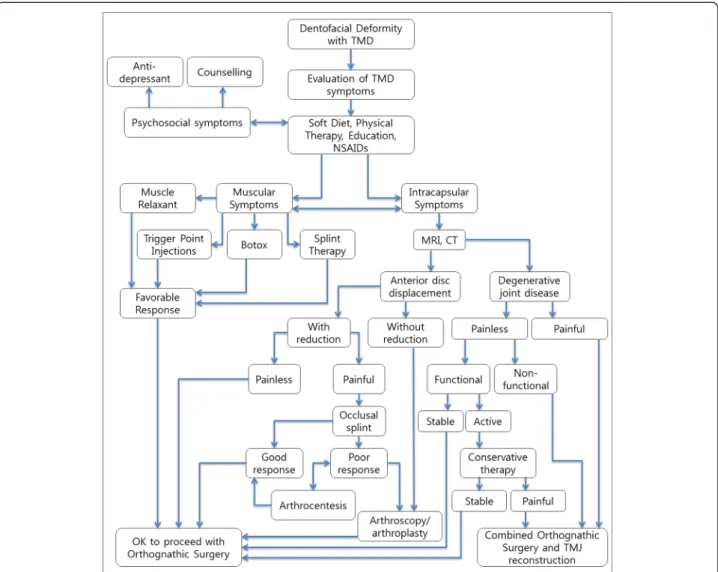
Once a correct diagnosis is made from detailed clinical data, initial treatment must be started with reversible op- tions including patient education, medications, physical therapy, and occlusal splint therapy. Minimally invasive options (eg, trigger point injections, Botox injections, arthrocentesis, or arthroscopy) are available for TMJ pain and dysfunction, and also open arthroplasty can be per- formed as later options when indicated (Table 3).
Most TMD symptoms (approximately 85-90%) are treated with noninvasive, nonsurgical, and reversible inter- ventions [13-15]. Patients with intra-articular disorder who has been refractory to nonsurgical treatment over 3 to 6 months with persistent pain and limited function would require a consideration for surgical interventions (Figure 4).
Influences of temporomandibular joint disorder to dentofacial deformities
Adaptive developmental changes on mandibular con- dyles [16] and post-developmental degenerative changes of mandibular condyles can create alteration on facial skeleton and occlusion [17,18]. Also, trauma or develop- mental deformity causing the changes in morphology and occlusion, can alter biomechanics of TMJ, conse- quently develop into TMJ internal derangement (TMD ID) [19].
Skeletal class II malocclusion in children has higher propensity for TMD symptoms [20]. Also, skeletal class II malocclusion, longer posterior facial height, and hyperdivergent profile tend to show increased severity of TMJ ID [21]. In a retrospective study including children younger than 14, the presence of TMD affects a normal development of facial bones, and can result in mandibu- lar asymmetry [18]. And Legrell PE and Isberg A also demonstrated a development of mandibular asymmetry from the condyle-disc complex disorder after surgically altering unilateral TMJ disc in animal study [22]. Thus, TMD manifesting in a variety of symptoms, also can apply abnormal stress to mandibular condyles and affect its growth pattern of mandible [23].
Table 1 Physical examination directed toward mandibular dysfunction
Examination Observations
Inspection Facial asymmetry, swelling, and masseter and temporal muscle hypertrophy Opening pattern (corrected and uncorrected deviations, uncoordinated movements, limitations)
Assessment of range of mandibular movement
Maximum opening with comfort, with pain, and with clinician assistance Maximum lateral and protrusive movements
Palpation examination Masticatory muscles Temporomandibular joints Neck muscles and accessory muscles of the jaw Parotid and submandibular areas Lymph nodes
Provocation tests Static pain test (mandibular resistance against pressure) Pain in the joints or muscles with tooth clenching Reproduction of symptoms with chewing (wax, sugarless gum)
Intraoral examination Signs of parafunction (cheek or lip biting, accentuated linea alba, scalloped tongue borders, occlusal wear, tooth mobility, generalized sensitivity to percussion, thermal testing, multiple fractures of enamel, restorations) From De Rossi S, Stern I, Sollecito TP. Disorders of the masticatory muscles. Dental clinics of North America 2013;57:449–64; and Data from references [4,6,111-115].
Figure 2 CT scan images of TMJ. Patients with history of trauma shows ankylosed TMJ on CT scan image, and 3D reconstruction demonstrates
the overall shape of TMJ.
Patients with bilateral TMJ ID tend to have a short ramus, clockwise rotation of mandible, and retrognathic mandible [24-27], while patients with unilateral TMJ ID present lateral displacement of mandible and deviated occlusal and mandibular plane [19]. The severity of TMJ ID is also associated with amount of displacement of Antegonion and Menton [28,29], and the growth pattern can be altered in maxilla as well as mandible [30]. Uni- lateral TMJ ID displays a deviation of mention to the af- fected side [28,29,31]. And reverse examination study also demonstrated positive correlations between short ramus and condyle as well as deviated mention being as- sociated with TMJ disc displacement and derangement [32,33]. Thus, the severity of TMD and disc displace- ment can lead to mandibular hypoplasia or facial asym- metry [18]. Also, degenerative changes and resorption of manduibular condyle beyond growth completion can lead to changes in skeletal shape [34].
Influence of orthognathic surgery to temporomandibular joint
Many DFD patients desire to improve stomatognathic function and esthetics, as well as TMJ symptoms [35].
However, current literatures on the relationship between OGS and TMJ complications are still debatable [36].
Some authors claim that TMJ dysfunction can be
improved after OGS, yet others claim deleterious effects on TMJ can occur after OGS [37,38].
Routine OGS procedure involves surgical movement of upper jaw via LeFort I osteotomy and lower jaw via ramus osteotomy. LeFort I osteotomy is not associated with direct trauma to TMJ or masticatory musculature, thus there are only minimal effects on TMJ dysfunction or mandibular movement [39]. Therefore, This review article focused on mandibular surgical modalities which directly affect the mandibular range of motion, mastica- tion, and TMJ symptom changes.
Sagittal split ramus osteotomy (SSRO)
SSRO is well known and very commonly used surgical technique worldwide for repositioning mandibular den- tal arch in both directions by advancement and setback movement of mandibular body [40]. SSRO provide a broad medullary contact between the bony segments that ensures stable healing capability. Internal fixation of bony segments eliminates or reduces the duration of intermaxillary fixation (IMF), plus a predictable immedi- ate postoperative occlusion is achievable. The risk of neurovascular bundle injury is higher compared to intraoral vertical ramus osteotomy (IVRO) [41], and the risk of unfavorable fracture during the split between the bony segments was reported at 0.9% [42]. The risk of
Figure 3 MRI Sagittal view showing disc displacement without reduction; A. Closed mouth; B. Open mouth.
Table 2 AAOP diagnostic classification of TMDs Diagnostic Category Diagnoses
Cranial bones (including the mandible)
Congenital and developmental disorders: aplasia, hypoplasia, hyperplasia, dysplasia (eg, first and second branchial arch anomalies, hemifacial microsomia, Pierre Robin syndrome, Treacher Collins syndrome, condylar hyperplasia, prognathism, fibrous dysplasia) Acquired disorders (neoplasia, fracture)
TMJ disorders Deviation in form Disc displacement (with reduction; without reduction) Dislocation Inflammatory conditions (synovitis, capsulitis) Arthritides (osteoarthritis, osteoarthrosis, polyarthritides) Ankylosis (fibrous, bony) Neoplasia Masticatory muscle disorders Myofascial pain Myositis spasm Protective splinting Contracture
Adapted from Leeuw Rd, Klasser GD, American Academy of Orofacial P. Orofacial pain : guidelines for assessment, diagnosis, and management. 5th edition.
Chicago: Quintessence Publishing; 2013.
complications is reduced when experienced surgeons perform the procedure. Reproducing the original con- dylar position is difficult, and too much pressure can be placed against the articular disc or unfavorable condylar position can be created during SSRO. These conditions can potentially result in joint noise or pain, and can worsen any pre-existing TMD symptoms [43-47].
In association with mandibular setback surgery using SSRO, Ueki et al. [48] reported TMJ symptom relief in 66.7% of patients after SSRO, and Hu J et al. [49] re- ported symptom improvement in 40% of patients, yet a development of new TMJ symptom in 8% after SSRO.
Kerstens et al. [50] reported 66% improvement of TMJ symptoms and 11.5% aggravation of symptoms while White and Dolwick [51] showed 89.1% improvement, 2.7% no changes, and 8.1% aggravation in TMJ symp- toms. Although small degree of postoperative posterior or lateral displacement of condyle can be made following SSRO in class III patients, however these minor changes do not create significant changes in TMJ disc position or postoperative pain [35,52-54].
TMJ remodeling is divided into functional and dys- functional remodeling. Dysfunctional remodeling has a significant alteration of the joint or occlusion and can cause reduction of condylar-ramus height, man- dibular setback leading to class II malocclusion [55,56]. Dysfunctional remodeling is also known as condylar resorption which can be induced from sys- temic and local arthritis or trauma. Because a clear etiology is not present, it is categorized as idiopathic condylar resorption (ICR).
Maxillomandibular complex counterclockwise rota- tion via LeFort I osteotomy and SSRO can increase the mechanical loading of TMJ, and can lead to post- operative relapse [57]. Patients with systemic diseases such as rheumatoid arthritis, scleroderma, systemic lupus erythematosus, and other vascular collagenous diseases are known as high risk factors for condylar resorption [58-61]. Predisposing factors for ICR are the presence of TMJ dysfunction, young woman, high mandibular plane angle, and posteriorly inclined con- dylar neck [59,61-66].
A large amount of mandibular advancement via SSRO should be avoided to prevent condylar resopr- tion occurring from the tension of stretched sur- rounding soft-tissue components [67-69]. Internal fixation with monocortical miniplates and screws (1.5- 8.9%) showed more favorable response to condylar resoprtion than using bicortical screws (2–50.3%) dur- ing SSRO [70]. This is likely due to a torques being created on condyles from the proximal segment dis- placement during bicortical fixation [71]. The use of computer-aided design/computer-aided manufactur- ing-made condyle positioning jig has been suggested to minimize a significant condylar displacement or torque [72].
The postoperative relapse of open bite from condylar resorption usually occurs between 6 months to 3 year, thus a regular follow up is important to intervene early in the process [73]. Anti-inflammatory medication, tumor necrosis factor inhibitor, or matrix metallopro- teinase inhibitor as pharmacotherapy [74], or an utilization of occlusal splints to reduce the joint load- ing [75] can help prevent resorption process. A total joint replacement option is also available if further ac- tive resorption process continues.
Zimmer et al. [76] reported that two-jaw surgery (maxillary advancement and mandibular setback sur- gery) had no influcence on mandibular mobility com- pared to a single-jaw surgery. Mandibular hypomobility is a common condition after mandibular advancement via SSRO especially with a prolonged IMF duration [77,78], degenerative changes during the periods of IMF [79], masticatory muscle unused atrophy, and de- crease in muscle energy reserves due to immobilization [80,81]. Atrophy of human skeletal muscles and a de- crease in strength and muscle energy reserves have also been associated with immobilization [80]. Aragon et al.
recommended a sound postoperative rehabilitation pro- gram following orthognathic procedures to prevent hypomobility [82].
Intraoral vertical ramus osteotomy (IVRO)
IVRO is one of mandibular osteotomy techniques com- monly used for mandibular setback procedure [83-86]. A Table 3 Treatment of TMD symptoms in patients with
dentofacial deformity
Reversible treatment of TMD Irreversible treatment of TMD
Patient Education Trigger Point Injections
Medications: Botox
Nonsteroidal anti-inflammatory drugs (anti-RA meds)
Arthrocentesis
Muscle relaxants Arthroscopy
Antidepressants Open Arthroplasty
Physical Therapy ROM exercised Passive stretching Spray and stretch Ultrasound
Transcutaneous electrical nerve stimulation
Occlusal Splint Therapy
From Nale JC. Orthognathic Surgery and the Temporomandibular Joint Patient.
Oral and maxillofacial surgery clinics of North America 2014;26:551 –64 [ 116].
vertical osteotomy is made posterior to lingula, and proximal segment is placed lateral to distal segment without internal rigid fixation. It is relatively simple pro- cedure and surgical time is much reduced compared to SSRO. Also, the risk of inferior alveolar nerve damage and neurologic deficit is lower [87,88]. Less than 1 mm of posterior relapse can occur after mandibular setback via IVRO, but the risk of further relapse is low and over- correction is not commonly indicated [89]. Increased in transverse facial width from laterally positioned proximal segment is less than 1% due to continuing remodeling process [90].
A drawback of IVRO is the requirement of IMF since internal rigid fixation is not performed, and some clinicians recommend more than 4 weeks of IMF postoperatively [91-94]. However, active physical therapy with less than 2 weeks of IMF demonstrated a stable occlusion and good bone healing [95], (Table 4) Some study reported just one-
day of IMF followed by early jaw exercise being sufficient [96]. When initial bite is unstable, an active physical ther- apy with close follow-ups, and re-IMF protocol is used to obtain improved occlusion, and 88% of patients achieve a stable occlusion after IVRO within 10 days of active phys- ical therapy and Maximum mouth opening (MMO) more than 30 mm [95].
IVRO requires a wide dissection of lateral aspect of ramus and medial aspect of proximal segment for muscle detachment. Freed proximal segment initially moves anterio-inferiorly and reduces the pressures on articular disc by physiologic equilibrium position and better condyle-disc relation [97]. Anterio-inferiorly moved condyle eventually returns back to its original position over time postoperatively [98].
Improvement of joint sound, pain, and other TMJ symptoms after IVRO is likely due to resting of TMJ and surrounding musculatures during IMF period as well as
Figure 4 Flow diagram and treatment algorithm for patients with dentofacial deformity and TMD symptoms. Adopted from Nale, J.C. Orthognathic
Surgery and the Temporomandibular Joint Patient. Oral and maxillofacial surgery clinics of North America 2014;26:551 –64 [116].
the condylotomy effects from anterio-inferior movement of condyle [39,88,97-100]. IVRO was reported to have 50-100% improvement of TMJ symptoms [101-104], and Jung et al. [105] reported 70.8% ~ 94.3% improvement of joint sounds and 89.4% improvement of TMJ pain after IVRO. Ueki et al. [48] reported TMJ symptom improve- ment in class III patients 88% after IVRO and 66.7%
after SSRO, but MRI study showed 50% of improvement of anteriorly displaced disc after IVRO and no improve- ment after SSRO.
Horizontal condylar axis tend to be medially rotated when TMJ disc displacement or degenerative joint dis- ease is present, and some authors suggested that medi- ally rotated condyle is the etiological factor for TMD [106,107]. From this point of view, lateral rotation of condyle after IVRO is very effective improving TMJ symptoms. Choi et al. [108] evaluated 200 patients'
postoperative changes in proximal segment and condyles on the transverse plane after IVRO using submentover- tex cephalogram. This study reported 15.05 (SD: 8.97)°
of postoperative lateral rotation of condyles which slowly returned towards the original position, yet 4.53 (SD:
6.03)° of lateral rotation remained at 1 year. This study included only class III malocclusion patients with low TMD prevalence and some patients were without known TMD. The condyles remained in laterally rotated pos- ition in all patients including the ones without known prior TMDs. Thus, laterally rotated condyles from IVRO improving TMD cannot be concluded from this study.
Condylar sagging in lateral or anterio-inferior direc- tion can occur after IVRO. Condylar sagging can be avoided with careful dissection during the ramus osteot- omy and not violating condylar capsules. In fact, the changes in the intercondylar distance on transverse plane after IVRO is not significant [108]. Excessive inter- ference between the segments can induce sagging, thus reduction of bony interference or using a modified oste- otomy design should be considered [109].
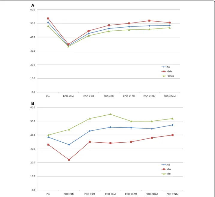
In order to prevent post OGS mandibular hypomobi- lity, implementation of a sound postoperative rehabilita- tion program is very important [82]. The incidence of mandibular hypomobility after IVRO is very low and re- covery of MMO is known to be 90-98% of pre-operative opening. Aragon et al. [82] showed 90% of recovery in 13 patients, Storum and Bell [110] showed 98% of recov- ery on 24 patients, Boyd et al. [39] showed 98% of recov- ery in 9 patients, and Jung et al. [95] reported 91.3% of recovery in 187 patients within 6 month and 95.7% re- covery at 24 month after the procedure (Figure 5A). Pa- tients with MMO less than 40 mm showed 112.5 to 123.2% recovery after IVRO procedure (Figure 5B).
Conclusion
The changes of facial skeleton in DFD patients after OGS have an impact on TMJ, masticatory musculature, and surrounding soft tissues. Patients with TMJ symp- toms requires a thorough evaluation including history taking, focused physical examination, and imaging mo- dalities such as CT or MRI as indicated in order to ob- tain correct diagnoses and treatments prior to OGS.
These evaluations are performed pre, intra, and post- operatively to determine the status of TMJ condition and managed appropriately. The changes of TMJ symp- toms after OGS are associated with multiple factors in- cluding masticatory and facial musculature, and improvement in disc-condyle relationship as well as psy- chological factor. Any surgical procedures involving mandibular osteotomy can directly affect TMD, thus, pre-existing TMJ symptoms and diagnoses should be considered prior to treatment planning and OGS.
Table 4 Active Physical Therapy instruction form for patients
Instructions for active physical therapy (Yonsei Protocol) It has been about 2 weeks after undergoing your jaw surgery. The purpose of this active physical therapy is to help your facial musculatures and jaws adapt into a new position from the surgery.
Please follow the instructions in order to recover your original jaw movement and stable result.
1. Open your mouth as big as possible : Repeat 3 times
A. During the opening, check the lower incisal midline and do not allow laterally
B. deviated movement.
C. Close your mouth and lower tooth must be positioned into the splint without gap. If lower teeth are not positioned into the splint, try to close gap by pushing the jaw with your hands.
2. Move your lower jaw anteriorly : Repeat 3 times. From the original position, move your lower jaw anteriorly and move back to its original position. Check the midline of the lower teeth and do not allow laterally deviated movement.
3. Move your lower jaw to the left side : Repeat 3 times.
4. Move your lower jaw to the right side : Repeat 3 times.
5. Above instruction is 1 cycle. Please follow the instruction in order.
6. You have to repeat above physical therapy protocol for 1 hour.
7. Then, you have to fix the lower jaw to upper jaw for 2 hours.
8. During the physical therapy, training elastics must be kept in the instructed site.
9. Please avoid relatively hard food and be careful not to break the splint.
The splint is removed after 1 to 2 weeks of physical therapy, depending on the progess. It is not easy, but please be patient until finishing the physical therapy. This physical therapy is continued about 1 month and this therapy makes stable functional results.
Department of Oral & Maxillofacial Surgery Dental Hospital, Yonsei Medical Center
Adopted from Jung HD, Jung YS, Park JH, Park HS. Recovery pattern of
mandibular movement by active physical therapy after bilateral transoral
vertical ramus osteotomy. J Oral Maxillofac Surg 2012;70:e431-7.
Competing interest
The authors declare that they have no competing interests.
Authors ’ contribution
Conception and design of study/review/case series: HDJ, YSJ Acquisition of data: laboratory or clinical/literature search: SYK, HSP Analysis and interpretation of data collected: HDJ, SYK, HSP Drafting of article and/or critical revision:
HDJ, SYK Final approval and guarantor of manuscript: HDJ, YSJ. All authors read and approved the final manuscript.
Acknowledgements
This article was supported by a faculty research grant of Yonsei University College of Dentistry for 2014 (6-2014-0101).
Author details
1