• 검색 결과가 없습니다.

Analysis of Keratocystic Odontogenic Tumor

N/A
N/A
Protected

Academic year: 2021

Share "Analysis of Keratocystic Odontogenic Tumor"

Copied!
5
0
0

로드 중.... (전체 텍스트 보기)

전체 글

(1)

1956년 Phillipsen에 의해 치성 각화낭(odontogenic keratocyst)으로 소개된 이후,

1)

국제보건기구(World Health Organization)에 의해 현재는 각화낭성 치성종양 (keratocystic odontogenic tumor; KCOT)으로 재분류되 었다. KCOT는 양성의 단방성 또는 다방성으로 나타나는 치성 기원의 골내종양으로서, 착각화된(parakeratinized) 중층 편평상피로 이장되어 있고 공격적이고 침습적인 성향 을 띄는 종양의 특징을 반영하여 WHO는 KCOT로 명명할 것을 추천하였으며 이 중 조직학적으로 정각화된(ortho- keratinized) 경우는 KCOT에 포함하지 않는다고 하였 다.

2)

이는 악골의 어느 부위에서도 발생할 수 있으나 대부

분은 하악의 후방부와 상행지부에 호발한다.

3)

방사선학적으 로 각화낭성 치성종양은 경계가 명확한 방사선 투과성 병소 로 나타나거나 부드럽고 치밀화된 경계를 갖는 단방성 혹은 다방성의 방사선투과성 병소로 나타나며, 약 25%에서 40%에서 병소에 미맹출 치아와 연관이 있다고 보고되고 있다. 조직학적으로 각화낭성 치성종양은 6-10층 정도의 세포 두께를 가지는 비교적 일정한 두께의 부전 각화 편평 상피, 원주 또는 입방형 기저 세포의 책상배열, 내강의 주름 진 부전 각화층, 상피돌기의 소실 등의 특징을 가지고 있 다.

4,5)

각화낭성 치성종양은 인접 조직의 침입 등의 공격적인 생 물학적 경향으로 인해 치성낭에서 낭성 종양으로 재분류되 었으며 재발률도 높아 임상적으로도 특이한 형태로 평가되 임형섭∙김수관∙문성용∙오지수∙문경남∙윤정훈

1

조선대학교 치의학전문대학원 구강악안면외과학교실,

1

구강병리학교실

각화낭성 치성종양에 관한 분석

Analysis of Keratocystic Odontogenic Tumor

Hyoung-Sup Lim, Su-Gwan Kim, Seong-Yong Moon, Ji-Su Oh, Kyung-Nam Moon, Jeong-Hoon Yoon

1

Department of Oral and Maxillofacial Surgery,

1

Department of Oral Pathology, School of Dentistry,

Chosun University, Gwangju, Korea

We reviewed 30 cases of keratocystic odontogenic tumor (KCOT) managed during the 8-year period between 2001 and 2008. This case report described the clinical, radiographic and histopathologic features of these KCOT. Of the 30 patients in whom KCOTs were diagnosed, 18 (60%) of the patients were male and 12 (40%) were female. The mean age of these patients was 34.2 years, with peak incidence occurring in the third decade of life. The lesions were mostly located in the mandible (74.2%) and in the maxilla (25.8%). There was a marked predilection to occur in the posterior mandible. Radiographically, 19 (63%) out of the 30 cases were unilocular type with a well demarcated border, while 11 cases (37%) were multi- locular in appearance. Histopathologically, 73% of the cysts were lined with parakeratinized stratified squamous epithelium, while only 3% of the cysts were lined with mixed parakeratinized of orthokeratinized epithelium. 22 cases (73%) contained keratin in the lumen. A satellite cyst was observed in 14 cases (47%). All cysts were treated by enucleation. The recurrence rate was shown as 10% for 3 patients with a follow up period and recurred lesions were treated by re-enucleation.

Key words: Odontogenic keratocyst, Keratocystic odontogenic tumor, Keratin, Satellite cyst, Enucleation

Abstract

(2)

고 있다.

2,5-8)

각화낭성 치성종양의 치료법으로는 조대술, 적 출술 등의 시술이 행해졌으며, 냉동요법 혹은 Carnoy 용 액, 변연절제술, radical resection 등도 함께 시행되기도

하였다.

7-14)

치성 각화낭의 재발률에 대해서는 여러 임상 연

구들에서 20%에서 62%까지 다양하게 보고되었다.

3,4,7,9,15)

이 연구는 저자 등이 경험한 각화낭성 치성종양의 증례를 통해 임상적, 방사선학적, 조직병리학적인 형태에 대한 분 류 및 평가를 위해 시행하였다.

연구 대상 및 방법

본 연구는 2001년부터 2008년까지 조선대학교 치과병원 구강악안면외과에서 치성 각화낭 및 각화낭성 치성종양으 로 진단되었던 30명의 환자를 대상으로 시행되었다.

임상기록지 및 방사선 사진을 근거로 환자의 나이, 성별, 발생 부위에 대해 세분화하여 기록하였으며, 방사선학적으 로 병소의 크기, 매복치의 포함여부를 평가하고 조직학적 분류를 시행하였다. 조직학적 기준에 따라 진단되었으며 6- 10층 정도의 세포 두께를 가지는 비교적 일정한 두께의 부 전 각화 편평상피, 원주 또는 입방형 기저 세포의 책상배열, 내강의 주름진 부전 각화층, 상피돌기의 소실을 기준으로 하였다.

16)

또한, WHO의 재분류에 따라 정각화된 경우는 제외하였으며 착각화된 각화낭성 치성종양에 한하여 평가 하였다.

연구 결과

1. Clinical findings

30명의 연구 대상은 성별에 따라 18명의 남성(60%)과 12명의 여성(40%)으로 분류되었고, 남성과 여성의 비율은 1.5 : 1로 나타났다. 연구 대상의 나이의 분포는 12세부터 83세까지 다양하였고, 평균 나이는 34.2세로 나타났다 (Table 1).

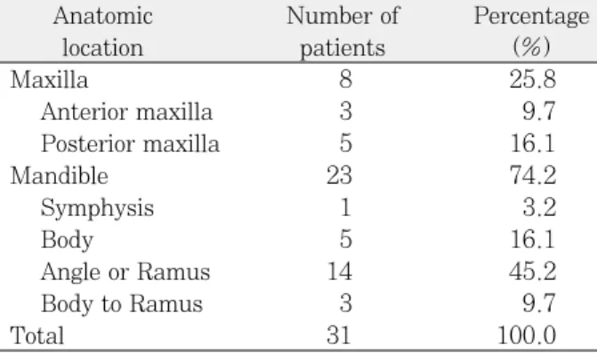
30명에서 31부위가 각화낭성 치성종양으로 진단되었으 며 이 중 23예(74.2%)가 하악에서 발생하였고, 8예 (25.8%)가 상악에 발생하여 하악과 상악에 따른 비율은 2.9 : 1로, 하악에서 3배정도 호발하였다. 이 중 하악 전방 부에서 1예, 하악 골체부 단독발생이 5예였으며 가장 호발 한 부위는 17예가 발생한 하악각 및 하악지 부위였다. 또한 이 중 하악체에서 하악지까지 이어진 큰 병소는 3예에서 나 타났다. 상악의 경우, 전방부에 3예, 후방부에 5예가 나타 났으며(Table 2). 매복된 치아가 병소와 연관된 경우가 8 예(25.8%)였으며, 연관되지 않은 경우가 22예로 나타났 고, 이중 하악에서 6예, 상악에서 2예로 조사되었다(Table 3). 병소와 관련된 매복치는 하악에서는 제3대구치가 5예,

제2대구치와 제3대구치가 모두 매복되어 있었던 예가 1예 였으며 상악에서는 2예 모두 매복 제3대구치와 관련이 되 어 있었다.

2. Radiographic findings

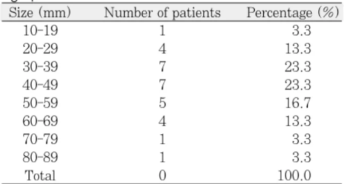
방사선학적으로 19예에서 경계가 명확한 단방성 낭의 형 태로 나타났으며, 12예에서 다방성 낭의 형태를 띄었다 (Fig. 1). 병소의 크기는 파노라마 방사선 사진에서 가장 긴 직경을 측정하여 결정하였다. 병소의 크기는 19 mm에서 85 mm까지 다양하게 측정되었고 평균 크기는 파노라마 상 에서 45.2 mm였다(Table 4).

Table 3. Association with an impacted tooth

Number of Percentage patients (%) Involved of impacted tooth 8 26.7

maxilla 2 6.7

mandible 6 20.0

Non-involved of impacted tooth 22 73.3

Total 30 100.0

Table 1. Distribution by age

Age (years) Number of patients Percentage (%)

10-19 10 33.3

20-29 6 20.0

30-39 2 6.7

40-49 5 16.7

50-59 3 10.0

60-69 3 10.0

70- 1 3.3

Total 30 100.0

Table 2. Distribution by anatomic locations

Anatomic Number of Percentage

location patients (%)

Maxilla 8 25.8

Anterior maxilla 3 9.7

Posterior maxilla 5 16.1

Mandible 23 74.2

Symphysis 1 3.2

Body 5 16.1

Angle or Ramus 14 45.2

Body to Ramus 3 9.7

Total 31 100.0

(3)

3. Histopathologic findings

22예에서 부전 각화 양상을 보였고, 8예에서는 부전 각화 와 정각화가 혼합되어 있는 양상을 보였다. 14예에서 한 개 이상의 딸낭이 관찰되었다. 또한, 낭의 내강에 부전각질 등

의 케라틴(keratin)이 함유된 경우가 22예에서 관찰되었다 (Fig. 2, 3).

4. Treatment and Recurrence

환자에 대한 치료는 모든 증례에서 외과적 적출술(enu- cleation)을 시행하였으며, 3명에서 재발되어 재적출술을 시행하였다.

본 연구에서 각화낭성 치성종양을 가진 환자의 나이와 성 별은 이전의 다른 연구들과 비교하여 비슷한 통계학적 수치 를 보였다.

4,17,18)

다소 넓은 범위의 나이 분포를 가졌으며, 평 균 나이는 34.2세였다.

남성 대 여성의 비율은 남성에서 다소 호발하는 경향이 있 다는 보고가 있으며

7,19)

남녀 성비율에 대한 차이가 없다고 보고되기도 했으나

20)

본 연구에서는 1.5 : 1로서 남성이 50%정도 더 많은 수치를 보였다.

호발 부위는 하악체 후방에서 상행지로 이어지는 하악후 방부에 호발한다고 보고되고 있으며,

18,21-23)

본 연구에서도 71%에서 하악 후방부에 발생한 것으로 조사되었다.

각화낭성 치성종양의 치료법은 낭 자체의 공격적인 성향

2,24)

20-62%로 다양하게 보고되어지고 있는 높은 재발률

4,7,9,15)

인해 현재까지 많은 논란이 있다. 본 연구에서는 모

든 증례에서 외과적 적출술을 시행하였으며 외과적 적출술 은 각화낭성 치성종양의 제거에 일반적인 치료법으로 이용 되고 있지만,

18,25,26)

적출술이 재발률을 높여 각화낭성 치성 종양의 치료에는 적절하지 않다는 보고도 많다.

9,10,15,27,28)

Fig. 1. Well-defined multilocular radiolucent lesion at right mandible seen in the panoramic radiograph.

Fig. 2. Photomicrograph showing uniform thickness of sur- face parakeratotic epithelial lining with a hyperchromatic and palisaded basal cell layer (X 200, H-E).

Fig. 3. Photomicrograph showing a daughter cyst filled with keratin in the cyst wall composed of collagenous connec- tive tissue (X 200, H-E).

Table 4. Distribution by size in the panoramic radi- ograph

Size (mm) Number of patients Percentage (%)

10-19 1 3.3

20-29 4 13.3

30-39 7 23.3

40-49 7 23.3

50-59 5 16.7

60-69 4 13.3

70-79 1 3.3

80-89 1 3.3

Total 0 100.0

(4)

한 재발률을 감소시키기 위해 적출술과 더불어 liquid nitrogen이나 Carnoy's solution의 이용과 주변골의 골절 제술을 시행하여 왔다.

28,29)

그러나 최근의 Stoelinga의 보고 에 의하면 주변골의 골절제술은 오히려 재발률을 높일 수 있다고 하였다.

29)

또한, 최근에 상피도(epithelial island) 나 microcyst의 대부분이 주변골보다는 낭을 덮고 있는 점 막에 있다는 것이 밝혀지면서 각화낭성 치성종양의 치료방 법으로 적출술과 더불어 Carnoy's solution으로 골결손부 를 처치하고 골천공이 일어난 경우에는 주변의 점막까지 Carnoy's solution으로 처치할 것을 추천하였다.

28,29)

근치 적 수술은 법랑아세포종이나 암종으로의 변화가 있거나 보 존적인 치료가 불가능한 반복된 재발일 경우에 한해서 시행 해야 한다고 하였다.

18)

각화낭성 치성종양은 높은 재발률을 보이며 재발은 수술 후 5년 이내가 많았다고 보고되고 있으며,

9,28,29)

어떠한 임상 적 증상이 없이 재발되는 경우가 많아 초기 5년 동안은 매 년 추적검사를 시행할 것을 추천하고 있다.

30)

또한 수술 10 년 후에 재발한 증례도 많이 보고되고 있어 각화낭성 치성 종양으로 진단된 경우 오랜 기간의 추적검사가 요할 것으로 생각된다.

2,31,32)

본 연구는 2001년부터 2008년까지 조선대학교 치과병원 구강악안면외과에서 치성각화낭 및 각화낭성 치성종양으로 진단받았던 30명의 환자를 대상으로 임상적, 방사선학적, 조직병리학적인 평가를 시행하였다.

1. 남성과 여성의 비율은 1.5 : 1이었으며 연령은 12-83 세로 평균 34.2세였다.

2. 31부위에서 하악이 23예(74.2%), 상악이 8예 (25.8%)였으며 2.9 : 1로 하악에서 호발하였다.

3. 17예가 발생한 하악각 및 하악지 부위가 가장 호발한 부위였다.

4. 매복치와 관련된 병소는 8예(25.8%)로 상악에서 2예, 하악에서 6예였으며 하악2대구치를 포함한 1예를 포 함하여 모든 예에서 매복 제3대구치와 관련이 있었다.

5. 19예에서 단방성의 형태로 나타났으며, 12예에서 다 방성의 형태를 보였다. 병소의 크기는 파노라마상에서 직경 19-85 mm로 평균직경은 45.2 mm였다.

6. 22예서 부전 각화 양상을 보였고, 14예에서 한 개 이 상의 딸낭이 관찰되었다. 케라틴을 함유한 경우가 22 예에서 관찰되었다.

7. 모든 증례에서 적출술을 시행하였으며 3명(10%)이 재발되어 재적출술을 시행하였다.

References

1. Philipsen HP : On “keratocysts”in the jaws[in Danish].

Tandlaegebladet 60 : 963, 1956.

2. Barnes L, Eveson JW, Reichart P et al : Pathology and genetics of head and neck tumours. Lyon : IARC Press, 2005. WHO classification of tumours series.

3. Ahlfors E, Larsson A, Sjogren S : The odontogenic kerato- cyst : a benign cystic tumor? J Oral Maxillofac Surg 42 : 10, 1984.

4. Kakarabtza-Angelopoulopu E, Nicolatou O : Odontogenic keratocysts : clinicopathologic study of 87 cases. J Oral Maxillofac Surg 48 : 593, 1990.

5. Partridge M, Towers JF : The primordial cyst (odontogenic keratocyst) : its tumor-like characteristics and behavior.

Br J Oral Maxillofac Surg 25 : 271, 1987.

6. Shear M : Developmental odontogenic cyst. An update. J Oral Pathol Med 23 : 1, 1994.

7. El-Hajj G, Anneroth G : Odontogenic keratocysts - a ret- rospective clinical and histologic study. Int J Oral Maxillofac Surg 25 : 124, 1996.

8. Stoelinga PJ : Long-term follow-up on keratocysts treated according to a defined protocol. Int J Oral Maxillofac Surg 30 : 14, 2001.

9. Blanas N, Freund B, Schwartz M et al : Systemic review of the treatment and prognosis of the odontogenic kerato- cyst. Oral Surg Oral Med Oral Pathol Oral Radiol Endod 90 : 553, 2000.

10. Zhao YF, Wei JX, Wang SP : Treatment of odontogenic keratocysts : a follow up of 255 Chinese patients. Oral surg Oral Med Oral Pathol Oral Radiol Endod 94 : 151, 2002.

11. Nakamura N, Mitsuyasu T, Mitsuyasu Y : Marsupiali- zation for odontogenic keratocysts : long-term follow up analysis of the effect and changes in growth characteris- tics. Oral Surg Oral Med Oral Pathol Oral Radiol Endod 94 : 543, 2002.

12. Williams TP, Connor FA : Surgical management of large odontogenic keratocysts: Aggressive approach. J Oral Maxillofac Surg 52 : 964, 1994.

13. Pogrel MA : The use of liquid nitrogen cryotherapy in the management of locally aggressive bone lesion. J Oral Maxillofac Surg 51 : 269, 1993.

14. Schmidit BL, Pogrel MA : The use of enucleation and liq- uid nitrogen cryotherapy in the management of odonto- genic keratocysts. J Oral Maxillofac Surg 59 : 720, 2001.

15. Myoung H, Hong SP, Hong SD et al : Odontogenic kerato- cyst : review of 256 cases for recurrence and clinicopatho- logic parameters. Oral Surg Oral Med Oral Pathol Oral Radiol Endod 91 : 328, 2001.

16. Pindborg JJ, Philipsen HP, Henriksen J : Studies on odontogenic cyst epithelium. American Association for the Advancement of Science, Washington (DC) 151, 1962.

17. Forssel K, Forssel H, Kahnberg KE : Recurrence of kera- tocysts in a long-term follow-up study. Int J Oral Maxillofac Surg 17 : 25, 1988.

18. Chow HT : Odontogenic keratocyst: a clinical experience in Singapore. Oral Surg Oral Med Oral Pathol Oral Radiol Endod 86 : 573, 1998.

19. Kondell PA, Wiberg J : Odontogenic keratocyst. A follow- up study of 29 cases. Swed Dent J 12 : 57, 1988.

20. Magnusson BC : Odontogenic keratocysts: a clinical and

histological study with special reference to enzyme histo-

chemistry. J Oral Pathol 7 : 8, 1978.

(5)

21. Meara JG, Shah S, Li KK et al : The odontogenic kerato- cyst : a 20-year clinicopathologic review. Laryngoscope 108 : 280, 1998.

22. Zachariades N, Papanicolaou S, Triantafyllou D : Odontogenic keratocysts : review of the literature and report of sixteen cases. J Oral Maxillofac Surg 43 : 177, 1985.

23. Keith DA : Macroscopic satellite cyst formation in the odontogenic keratocyst. Report of two cases. Oral Surg Oral Med Oral Pathol 35 : 21, 1973.

24. Jackson IT, Potparic Z, Fasching M et al : Penetration of the skull base by dissecting keratocyst. J Oral Maxillofac Surg 21 : 319, 1993.

25. Irvine GH, Bowerman JE : Mandibular keratocysts; surgi- cal management. Br J Oral Maxillofac Surg 23 : 204, 1985.

26. Eyre J, Zakrzewska JA : The conservative management of large odontogenic keratocysts. Br J Oral Maxillofac Surg 23 : 195, 1985.

27. Chirapathomsakul D, Sastravaha P, Jansisyanont P : A review of odontogenic keratocysts and the behavior of recurrences. Oral Surg Oral Med Oral Pathol Oral Radiol Endod 101 : 5, 2006.

28. Tolstunov L, Treasure T : Surgical treatment algorithm for

odontogenic keratocyst : combined treatment of odonto- genic keratocyst and mandibular defect with marsupializa- tion, enucleation, iliac crest bone graft, and dental implants. J Oral Maxillofac Surg 66 : 102, 2008.

29. Stoelinga PJ : The treatment of odontogenic keratocysts by excision of the overlying, attached mucosa, enucleation, and treatment of the bony defect with Carnoy solution. J Oral Maxillofac Surg 63 : 1662, 2005.

30. Pitak-Arnnop P, Chaine A, Oprean N et al : Management of odontogenic keratocysts of the jaws : A ten-year experi- ence with 120 consecutive lesions. J Craniomaxillofac Surg 38 : 358, 2010.

31. Oikarnen VJ : Keratocyst recurrences at intervals of more than 10 years : case reports. Br J Oral Maxillofac Surg 28 : 47, 1990.

32. Crowley TE, Kaugars GE, Gunsolley JL : Odontogenic keratocysts : a clinical and histologic comparison of the parakeratin and orthokeratin variants. J Oral Maxillofac Surg 50 : 22, 1992.

33. Meara JG, Li KK, Shah SS et al : Odontogenic keratocysts in the pediatric population. Arch Otolaryngol Head Neck Surg 122 : 725, 1996.

34. Chuong R, Donoff RB, Guralnick W : The odontogenic ker- atocyst. J Oral Maxillofac Surg 40 : 797, 1982.

저자 연락처

우편번호 501-759

광주광역시 동구 서석동 375번지

조선대학교 치과대학 구강악안면외과학교실 김 수 관

원고 접수일 2010년 03월 22일 게재 확정일 2010년 06월 30일

Reprint Requests Su-Gwan Kim

Department of Oral and Maxillofacial Surgery, School of Dentistry, Chosun University

375, Seosuk-dong, Dong-gu, Gwangju, 501-759, Korea Tel: +82-62-220-3815 Fax: +82-62-228-7316 E-mail: [email protected]

Paper received 22 March 2010

Paper accepted 30 June 2010

수치

Table 1. Distribution by age
Fig. 2. Photomicrograph showing uniform thickness of sur- sur-face  parakeratotic  epithelial  lining  with  a  hyperchromatic and palisaded basal cell layer (X 200, H-E).

참조

관련 문서

Key words: False aneurysm, Inferior alveolar artery, Angiography, Therapeutic embolization, Keratocystic odontogenic tumor.. 서

두 번째 물질흐름분석 (material flow analysis) 은 초기에는 전세계 적 단위 , 혹은 국가적 단위로 물질의 흐름에 대한 데이터를 수집하 고 이를 계산함으로써

Epidermal inclusion cysts are formed by inclusion of keratinizing squamous epithelium within the dermis, resulting in a cyst filled with lamellated keratin.. These benign cysts

Adenomatoid odontogenic tumor (AOT) usually presents as a unilocular, pericoronal radiolucency in the maxillary anterior region in adolescent females. Very few conditions occur in

Philipsen HP, Srisuwan T, Reichart PA : Adenomatoid odon- togenic tumor mimicking a periapical (radicular) cyst: a case report.. Mendis BR, MacDonald DG : Adenomatoid

이외에도 거점과 수배송의 용량(Capacity)의 제약과 단일 조달 등의 고려여부, 수요가 공급거리에 반비례하 여 공급거리가 멀어지면 수요가 줄어든다는 것 등과 같이

In several studies, Ki-67 was expressed higher in the epithelium of KCOT than in the radicular cyst or oral mucosa, indicating that the epithelium of KCOT has higher

시스템 설계 인자에 대한 성능 변화를 분석 하기 위하여 냉․난방기( 1월 및 8 월) 를 평균 기상 조건을 기준으로 일사량,중공층 높이, 중공층 두께 등의