홍삼가수분해추출물의 db/db 마우스에서 신장 손상 예방효과
김찬식1#, 조규형1, 표미경2, 김진숙1, 김정현1,3*
1: 한국한의학연구원, 2: (재)금산국제인삼약초연구소, 3: 전북대학교 치과대학 구강병리학실
Preventive Effects of Pectin Lyase-Modified Red Ginseng Extract on renal injury in db/db mice
Chan-Sik Kim
1#, Kyuhyung Jo
1, Mi Kyung Pyo
2, Jin Sook Kim
1, Junghyun Kim
1,3*1: Korea Institute of Oriental Medicine, 2: International Ginseng & Herb Research Institute 3: Department of Oral Pathology, College of Dentistry, Chonbuk National University
ABSTRACT
Objectives : Diabetic nephropathy is one of the most significant chronic complications of diabetes. Advanced glycation end products (AGEs) have been implicated in the development of diabetic nephropathy. GS-E3D is an enzymatic modified red ginseng extract by pectin lyase and has an increased concentration of the ginsenoside Rd compared to an unmodified red ginseng extract. In this study, we evaluated the preventive effects of GS-E3D on renal dysfunction in the type 2 diabetic db/db mice.
Methods : GS-E3D (100 or 250 ㎎/㎏ body weight per day) was given to db/db mice through oral gavage for 6 weeks.
Body weight and blood glucose levels were examined. At the end of the experiment, albuminuria was measured. The renal tissues were collected for histological examination, and immunohistochemical staining was used to detect renal accumulation of AGEs and podocyte loss
Results : In the db/db mice, severe hyperglycemia developed, and albuminuria was significantly increased. Diabetes induced markedly morphological alterations to the renal glomerular cells. AGE accumulations and podocyte loss were detected in renal glomeruli. No difference in blood glucose levels was noted between GS-E3D-treated and vehicle- treated diabetic db/db mice. However, GS-E3D treatment significantly reduced albuminuria and AGE accumulations in diabetic mice. Moreover, the loss of podocytes was restored by GS-E3D treatment.
Conclusions : GS-E3D might be beneficial for the treatment of diabetic nephropathy. The ability of GS-E3D on to attenuate albuminuria and podocyte dysfunction in the db/db mice may be mediated by the inhibition of AGE accumulation.
1)
Key words : Diabetic nephropathy, GS-E3D, Red ginseng
Ⅰ. 서 론
당뇨병성 신증은 만성 당뇨환자들에서 흔히 나타나는 합병 증 중 하나이다
1). 만성 당뇨에 의한 지속적인 고혈당은 산화 스트레스를 야기하고 염증을 유발하여 신장손상을 유발 할 수
있다. 당뇨병성 신증의 임상적인 특징은 점진적인 신장 기능 저하 및 신장세포의 손상에 의한 알부민뇨의 발생이다
2). 신장 사구체에 존재하는 세포중 족세포(podocyte)는 신장의 여과 장벽(filtration barrier)을 구성하는 중요한 요소 중 하나로서, 족세포의 소실은 신장의 여과기능에 문제를 야기하여 알부민
*Corresponding author : Junghyun Kim, Department of Oral Pathology, School of Dentistry, Chonbuk National University, 567 Baekje-daero, Jeonju, Jeollabuk-do, 54896, South Korea.
·Tel : +82-63-270-4032 ·Fax : +82-63-270-4025 ·E-mail : [email protected]
#First author : Chan-Sik Kim, Korea Institute of Oriental Medicine, 1672 Yuseongdaero, Yuseong-gu, Daejeon 305-811, South Korea.
·Tel : +82-42-868-9473 ·Fax : +82-42-868-9471 ·E-mail : [email protected] ·Received : 09 April 2018 ·Revised : 21 May 2018 ·Accepted : 25 July 2018
뇨를 유발하게 되는 것이다
3). 당뇨병성 신증에서 족세포의 소 실은 가장 초기에 관찰할 수 있는 병변으로서, 족세포의 소실은 당뇨병성 신증의 정도 및 예후를 판단하는데도 중요한 마커가 될 수 있다
4, 5).
당뇨환자들에 있어 엄격하게 혈당을 낮게 관리할 경우에는 이러한 당뇨병성 신증의 발병을 지연시킬 수 있는 것으로 알 려져 있다
6). 하지만 혈당을 낮춰주는 여러 종류의 당뇨병 약이 임상에서 사용되고 있지만, 당뇨병성 신증의 유병률은 전세계 적으로 여전히 증가하고 있는 실정이다
7). 따라서 근본적으로 당뇨병성 신증을 치료할 수 있는 의약품의 개발이 필요한 상황 이다.
최종당화산물 (advanced glycation end-products, AGEs)은 포도당과 단백질이 반응하여 생성되는 물질로 당뇨병과 같은 고혈당 조건에서는 최종당화산물의 생성 속도가 촉진되어 있 으며, 더욱이 최근의 여러 연구결과에 따르면 최종당화산물은 당뇨병성 신증의 발병에 있어 중요한 원인물질 중 하나로 밝혀
졌다
8-12). 최종당화산물은 세포에 존재하는 수용체인 RAGE
(receptor for AGEs)와 결합하여 활성산소종(reactive oxygen species, ROS)의 생성 및 염증성 싸이토카인 (cytokine)의 생성과 같이 세포의 손상을 줄 수 있는 물질의 생성을 촉진한 다
13). 실제로 몇몇 세포실험에서 최종당화산물 및 RAGE의 상호작용에 의해 사구체 세포
14)및 족세포의 apoptosis가 유 발되는 것이 확인되었고
15), 최종당화산물 억제제는 당뇨 모델 동물에서 신장세포의 손상 및 알부민뇨를 억제하는 효과가 있 음이 확인되었다
16-18). 최근에는 여러 한약재들이 최종당화산
물 억제 효능을 보이는 것으로 확인되었다
19).
홍삼 (Red ginseng)은 인삼 (
Panax ginsengC. A. Meyer) 을 반복적인 열처리와 건조과정을 거쳐 가공한 형태로 우리나라 에서 전통적으로 사용되어져 왔던 전통 약물로 면역력 증진, 항피로, 항산화, 혈당강하 등 다양한 생리활성 효능이 밝혀지 면서 식의약품 소재로 널리 이용되고 있다
20-24). 고려인삼의 대표적인 약리활성 물질로는 ginsenoside가 알려져 있으며, 이중 ginsenoside Rd는 항산화, 항염증 및 신경세포 보호 작 용 등이 알려져 있다
25-27). GS-E3D는 홍삼 추출물을 효소 가수분해하여 ginsenoside Rd의 함량을 증가시킨 표준화된 인삼 추출물로서 본 연구에서는 GS-E3D 소재의 2형당뇨모델 동물에서 신장보호효과를 확인해 보았다.
Ⅱ. 재료 및 방법
1. GS-E3D 제조
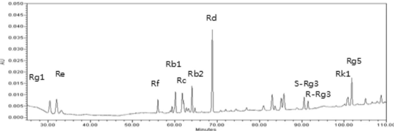
실험에 사용된 홍삼은 4년근을 우신산업 (충청남도, 금산, 대한민국)에서 구입하여 추출한 후, GS-E3D를 제조하기 위 하여 5 brix 홍삼 추출물에 중량대비 10%의 pectin lyase를 넣고 50℃에서 5일간 반응시킨 후 95℃에서 효소를 불활성화 한 다음 진공 건조하여 분말화하여 시료로 사용하였다. GS- E3D에 포함된 총 사포닌 (saponin) 함량은 120.2 ㎎/g이였고, ginsenoside 함량은 Fig. 1 및 Table 1에 정리하였다.
Figure 1. The HPLC chromatogram of GS-E3D with UV detection at 203 nm.
Ginsenoside Content Ginsenoside Content
Rg1 5.9 ㎎/g Rb3 2.5 ㎎/g
Re 12.6 ㎎/g Rd 27.7 ㎎/g
Rf 4.7 ㎎/g 20(S)-Rg3 1.3 ㎎/g
Rb1 30.2 ㎎/g 20(R)-Rg3 1.4 ㎎/g
Rc 14.0 ㎎/g Rk1 0.8 ㎎/g
Rb2 17.6 ㎎/g Rg5 1.5 ㎎/g
Table 1. Materials and Vouchers
2. 실험동물의 사육 및 실험군
6주령 웅성 db/db 및 db/+ 마우스를 중앙실험동물 (서울, 대한민국)에서 구입 하였으며 물과 고형사료를 충분히 공급하 여 1주일간 실험실 환경에 적응시킨 후 실험에 사용하였다.
군분리는 다음과 같이 무작위로 각군당 10마리씩 총 4군으로 나누었다. (1) 정상군 (db/+ 마우스, NOR), (2) 당뇨군 (db/db 마우스, DM), (3) GS-E3D 100 ㎎/㎏ 투여군 (db/db 마우스 + GS-E3D 100 ㎎/㎏, GS-E3D-100), (4) GS-E3D 250 ㎎/㎏ 투여군 (db/db 마우스 + GS-E3D 250
㎎/㎏, GS-E3D- 250). GS-E3D는 멸균 증류수에 용해시 켜 매일 1회씩 6주동안 경구투여 (5 ㎖/㎏) 하였으며, 정상군 및 당뇨군에는 동량의 증류수만 경구 투여하였다. 매주 1회씩 체중을 측정하였으며, 정기적으로 미정맥으로부터 채혈하여 혈당을 측정하였다.
3. 알부민뇨 측정
부검 하루 전에 대사케이지를 이용하여 24hr 뇨(urine)를 채취하였고 분순물을 제거하기 위해 spin-down시킨 뒤, 상 층액을 이용하여 실험을 수행하였다. Urine 100㎕를 96- well plate에 분주한 다음 coating을 위해서 37℃ 에서 2시간 배양하였다. 다음 PBS로 세척을 3번 수행하고 blocking을 위해 casein blocking buffer를 37℃에서 40분 정도 배양하 였고 세척한 다음 anti-albumin antibody (Santa Cruz, CA, USA)를 1:2000배 PBS에 희석하여 100㎕를 분주하여 37℃에서 2시간 반응 시켰다. 이후 각 well을 PBS로 세 번 세척한 뒤 각 해당하는 HRP conjugated second antibody (1:2000) 100㎕를 분주하여 반응시켰다. 반응 후 3번 세척한 뒤 TMB(3.3'.5.5'-tetramethylbenzidine)로 발색하고 반응은 0.5N H2SO4를 첨가하여 정지시킨 뒤 450㎚ 흡광도를 측정 한다.
4. 조직병리학적 검사
부검 시 장기를 적출하여 10% 중성화 포르말린에 하룻밤 고정한 후 탈수과정 후 xylene으로 3회 치환한 후 파라핀으로 포매하였다. 포매된 조직 블록을 4㎛ 두께로 연속 절편을 제 작하여 슬라이드에 올려 사용하였다. 형태학적 변화를 관찰하 기 위하여 Periodic Acid-Schiff (PAS) 염색을 실시하였다.
염색이 끝난 slide는 광학현미경 하에서 관찰하고, 사구체의 크기를 Image J 소프트웨어 (NIH, MD, USA)를 이용하여 분석하였다.
5. 면역조직화학 염색
파라핀 포매 조직을 5 ㎛ 두께로 박절하여 자일렌과 에탄 올에서 탈파라핀 한 후 10 mM citrate buffer (pH 6.0)에 담가 10분간 microwave로 처리한 다음 수세하여 비특이적 반응을 제거하기 위하여 blocking한 후, anti-synaptopodin antibody (Santa Cruz, CA, USA) 및 anti-AGEs antibody (Transgenic Inc., Kobe, Japan)를 1:1,000으로 희석하여
overnight 반응하였다. PBS로 3회 세척한 후 synaptopodin 의 검출을 위해서는 rhodamine labeled anti-rabbit IgG antibody를 반응 후 형광현미경 하에서 관찰하였으며, AGEs의 검출을 위해서는 HRP labeled streptoavidin biotin (LSAB) kit (Dako, USA)를 반응한 후 DAB로 발색하여 광학현미경 하에서 관찰하였다. 정량 분석을 위해 염색의 강도는 Image J 소프트웨어 (NIH, MD, USA)를 이용하여 분석하였다.
6. 통계처리
각 분석값은 평균 ± 분산으로 표시하였으며, 각 실험군 간 의 유의성 검증은 GraphPad Prism 4.0 software (Graph pad, CA, USA)를 사용하여 one-way analysis of variance (ANOVA) followed by Tukey’s multiple comparison test를 이용하여 p<0.05 수준에서 실시하였다.
Ⅲ. 결 과
1. 체중 및 혈당
6주간의 시험기간 동안 체중의 변화는 정상군에 비해 당뇨 마우스가 체중이 현저히 증가하였으나 (p<0.05), GS-E3D 투여에 의한 영향은 관찰되지 않았다. 혈당 변화 또한 당뇨군 은 440.8 ± 101.1 ㎎/dl 였으며, GS-E3D 투여 군에서는 100 ㎎/㎏ 농도에서 430.6 ± 75.5 ㎎/dl, 250 ㎎/㎏ 농도 에서 402.2 ± 130.0 ㎎/㎏으로 혈당에 미치는 영향도 관찰 되지 않았다.
Blood glucose
(㎎/dL) Body weight (g)
NOR 27.1±1.5 137.6±20.1
DM 47.9±4.1* 440.8±101.1*
GS-E3D-100 46.3±3.4 430.6±75.5
GS-E3D-250 44.1±.4.0 402.2±130.0
NOR, normal mice; DM, diabetic mice; GS-E3D-100, DM treated with GS-E3D (100 ㎎/㎏); GS-E3D-250, DM treated with GS-E3D (250 ㎎/
㎏). All data were expressed as mean ± SD (n=10). *P <0.05 vs. NOR group.
Table 2. Body weight and blood glucose levels.
2. 조직병리 분석
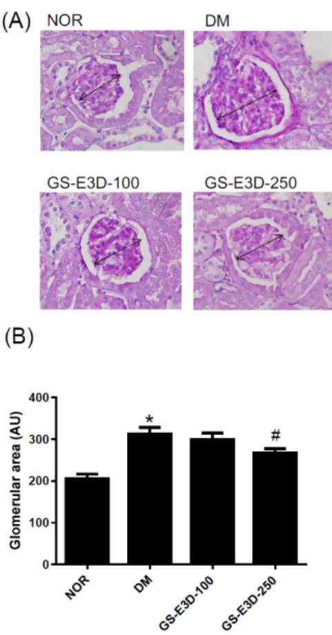
당뇨 마우스의 신장 사구체에서는 국소적인 사구체 기질의
확장 (mesangial matrix expansion) 및 tubulointerstitial
부위의 손상이 관찰되었다 (Fig. 2A). 또한 사구체의 크기도
정상 마우스에 비해 현저히 커져 있었다 (Fig. 2B). 그러나
GS-E3D 투여 마우스의 경우에는 사구체의 형태학적 변화가
관찰되지 않았으며, 사구체의 크기 또한 250 ㎎/㎏ 투여군에
서 당뇨군에 비해 유의적으로 감소해 있었다 (p<0.05).
Figure 2. Renal histopathology. (A) Periodic acid-Schiff staining of glomeruli. Arrows indicate glomeruli of various sizes. ×400 magnification. (B) Quantitative analysis of glomerular size. All data are expressed as the mean ± SD. (n=10). * P<0.05 vs.
NOR group; # P<0.05 vs. DM group.
3. 알부민뇨 분석
신기능을 평가하기 위해 대사케지를 이용하여 24시간 뇨를 이용하여 알부민 농도를 분석하였다. Fig. 3에서 보이는 바와 같이 정상군의 알부민뇨는 0.56 ± 0.29 ㎍/㎎ protein에 비해 당뇨군은 8.88 ± 3.68 ㎍/㎎ protein으로 10배 이상 유의 적으로 증가해 있었으며, GS-E3D 100 ㎎/㎏ 투여군은 5.62
± 4.02 ㎍/㎎ protein, 250 ㎎/㎏ 투여군은 4.17 ± 3.69
㎍/㎎ protein으로 농도 의존적으로 알부민뇨를 감소시키는 효능이 관찰되었다.
NOR
DM GS-E3D-100
GS-E3D-250
Figure 3. Renal function. Albuminuria were assayed using an ELISA. All data are expressed as the mean ± SD. (n=10). * P<0.05 vs. NOR group; # P<0.05 vs. DM group.
4. 족세포(podocyte)의 소실 분석
당뇨병성 신증은 족세포의 손상 및 소실을 특징으로 하며, 이러한 족세포의 소실정도를 분석하기 위해 족세포의 마커인 synaptopodin
28)을 이용하여 면역조직화학염색을 실시하였다.
Fig. 4에서 보이는 바와 같이 당뇨군에서는 정상군에 비해 족 세포의 소실에 의해 synaptopodin에 염색된 강도가 현저히 감소해 있었으며, GS-E3D 투여에 의해서 족세포 소실이 억 제되어 synaptopodin의 염색강도 또한 250 ㎎/㎏ 농도 투여 군에서 유의적으로 증가해 있었다 (p<0.05).
Figure 2. Renal histopathology. (A) Periodic acid-Schiff staining of glomeruli. Arrows indicate glomeruli of various sizes. ×400 magnification. (B) Quantitative analysis of glomerular size. All data are expressed as the mean ± SD. (n=10). * P<0.05 vs.
NOR group; # P<0.05 vs. DM group.
5. 신장 조직내 최종당화산물의 축적 분석
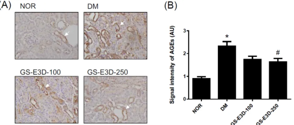
신장 조직내 최종당화산물의 축적 정도를 면역조직화학염 색을 통해 분석하였다. Fig. 5에서 보는 바와 같이 당뇨군에 서는 신장 조직내 최종당화산물이 과량으로 축적되어 염색된 강도가 증가해 있었으며, 특히 세뇨관 부위에 최종당화산물의 축적이 현저히 증가해 있었다. 그러나 GS-E3D 250 ㎎/㎏
투여군에서는 염색의 강도가 감소해 있었으며 (p<0.05), 이를
통해 GS-E3D가 최종당화산물의 축적을 억제하는 효능이 있
음을 확인할 수 있었다.
Figure 5. Effect of GS-E3D on AGE accumulation in the renal tissues. (A) Immunohistochemical staining of AGEs. Arrows indicate cells with positive staining for AGEs. X100 magnification. (B) Quantitative analysis of the AGE signals in histological sections. All data are expressed as the mean ± SD. (n=10). * P<0.05 vs. NOR group; # P<0.05 vs. DM group.
Ⅳ. 고 찰
당뇨병의 발병원인은 아직 정확하게 규명이 되어 있지는 않지만 현재까지 밝혀진 바에 의하면 유전적 요인 뿐만 아니라 식생활, 비만, 스트레스 및 운동 부족 등 후천적 환경 요인으 로도 영향을 받는 대표적인 대사성 이상 질환이다
29). 인슐린의 발견으로 당뇨병 환자의 수명은 극적으로 연장은 되었으나 아직 까지 현대 의학적으로 당뇨병 치료 방법은 혈당을 정상적인 수준으로 유지되도록 하는 것이 최선의 치료법으로 근본적인 치료방법은 아직 개발되지 못하고 있는 실정이며, 혈당 강하제 등으로 혈당을 정상 수치로 유지한다 하더라도 당뇨병성 신증과 같은 합병증까지 막는 데에는 한계가 있다
30, 31). 고혈당으로 인해 형성된 최종당화산물은 체내의 단백질과 비가역적인 교차 결합 (cross-link)을 형성하여 기능 이상을 초래하게 된다.
그러므로, 최종당화산물과 관련된 당뇨병성 신증을 예방하기 위해서는 최종당화산물의 생성 및 체내 축적을 억제해야 한다.
Aminoguanidine, LR-90과 같은 최종당화산물 억제제가 당뇨 모델동물에서 사구체 경화증 및 단백뇨를 억제하는 효능이 있음이 보고되었으며
16-18). 까마귀쪽나무 (
Litsea japonica)와 같이 한약재 유래 최종당화산물 억제 효능이 있는 물질이 2형 당뇨모델 동물에서 신장기능 저하를 억제하고 족세포의 손상을 억제하는 효능이 있는 것도 보고되었다
32). 본 실험에서는 2형 당뇨모델 동물인 db/db 마우스의 신장기능 저하 및 족세포의 손상을 확인하였으며, 최종당화산물의 신장 조직내 축적을 확 인하였다. GS-E3D 6주 투여에 의해 최종당화산물의 신장 조직내 축적을 억제하여 신장기능 회복 및 족세포 보호 효과 가 있는 것을 확인할 수 있었다.
인삼의 항당뇨 효능은 보고되었으며
3), 홍삼은 cyclosporine 으로 유도된 신장 손상을 억제하는 효과가 있었다
34). 특히 홍 삼의 유효성분 중 하나인 ginsenoside Rd의 경우 cephaloridine 으로 유도하는 급성 신부전 동물모델 및 ischemic-reperfused 신부전 동물 모델에서 산화스트레스를 억제하고 신장기능 저 하를 예방하는 효과를 보였다
35-37). 더욱이 Quan 등은 홍삼이 당뇨모델 동물의 신장 조직에서 염증을 억제하여 신장 손상을
예방하는 효과가 있다고 보고하였다
38). 이러한 선행 연구결과를 바탕으로 GS-E3D 또한 2형 당뇨모델 동물에서 신장보호 효 과가 있을 것으로 추정할 수 있었으며, GS-E3D는 홍삼을 pectin lyase로 생물전환하여 ginsenoside Rd의 함량을 강 화시킨 물질로 홍삼에 비해 더 우수한 신장 보호효과를 기대 할 수 있다. 이전 보고에 따르면, GS-E3D는 ginsenoside Rd 함량 증가를 통하여 고지방식이로 유도한 비만 동물모델에서 체중증가를 억제하고, 간조직내 지방축적을 억제하였으며, 혈 당을 낮추는 효과가 관찰되었다
39). 또한 최근에는 GS-E3D가 1형 당뇨모델동물에서 신장보호 효과가 있음이 보고되었다
40). 비록 본 연구에서 GS-E3D와 홍삼간에 직접적인 신장 보호 효능을 비교하지는 않았다. 하지만, Quan 등이 보고한 결과에 따르면 streptozotocin으로 유도한 당뇨동물에 4주간 홍삼 추출물을 투여한 결과 250 ㎎/㎏의 농도로 홍삼을 투여한 군 에서 신장기능을 개선하고 신장조직의 손상을 예방하였지만, 뇨로 배출되는 최종당화산물의 양을 감소시키지 못하였고, 신 장조직내에 최종당화산물의 한 종류인
N-(carboxymethyl) lysine의 축적을 유의적으로 억제하지는 못하였다
38). 그러나 최근에 보고된 연구결과에 따르면 GS-E3D는 최종당화산물 의 형성을 억제하며 (IC
50= 19.65 ± 4.35 ㎍/㎖), 홍삼의 최종당화산물 억제 효능 (IC
50= 139.46 ± 68.18 ㎍/㎖)에 비해 약 7배 가량 강한 억제 효능을 확인할 수 있었다
41). 본 연구결과에서도 GS-E3D는 신장 조직내 최종당화산물의 축 적을 현저히 억제하였으며, 이러한 결과는 GS-E3D는 홍삼 추출물과 달리 최종당화산물 억제 기전을 통해 신장보호 효과를 보이는 것을 알 수 있으며, GS-E3D가 홍삼보다 더 우수한 당뇨병성 신증 예방효과가 있을 것으로 추정할 수 있다.
Ⅴ. 결 론
2형당뇨 모델동물인 db/db 마우스에서 pectin lyase로 가
수분해하여 ginsenoside Rd 함량을 강화시킨 GS-E3D 추출
물의 신장 보호효능을 평가하였다.
1. GS-E3D 6주 투여에 의해 당뇨병에 의한 알부민뇨 증 가를 억제하였고, 신장 사구체 형태 변화 및 신장 족세 포의 소실을 억제하였으며,
2. 이러한 신장 보호 효과는 최종당화산물의 축적억제 기 전을 통해 나타나는 것을 확인하였다. 이상의 결과를 토 대로 GS-E3D를 이용한 당뇨병성 신증 억제제의 개발 가능성이 있을 것으로 사료된다.
감사의 글
본 연구는 농림축산식품부의 재원으로 농림수산식품기술기 획평가원의 수출전략기술개발사업 (315049-05-2-CG000)의 지원을 받아 연구되었으며, 본 연구의 일부는 산림청(한국임업 진흥원) 산림생명자원소재발굴연구사업 (2017040B00-1819- BA01)의 지원에 의하여 수행되었으며 이에 감사드립니다.
References