• 검색 결과가 없습니다.

Allium Hookeri Extract Enhances Glucose Uptake through GLUT4 Up-regulation in 3T3-L1 Cells

N/A
N/A
Protected

Academic year: 2021

Share "Allium Hookeri Extract Enhances Glucose Uptake through GLUT4 Up-regulation in 3T3-L1 Cells"

Copied!
6
0
0

로드 중.... (전체 텍스트 보기)

전체 글

(1)

Allium Hookeri Extract Enhances Glucose Uptake through GLUT4 Up-regulation in 3T3-L1 Cells

Young Eun Kang

, Kyeong-Mi Choi

, Eunjin Park, Won-Beom Jung, Heejin Jeong and Hwan-Soo Yoo*

College of Pharmacy, Chungbuk National University, Cheongju 28644, Korea

Received January 2, 2017 /Revised February 27, 2017 /Accepted March 23, 2017

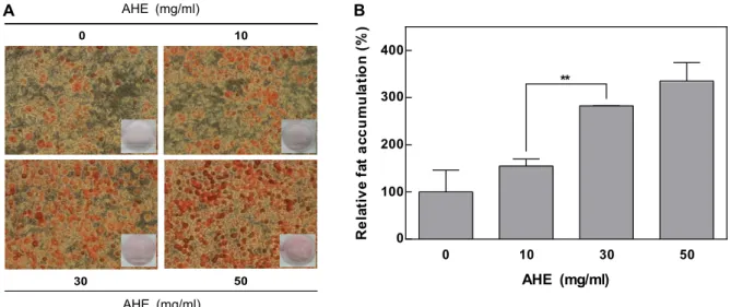
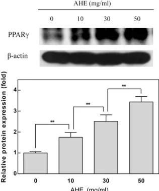
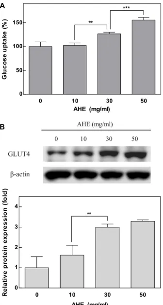
Diabetes mellitus is associated with insulin resistance, which leads to down-regulation of insulin sig- naling and the decreased glucose uptake. Adipocytes are sensitive to insulin, and closely implicated in insulin resistance and diabetes. Insulin stimulates differentiation of preadipocytes to adipocytes, and increases glucose transport. Allium species have been used as traditional medicine and health-pro- moting foods. Allium hookeri (A. hookeri) is reported to improve the pancreatic β-cell damage and ex- hibit pancreatic anti-inflammatory activity in streptozotocin-induced diabetic rats. We investigated whether A. hookeri extract (AHE) may stimulate glucose uptake in adipocytes through increasing in- sulin sensitivity. AHE enhanced fat accumulation, a differentiation biomarker, under the partial in- duction of differentiation by insulin. PPARγ, a transcription factor highly expressed in adipocytes, promotes adipocyte differentiation and insulin sensitivity. AHE increased the differentiation of pre- adipocytes through up-regulation of PPARγ. The activation of PPARγ increases the GLUT4 expression during adipocyte differentiation. GLUT4 is responsible for glucose uptake into the adipocytes. AHE increased the expression of GLUT4 in adipocytes, and subsequently enhanced the insulin-stimulated glucose uptake. These results suggest that AHE promotes adipocyte differentiation through activation of PPARγ, and leads to enhance glucose uptake in adipocytes along with GLUT4 up-regulation. Thus, AHE may be effective for the insulin-sensitizing and anti-diabetic activities.

Key words : Allium hookeri, adipocyte differentiation, glucose uptake, PPARγ, 3T3-L1 cells

Authors contributed equally.

*Corresponding author

*Tel : +82-43-261-3215, Fax : +82-43-268-2732

*E-mail : [email protected]

This is an Open-Access article distributed under the terms of the Creative Commons Attribution Non-Commercial License (http://creativecommons.org/licenses/by-nc/3.0) which permits unrestricted non-commercial use, distribution, and reproduction in any medium, provided the original work is properly cited.

Journal of Life Science 2017 Vol. 27. No. 3. 289~294 DOI : https://doi.org/10.5352/JLS.2017.27.3.289

Introduction

Adipocytes have been studied as a potential target for diabetes mellitus as well as obesity [9, 14]. Dysfunctional adipocytes induce insulin resistance and inflammation [2].

Insulin is an essential regulator for stimulating the adipo- cyte differentiation which is induced through transcription factors such as peroxisome proliferator-activated receptor γ (PPARγ) and CCAAT/enhancer-binding protein α [1, 5, 10].

PPARγ plays the major role in the adipogenic transcrip- tional cascade, and activates the expression of various genes involved in glucose and lipid metabolism [5, 13, 20]. PPARγ activates expression of glucose transporter 4 (GLUT4) dur- ing adipogenesis which is responsible for glucose uptake into adipocytes [13, 20].

Allium species have been used as traditional medicine and health-promoting foods [3, 4, 11]. Allium hookeri (A. hookeri), a member of Liliaceae family, is widely found in Sri Lanka, India, China and Bhutan, and has anti-oxidant and anti-in- flammatory activities [3, 7, 12]. Water extract of A. hookeri protected oxidative stress-mediated inflammatory responses and pancreatic β-cell damage in streptozotocin-induced dia- betic rats [12]. In addition, A. hookeri leaf or root decreased blood glucose level, and increased plasma insulin level in type 2 diabetic mice [8].

In this study, we investigated whether A. hookeri extract may modulate adipocyte differentiation under sub-optimal concentrations of insulin, and elucidated its mechanism for increasing insulin sensitivity.

Materials and Methods

Materials

3T3-L1 cells were purchased from the American Type

Culture Collection (ATCC; Manassas, VA, USA). Dulbecco’s

Modified Eagle’s Medium (DMEM), bovine calf serum (BCS)

and fetal bovine serum (FBS) were purchased from

Invitrogen (Carlsbad, CA, USA). Insulin and bovine serum

(2)

10 0

30 50

AHE (m /mL)

AHE (m /mL)

AHE (mg/ml)

AHE (mg/ml)

A

0 10 30 50

0 100 200 300 400

AHE (mg/mL)

R el ati v e fa t a cc u m ul a ti o n ( % )

**

B

AHE (mg/ml)

Fig. 1. Enhancing effect of AHE on adipocyte differentiation. Adipocyte differentiation was induced with a mixture of insulin (0.1 μg/ml), IBMX (0.5 mM) and dexamethasone (1 μM). 3T3-L1 cells were treated with AHE (0, 10, 30 or 50 mg/ml) every other day during the first 4 days of differentiation. (A) On day 6, cells were stained with Oil Red O, and visualized under a light microscope (×100). (B) Intensities of Oil Red O staining were quantified. Results are expressed as mean ± SD of triplicate experiments. Statistical significance: **p<0.01.

albumin (BSA) were obtained from Roche Diagnostics (Mannheim, Germany). Dexamethasone, 3-isobutyl-1-meth- ylxanthine (IBMX) and Oil Red O were purchased from Sigma Chemical Co. (St. Louis, MO, USA). 2-deoxy-D- [1-

3

H(N)] glucose was obtained from American Radiolabeled Chemicals, Inc. (Saint Louis, MO, USA). Antibodies against PPARγ, GLUT4 and β-actin were purchased from Santa Cruz Biotechnology, Inc. (Santa Cruz, CA, USA).

Preparation of AHE

All parts of A. hookeri were harvested before it was grow- ing 30 cm by height, and then dried. Ten kg of A. hookeri was extracted with 27 liters of hot water overnight. The AHE was sterilized with 0.2 μm Millipore filter kit (Merck Millipore; Darmstadt, Germany). The AHE at the concen- trations of 10~50 mg/ml was directly used for further study.

Cell culture and adipocyte differentiation 3T3-L1 preadipocytes were cultured and differentiated in- to adipocytes using a modified protocol [15]. Briefly, 3T3-L1 preadipocytes originating from Swiss mouse embryos were cultured in DMEM containing 10% BCS at 37°C in a 5%

CO

2

incubator. To induce differentiation, 2 day post- confluent preadipocytes were cultured for 2 days in differ- entiation medium containing 0.1 μg/ml insulin, 0.5 mM IBMX, 1 μM dexamethasone and 10% FBS. The medium was then changed to DMEM containing 0.1 μg/ml insulin

and 10% FBS, and cells were cultured for a further 2 days.

Subsequently, cells were cultured in DMEM supplemented with 10% FBS for an additional 2 days.

Oil Red O staining

After induction of adipocyte differentiation, cells were washed with phosphate-buffered saline (PBS), fixed at room temperature with 10% formalin for 1 hr, stained at room temperature with Oil Red O for 1 hr, and washed three times with distilled water. For quantitative analysis, Oil Red O dye was dissolved in isopropanol and the optical density was measured at 490 nm using a microplate reader (Molecular Devices, LLC; Sunnyvale, CA, USA).

Glucose uptake assay

Adipocytes were incubated in DMEM containing 0.2%

BSA for 4 hr and washed two times with Krebs-Ringer

Hepes buffer (KRH buffer; 136 mM NaCl, 4.7 mM KCl, 1.25

mM CaCl

2

, 1.25 mM MgSO

4

, 20 mM Hepes, pH 7.4). The

cells were incubated in KRH buffer with 10 ng/ml insulin

at 37°C for 15 min. Glucose uptake was initiated by addition

of 0.5 μCi/ml 2-deoxy-D-[1-

3

H(N)] glucose as the final con-

centration in KRH buffer. After 10 min, the cells were quick-

ly washed two times with ice-cold KRH buffer to terminate

the reaction. The cells were lysed with 0.5 N NaOH, and

the radioactivity was counted using a liquid scintillation

analyzer (PerkinElmer, Inc.; Waltham, MA, USA).

(3)

AHE (mg/ml)

Fig. 2. Increased effect of AHE on PPARγ expression in adipo- cytes. Adipocyte differentiation was induced with a mix- ture of insulin (0.1 μg/ml), IBMX (0.5 mM) and dex- amethasone (1 μM). 3T3-L1 cells were treated with AHE (0, 10, 30 or 50 mg/ml) every other day during the first 4 days of differentiation. On day 6, cells were harvested and lysates were subjected to Western blot analysis for PPARγ. Results are expressed as mean ± SD of triplicate experiments. Statistical significance: **p<0.01.

Western blot analysis

3T3-L1 cells were collected and lysed in a buffer contain- ing 62.5 mM Tris-HCl (pH 6.8), 2% SDS, 10% glycerol, 50 mM dithiothreitol, and a protease inhibitor cocktail tablet (Roche Diagnostics; Mannheim, Germany). The total protein concentration of the lysates was determined with the BCA Protein Assay Reagent (Pierce). Proteins in the lysates were separated on a 10% SDS polyacrylamide gel and transferred to a polyvinylidene difluoride membrane (GE Healthcare Life Sciences; Piscataway, NJ, USA). Membranes were blocked in 5% BSA overnight at 4°C and then incubated overnight at 4°C with the following primary antibodies:

PPARγ, GLUT4 and β-actin. Membranes were incubated with horseradish peroxidase-conjugated secondary anti- bodies overnight at 4°C. The bands were visualized with enhanced chemiluminescence (Amersham Pharmacia Bio- tech; Buckinghamshire, UK) and exposed to X-ray film (Eastman Kodak; Rochester, NY, USA).

Statistical analysis

All values are presented as mean ± standard deviation (SD). Statistical significance was determined by one-way analysis of variance with Newman-Keuls multiple compar- ison test. P<0.05 was considered statistically significant.

Results and Discussion

AHE enhanced adipocyte differentiation in 3T3-L1 cells

Insulin stimulates differentiation of preadipocytes to adi- pocytes, and increases glucose transport and lipid synthesis [1, 17]. Adipocytes are sensitive to insulin and implicated in insulin resistance [16]. In this study, we examined wheth- er AHE modulates the differentiation of 3T3-L1 preadipocytes.

Differentiation of preadipocytes was induced by 0.1 μg/ml insulin plus 0.5 mM IBMX and 1 μM dexamethasone, and the cells were simultaneously treated with AHE (0, 10, 30 or 50 mg/ml) during the first 4 days of differentiation. AHE promoted adipocyte differentiation under sub-optimal con- centrations of insulin (Fig. 1). The fat content of adipocytes treated with 10, 30 or 50 mg/ml AHE was increased by ap- proximately 0.6-, 1.8- or 2.4-fold, respectively, compared with controls. These results suggest that AHE may promote the adipocyte differentiation by increasing insulin sensitivity.

AHE increased PPARγ expression in adipocytes PPARγ is highly expressed in adipocytes, and promotes adipocyte differentiation and insulin sensitivity [5, 13, 18].

AHE treatment significantly enhanced PPARγ expression in adipocytes in a concentration-dependent manner (Fig. 2).

The expression of PPARγ in adipocytes treated with AHE at 10, 30 or 50 mg/ml was increased by approximately 0.7-, 1.5- or 2.4-fold, respectively, compared with control culture without AHE. These results suggest that AHE increases the differentiation of 3T3-L1 preadipocytes by activating PPARγ.

AHE enhanced insulin-stimulated glucose uptake in adipocytes

PPARγ increases the GLUT4 expression during adipocyte

differentiation which is followed by enhancing glucose up-

take [13, 20]. We examined whether AHE enhances the glu-

cose uptake in adipocytes consistent with the increased adi-

pocyte differentiation by AHE. The glucose uptake was

measured in adipocytes exposed to AHE during adipo-

(4)

AHE PPARγ

Adipocyte differentiation ▲

GLUT4 ▲

Glucose uptake ▲

Fig. 4. Proposed mechanism for enhancement of adipocyte dif- ferentiation by AHE. AHE promotes adipocyte differ- entiation through up-regulation of PPARγ, and enhances glucose uptake in adipocytes by an increasing expression of GLUT4 during adipogenesis. The symbol ▲ repre- sents up-regulation.

0 10 30 50

0 1 2 3 4

R e la ti ve p ro tei n exp ress io n (f o ld )

**

0 10 30 50

0 50 100 150

G luc o s e up ta ke ( % )

**

***

AHE (mg/ml)

AHE (mg/ml)

A

B

Fig. 3. Stimulatory effect of AHE on glucose uptake in adipo- cytes. Adipocyte differentiation was induced with a mix- ture of insulin (0.1 μg/ml), IBMX (0.5 mM) and dex- amethasone (1 μM). 3T3-L1 cells were treated with AHE (0, 10, 30 or 50 mg/ml) every other day during the first 4 days of differentiation. (A) Glucose uptake was per- formed in adipocytes on day 6. (B) The cells were har- vested on day 6 and lysates were subjected to Western blot analysis for GLUT4. Results are expressed as mean

± SD of triplicate experiments. Statistical significance:

**p<0.01, ***p<0.001.

genesis. AHE significantly enhanced insulin-stimulated glu- cose uptake in 3T3-L1 adipocytes (Fig. 3A). In particular, 50 mg/ml AHE increased the insulin-stimulated glucose up- take in adipocytes by approximately 56% compared with control culture without AHE. Similarly, A. hookeri signifi- cantly reduced blood glucose level, and increased plasma

insulin level in the type 2 diabetic mice [8]. GLUT4 is a major insulin-responsive glucose transporter, which is re- sponsible for glucose uptake into the adipocytes [1, 6, 19].

AHE significantly increased the GLUT4 expression in adipo- cytes (Fig. 3B). The expression of GLUT4 in adipocytes treat- ed with AHE at 10, 30 or 50 mg/ml was increased by ap- proximately 0.6-, 2.0- or 2.3 folds, respectively, compared with controls. These results indicate that AHE may enhance insulin-mediated glucose uptake in adipocytes by increasing GLUT4 expression during adipogenesis.

In conclusion, AHE promotes adipocyte differentiation through activation of PPARγ, and leads to enhance glucose uptake in adipocytes along with GLUT4 up-regulation (Fig.

4). Thus, AHE may be effective for the insulin-sensitizing and anti-diabetic activities.

Acknowledgments

This research was supported by the Ministry of Trade, Industry & Energy (MOTIE, 1415126993) through the foster- ing project of Osong Academy-Indudtry Convergence (BAIO) and the National Research Foundation of Korea [NRF] grant funded by the Korea government (MSIP) (No.

MRC, 2008-0062275) and the National Research Foundation

of Korea (NRF) grant funded by the Korea government

(MOE) (No. 2015R1D1A1A01060790) and the intramural re-

search grant of Chungbuk National University in 2014.

(5)

References

1. Arner, P. 2003. The adipocyte in insulin resistance: key mol- ecules and the impact of the thiazolidinediones. Trends Endocrinol. Metab. 14, 137-145.

2. Bays, H., Mandarino, L. and Defronzo, R. A. 2004. Role of the adipocyte, free fatty acids, and ectopic fat in patho- genesis of type 2 diabetes mellitus: peroxisomal pro- liferator-activated receptor agonists provide a rational ther- apeutic approach. J. Clin. Endocrinol. Metab. 89, 463-478.

3. Cho, H. S., Park, W., Hong, G. E., Kim, J. H., Ju, M. G.

and Lee, C. H. 2015. Antioxidant Activity of Allium hookeri Root Extract and Its Effect on Lipid Stability of Sulfur-fed Pork Patties. Kor. J. Food Sci. Anim. Resour. 35, 41-49.

4. Dziri, S., Hassen, I., Fatnassi, S., Mrabet, Y., Casabianca, H., Hanchi, B. and Hosni, K. 2012. Phenolic constituents, antioxidant and antimicrobial activities of rosy (Allium roseum var. odoratissimum). J. Funct. Food 4, 423-432.

5. Gregoire, F. M., Smas, C. M. and Sul, H. S. 1998. Under- standing adipocyte differentiation. Physiol. Rev. 78, 783-809.

6. Hunter, S. J. and Garvey, W. T. 1998. Insulin action and insulin resistance: diseases involving defects in insulin re- ceptors, signal transduction, and the glucose transport ef- fector system. Am. J. Med. 105, 331-345.

7. Kim, C. H., Lee, M. A., Kim, T. W., Jang, J. Y. and Kim, H. J. 2012. Anti-inflammatory Effect of Allium hookeri Root Methanol Extract in LPS-induced RAW264.7 Cells. J. Kor.

Soc. Food Sci. Nutr. 41, 1645-1648.

8. Kim, N. S., Choi, B. K., Lee, S. H., Jang, H. H., Kim, J.

B., Kim, H. R., Kim, D. K., Kim, Y. S., Yang, J. H., Kim, H. J. and Lee, S. H. 2015. Effects of Allium Hookeri on Glucose Metabolism in Type II Diabetic Mice. Kor. J. Phar- macogn. 46, 78-83.

9. Nawrocki, A. R. and Scherer, P. E. 2005. Keynote review:

the adipocyte as a drug discovery target. Drug Discov. Today 10, 1219-1230.

10. Ntambi, J. M. and Young-Cheul, K. 2000. Adipocyte differ- entiation and gene expression. J. Nutr. 130, 3122S-3126S.

11. Radovanović, B., Mladenović, J., Radovanović, A., Pavlović,

R. and Nikolić, V. 2015. Phenolic composition, antioxidant, antimicrobial and cytotoxic activites of allium porrum L.

(Serbia) extracts. J. Food Nutr. Res. 3, 564-569.

12. Roh, S. S., Kwon, O. J., Yang, J. H., Kim, Y. S., Lee, S.

H., Jin, J. S., Jeon, Y. D., Yokozawa, T. and Kim, H. J. 2016.

Allium hookeri root protects oxidative stress-induced in- flammatory responses and beta-cell damage in pancreas of streptozotocin-induced diabetic rats. BMC Complement. Altern.

Med. 16, 63.

13. Rosen, E. D. and Spiegelman, B. M. 2001. PPARgamma : a nuclear regulator of metabolism, differentiation, and cell growth. J. Biol. Chem. 276, 37731-37734.

14. Rosen, E. D. and Spiegelman, B. M. 2006. Adipocytes as reg- ulators of energy balance and glucose homeostasis. Nature 444, 847-853.

15. Shin, D. M., Choi, K. M., Lee, Y. S., Kim, W., Shin, K. O., Oh, S., Jung, J. C., Lee, M. K., Lee, Y. M., Hong, J. T., Yun, Y. P. and Yoo, H. S. 2014. Echinacea purpurea root extract enhances the adipocyte differentiation of 3T3-L1 cells. Arch. Pharm. Res. 37, 803-812.

16. Smith, U., Axelsen, M., Carvalho, E., Eliasson, B., Jansson, P. A. and Wesslau, C. 1999. Insulin signaling and action in fat cells: associations with insulin resistance and type 2 diabetes. Ann. N. Y. Acad. Sci. 892, 119-126.

17. Taha, C. and Klip, A. 1999. The insulin signaling pathway.

J. Membr. Biol. 169, 1-12.

18. Tontonoz, P., Hu, E. and Spiegelman, B. M. 1995. Regulation of adipocyte gene expression and differentiation by perox- isome proliferator activated receptor gamma. Curr. Opin.

Genet. Dev. 5, 571-576.

19. Watson, R. T., Kanzaki, M. and Pessin, J. E. 2004. Regulated membrane trafficking of the insulin-responsive glucose transporter 4 in adipocytes. Endocr. Rev. 25, 177-204.

20. Wu, Z., Xie, Y., Morrison, R. F., Bucher, N. L. and Farmer, S. R. 1998. PPARgamma induces the insulin-dependent glu- cose transporter GLUT4 in the absence of C/EBPalpha dur- ing the conversion of 3T3 fibroblasts into adipocytes. J. Clin.

Invest. 101, 22-32.

(6)

초록:GLUT4 상향조절을 통한 Allium hookeri 추출물의 3T3-L1 세포 내 포도당 흡수 증진 효과

강영은

․최경미

․박은진․정원범․정희진․유환수*

(충북대학교 약학대학)

삼채(Allium species)는 전통적인 약재나 건강 증진 식품으로 사용되어 왔다. 특히, Allium hookeri (A. hookeri)는 제 2형 당뇨병 모델 마우스에서 혈당 감소 효과가 보고되었다. 본 연구에서는 A. hookeri 추출물이 3T3-L1 세포에 서 인슐린 민감성을 증진시키는지 시험하였다. 3T3-L1 지방세포분화가 불완전하게 유도되는 저농도의 인슐린 조 건에서, A. hookeri 추출물은 세포 내 지방 함량을 증가시키고, 분화 유도 전사인자인 PPARγ의 발현을 상승시켰다.

또한, A. hookeri 추출물은 포도당 수송체 4(GLUT4)의 발현을 증가시킴으로써 세포 내 포도당 흡수(glucose up-

take)를 향상시켰다. 이러한 결과들은 A. hookeri 추출물이 인슐린 민감성을 증진시켜 PPARγ와 GLUT4를 활성화

하고, 세포 내 포도당 흡수를 촉진한다는 사실을 보여준다. 따라서, A. hookeri 추출물은 당뇨병의 예방 및 치료에

임상적으로 응용될 수 있을 것으로 생각된다.

수치

Fig.  1.  Enhancing  effect  of  AHE  on  adipocyte  differentiation.  Adipocyte  differentiation  was  induced  with  a  mixture  of  insulin  (0.1  μg/ml),  IBMX  (0.5  mM)  and  dexamethasone  (1  μM)
Fig.  2.  Increased  effect  of  AHE  on  PPARγ expression  in  adipo- adipo-cytes.  Adipocyte  differentiation  was  induced  with  a   mix-ture  of  insulin  (0.1  μg/ml),  IBMX  (0.5  mM)  and   dex-amethasone  (1  μM)
Fig.  3.  Stimulatory  effect  of  AHE  on  glucose  uptake  in  adipo- adipo-cytes.  Adipocyte  differentiation  was  induced  with  a   mix-ture  of  insulin  (0.1  μg/ml),  IBMX  (0.5  mM)  and   dex-amethasone  (1  μM)

참조

관련 문서

In this study, we found that Pin1 plays a pivotal role in insulin- induced AP-1 activation through its interaction with p70S6K and prolongs activity of

Glutamate excitotoxicity induced by excessive activation of NMDA receptor causes various damage to cells, which leads to cell death.. In previous studies, increased ROS

PI3K inhibition decreased antioxidants/GD-induced apoptosis in A549 cells, and PI3K inhibitor LY294002 had inhibitory effect on antioxidants/GD-induced caspase-3

Activation of Autophagy by Mangiferin Protects Auditory Hair Cells from Oxidative stress Induced Ototoxicity.. Gyeongmin

• Theory can extent to molten polymers and concentrated solutions.. The single-molecule bead spring models.. a)

Taken together, these results suggested that latex containing the ficin inhibited the cell growth and induced apoptosis by caspase and Bcl-2 family signaling pathway in

V notched failure induced by high in situ stress, ~400 m deep, Winnipeg, V notched failure induced by high in situ stress, 400 m deep, Winnipeg, Canada

In the present study, consistent to that report, up-regulation of VEGF mRNA was observed in the colon cancer cell with the acquired resistance to 5-FU and this