363
Spontaneous Pulmonary Hematoma with No Underlying Causes: A Case Report
Eun Joo Lee, M.D.
1, Sang Hoon Park, M.D.
1, Ho Hyun Park, M.D.
1, Seung Heon Park, M.D.
1, Jung Yeon Lee, M.D.
2, Woo Surng Lee, M.D., Ph.D.
3and Sun-Young Yoon, M.D., Ph.D.
41
Department of Internal Medicine,
2Division of Pulmonary and Critical Care Medicine, Department of Internal Medicine,
3
Department of Thoracic and Cardiovascular Surgery,
4Division of Allergy and Pulmonology, Department of Internal Medicine, Konkuk University Chungju Hospital, Chungju, Korea
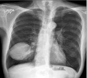
A 57-year-old male patient was admitted to our center because of a cystic mass on the lower portion of the right major fissure that was found incidentally by chest X-ray. He did not have a history of trauma or anticoagulant use. The lesion was removed by video-assisted thoracoscopic surgery. Pathological examination revealed an organizing pulmonary hematoma without any complications, and a follow-up chest X-ray after 1 year showed no recurrence.
Keyword: Hematoma
subclavian vein catheterization
3,4. One study reported a total of 38 cases of pulmonary hematoma, and all of these cases occurred after thoracic injury
5. To the best of our knowledge, no case of pulmonary hematoma has manifested without any underlying causes. Here, we report a case of spontaneous pulmonary hematoma where the patient did not have any identifiable underlying causes. Pulmonary hematoma was fi- nally diagnosed after histopathological review of the surgically removed mass.
Case Report
A 57-year-old male patient was admitted to our center because of a cystic mass in the right upper lung field that was discovered incidentally by chest X-ray during a routine health checkup. He was a current smoker with more than 30 pack years of cigarette exposure. He had been diagnosed with chronic obstructive pulmonary disease (COPD) 20 years previously and had been using an inhaled corticosteroid and bronchodilator. The patient has been working at the con- venience store, and had no history of trauma or use of any anticoagulants. He had no chest pain, fever, cough, or sputum.
There were no other episodes of hemoptysis. His physical ex- amination showed decreased lung sounds in both lung fields without wheezes or crackles. There was no palpable lymph node enlargement. Routine laboratory studies including a Copyright © 2015
The Korean Academy of Tuberculosis and Respiratory Diseases.
All rights reserved.
Introduction
Pulmonary hematomas are collections of blood within the alveolar and interstitial spaces
1. Usually, they are resolved within two to four weeks. However, if secondary infection accompanies a hematoma, it can progress into an abscess re- quiring drainage
2.
Non-penetrating injury of the thorax, by either direct blunt trauma or indirect forces, is generally known to be the ma- jor cause of pulmonary hematoma. There have been a few reports of spontaneous pulmonary hematoma as a com- plication of anticoagulant therapy or a rare complication of
CASE REPORT
http://dx.doi.org/10.4046/trd.2015.78.4.363ISSN: 1738-3536(Print)/2005-6184(Online) • Tuberc Respir Dis 2015;78:363-365
Address for correspondence: Sun-Young Yoon, M.D., Ph.D.
Division of Allergy and Pulmonology, Department of Internal Medicine, Konkuk University Chungju Hospital, 82 Gugwon-daero, Chungju 27376, Korea
Phone: 82-43-840-8691, Fax: 82-43-840-8961 E-mail: [email protected]
Received: Jun. 2, 2015 Revised: Jul. 1, 2015 Accepted: Jul. 6, 2015
cc