R E S E A R C H A R T I C L E Open Access
Clinical prognosis of isolated anterior cerebral artery territory infarction: a retrospective study
Hyungjong Park 1 , Young Seok Jeong 1 , Seo Hyeon Lee 1 , Seong Hwa Jang 1 , Doo Hyuk Kwon 1,2 , Jeong-Ho Hong 1 , Sung-Il Sohn 1 and Joonsang Yoo 1,3*
Abstract
Background: Isolated anterior cerebral artery territory (ACA) infarction is a rare phenomenon, and is known to have distinctive clinical features. Little is known regarding the clinical prognosis of isolated ACA territory infarction with associated factors, and its impact on dwelling and job status. We investigated the short- and long-term outcomes of anterior cerebral artery (ACA) territory infarction, and the associated factors involved in the development of the distinctive symptoms.
Methods: This retrospective study in a prospective cohort of acute ischaemic stroke patients included
consecutively enrolled patients with isolated ACA territory infarction. We investigated the functional status using the modified Rankin scale (mRS) score at discharge, three months ’ post-discharge, and one-year post-discharge. We also investigated the occlusion site of the ACA (proximal vs. distal); presence of distinctive symptoms of ACA territory infarction including behaviour changes, indifference, aphasia, and urinary incontinence; and the effect of these symptoms on dwelling and job status one year after discharge.
Results: Between April 2014 and March 2019, 47 patients with isolated ACA territory infarction were included.
Twenty-nine patients (61.7 %) had good outcomes (mRS ≤ 2) at discharge; however, the mRS score increased at three months (40; 85.1 %, p < 0.001) and one year (41; 87.2 %) post-discharge. Occlusion of the ACA proximal segment was independently associated with the development of distinctive symptoms (adjusted odds ratio, 17.68;
95 % confidence interval: 2.55 –122.56, p < 0.05). Twenty-one (48.8 %) patients with good outcomes at one year experienced a change in dwelling status and job loss; 20 (95.2 %) of them had distinctive ACA territory symptoms with proximal ACA occlusion.
Conclusions: Short- and long-term outcomes of isolated ACA territory infarction were favourable. However, proximal segment occlusion was associated with the development of distinctive symptoms, possibly related to future dwelling and job status.
Keywords: Anterior cerebral artery, Infarction, Dwelling status
© The Author(s). 2021 Open Access This article is licensed under a Creative Commons Attribution 4.0 International License, which permits use, sharing, adaptation, distribution and reproduction in any medium or format, as long as you give appropriate credit to the original author(s) and the source, provide a link to the Creative Commons licence, and indicate if changes were made. The images or other third party material in this article are included in the article's Creative Commons licence, unless indicated otherwise in a credit line to the material. If material is not included in the article's Creative Commons licence and your intended use is not permitted by statutory regulation or exceeds the permitted use, you will need to obtain permission directly from the copyright holder. To view a copy of this licence, visit http://creativecommons.org/licenses/by/4.0/.
The Creative Commons Public Domain Dedication waiver (http://creativecommons.org/publicdomain/zero/1.0/) applies to the data made available in this article, unless otherwise stated in a credit line to the data.
* Correspondence: [email protected]
1
Department of Neurology, School of Medicine, Keimyung University, Daegu, Korea
3
Department of Neurology, Yonsei University College of Medicine, Yongin Severance Hospital, 363 Dongbaekjukjeon-daero, Giheung-gu, 16995 Yongin, Korea
Full list of author information is available at the end of the article
Background
Isolated anterior cerebral artery (ACA) territory infarc- tion is a rare phenomenon [1], and accounts for only 0.5–3 % of all cerebral infarctions [2–4]. The relative rar- ity of isolated ACA territory infarction compared to middle cerebral artery (MCA) territory infarction may be related to the fact that the diameter of the A1 segment of the ACA is only approximately half that of the MCA; furthermore, the presence of the anterior communicating artery possibly plays an important role in emboli clearance [5].
Owing to their rarity, there have been few reports re- garding the natural prognosis of isolated ACA territory infarctions [2, 3, 6, 7]. Further, in most previous studies, ACA territory infarction was included in the group of hemispheric infarctions, despite its independent vascular topography [8]. ACA territory infarction also has diverse clinical features that are distinct from those of other cerebral vascular territory infarctions, which are not well defined by conventional functional outcome measures focusing on motor function [3, 4, 9, 10]. Therefore, nei- ther the natural prognosis of isolated ACA territory in- farction nor the influence of its distinctive symptoms on patients’ outcomes have been revealed.
Thus, we aimed to retrospectively investigate the fol- lowing: (1) the natural prognosis of patients with isolated ACA territory infarction in short (3-month) and long (1- year) time frames, (2) the factors associated with the de- velopment of distinctive symptoms in isolated ACA ter- ritory infarction, and (3) the influence of those symptoms of ACA territory infarction on dwelling and job status.
Materials and methods Study population
This study was a retrospective analysis of prospectively registered consecutive patients in a comprehensive stroke center in Korea. All patients experienced an is- chaemic stroke within seven days of symptom onset.
They were managed according to a standardised proto- col and care pathway, based on current guidelines. The inclusion criteria for this study were as follows: (1) iso- lated ACA territory infarction without involvement of any other cerebral vascular territory on magnetic reson- ance imaging (MRI); (2) modified Rankin Scale (mRS) ≤ 2 before the qualifying stroke; and (3) followed up at three months and one year after discharge.
During admission, brain MRI, computed tomography (CT) imaging, and cerebral angiography (magnetic res- onance angiography, CT angiography, or digital subtrac- tion angiography) were performed for all patients.
Systemic evaluations, including 12-lead electrocardiog- raphy, chest radiography, standard blood test, and lipid profile, were performed. For detecting the cardiac
embolic source, 24-h Holter or electrocardiographic monitoring was performed at the stroke unit. Echocardi- ography was performed for evaluating the presence of cardiac structural abnormalities or any evidence of car- diac thrombi. Based on these analyses, the stroke aeti- ology was retrospectively determined according to the Trial of ORG 10,172 in Acute Stroke Management (TOAST) [11] using neuroradiologists’ reports and the consensus of ≥ 2 stroke specialists in weekly stroke conferences.
Clinical variables and imaging analysis
Data on demographics and stroke risk factors including hypertension, diabetes, smoking status, hyperlipidaemia, and history of coronary artery disease or stroke were ob- tained. The severity of stroke was determined using the National Institutes of Health Stroke Scale (NIHSS) score at admission.
Involved vascular territory was determined using high signal intensities on diffusion-weighted images from brain MRIs taken during admission. The location of oc- clusion or stenosis was also determined by CT angiog- raphy or MR angiography. ACA territory was determined according to previous studies [5, 12] (shown in Fig. 1). If cerebral infarction was only involved in the ACA territory without involving any other cerebral vas- cular territories, the case fulfilled the enrolment criteria.
The ACA territory was defined as follows: (1) rostrum, genu, and splenium of the corpus callosum, cingulate gyrus, and frontal pole supplied by the frontopolar and orbitofrontal arteries; (2) medial aspect of the superior frontal gyrus supplied by the anterior and middle in- ternal frontal artery; (3) supplementary motor area sup- plied by the posterior internal frontal artery; and (4) paracentral lobule and cuneus supplied by the paracen- tral artery and superior parietal artery. To determine the differences depending on the location more clearly, we divided the ACA region into proximal and distal por- tions. The proximal portion covered the A1 (precommu- nicating) to A3 segments (around the genu of the corpus callosum genu), shown as a red line (shown in Fig. 1), and the distal portion covered A4 (terminal branch of A4) and its distal part. The ACA segment was defined according to the Fischer’s classification.
Outcome measurement
We obtained data regarding the mRS at discharge, three months after discharge, and one year after discharge.
The mRS at three months and one year were performed
at the outpatient clinic. If the patient missed a scheduled
visit, we obtained the information from them or their
guardians via telephonic interviews based on a struc-
tured questionnaire regarding mRS. Good outcome was
defined as mRS 0–2, which is considered to be function- ally independent, at three months and one year after dis- charge. In ACA territory infarction, the cerebral infarction is often not severe; thus, we also investigated the functional status of patients with mRS 0–1. In addition, we also checked other distinctive symptoms re- lated to ACA territory infarction, that could not be mea- sured using mRS [3, 4, 10]. These symptoms included altered consciousness; features of indifference including abulia, akinetic mutism, and apathy; aphasia; behavioral changes such as utilisation, perseveration, and social in- hibition; and urinary incontinence [3, 4]. We also inves- tigated the dwelling and job status at three months and one year after discharge.
Statistical analyses
Data are presented as the mean ± standard deviation, median (interquartile range), or a number (percentage), as appropriate. Differences according to the presence of distinctive symptoms were compared using the chi- square, Fisher’s exact, Student’s t-, and Mann-Whitney U tests, as appropriate. Temporal changes in mRS at dis- charge, three months after discharge, and one year after discharge were assessed using the chi-square test for trends. To determine the independently associated fac- tors for the development of distinctive symptoms in iso- lated ACA territory infarction, binary logistic regression analysis was performed, and the results are summarised as odds ratios and 95 % confidence intervals. For multi- variable analysis, variables with p < 0.1 on univariate ana- lyses were included in the multivariable binary logistic
regression analysis. All tests were two-sided, and p < 0.05 was considered statistically significant. The final model was fitted using the Hosmer–Lemeshow goodness-of-fit analysis and p > 0.05 was considered as the model fit to the binary logistic regression analysis. All statistical ana- lyses were performed using R software version 3.6.1 (R foundation for Statistical Computing, Vienna, Austria).
Results
Patients ’ characteristics
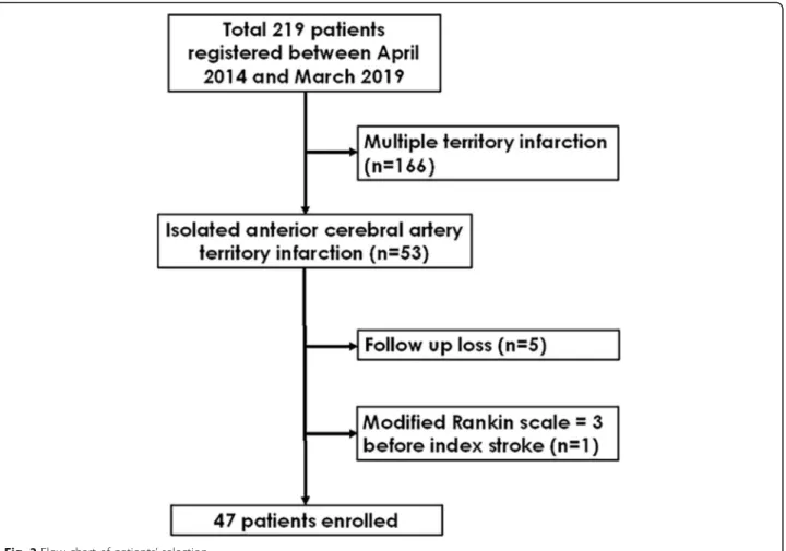
Between April 2014 and March 2019, a total of 219 pa- tients with cerebral infarction in the ACA territory were admitted in our hospital. Among them, 166 had infarc- tions in multiple cerebral artery territories. After exclud- ing those patients, 53 with isolated ACA infarction remained. Among these, five patients who were lost to follow-up and one with mRS 3 before onset of stroke were excluded. Finally, 47 patients who fulfilled the in- clusion criteria were included in this study (shown in Fig. 2). Among included patients, none underwent endo- vascular thrombectomy or intravenous tissue plasmino- gen activator administration. In the acute stage, 35 (74.5 %) and 14 (29.8 %) patients were treated with anti- platelet drugs and anti-coagulants, respectively. Brain protective drugs such as donepezil or choline alfoscerate were used in 13 (27.7 %) patients in the acute stage. In the chronic stage (3 months after stroke onset), new on- set atrial fibrillation was detected in 1 patient, and anti- platelet drugs were changed to anticoagulants. The mean age of the included patients was 71.7 ± 11.6 years, and 18 (38.3 %) were male; the initial NIHSS score was 2 (1–
Fig. 1 A schematic drawing of anterior cerebral artery (ACA) territory and branches of ACA
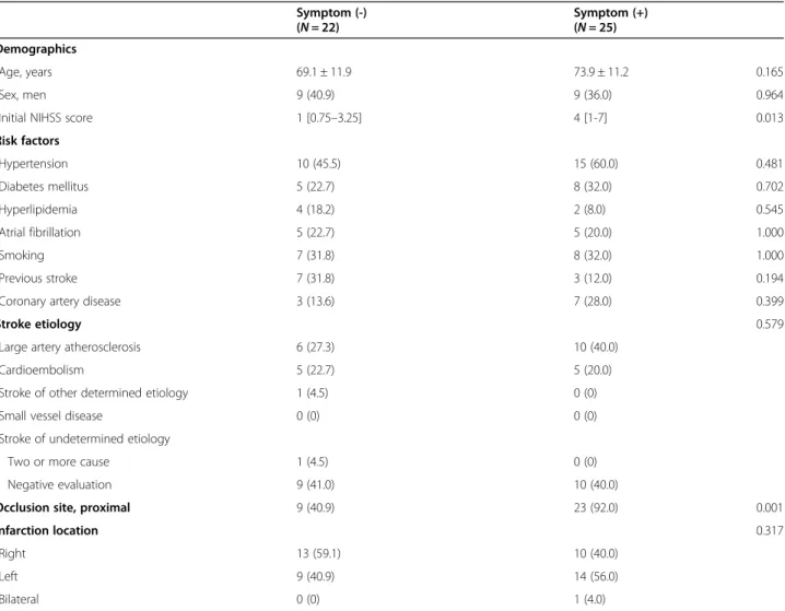
5). The most common symptom was weakness of the ex- tremities (33 patients, 70.2 %). The median NIHSS sub- score for motor function in the leg was 2.5, while the median NIHSS sub-score for motor function in the arm was 1. All patients underwent continuous electrocardio- graphic monitoring, and 39 patients (90.7 %) underwent echocardiography. The most common stroke aetiology was large artery atherosclerosis (n = 17, 36.1 %). Patients with distinctive symptoms of ACA infarction had higher initial NIHSS scores and more occlusion of the proximal ACA segment (Table 1).
Outcomes at discharge, three months after discharge, and one year after discharge
The median mRS at discharge was 2 (1–2); at that point, 29 (61.7 %) patients had good mRS. Short-term (three months) and long-term (one year) mRS after discharge were 1 (0–2) and 0 (0–1), respectively. The number of patients with good mRS increased to 40 (85.1 %) at three months (p < 0.001), and one patient (2.1 %) died within three months after discharge. Finally, 41 patients (87.2 %) achieved a good outcome at one year. Com- pared to mRS 0–1 at three months (68.1 %), the
proportion of mRS 0–1 was 76.6 % at one year; there were no additional deaths during this period. The tem- poral changes in mRS at discharge, three months after discharge (short-term), and one year after discharge (long-term) are shown in Fig. 3.
Associated factors for the development of distinctive symptoms in ACA territory infarction
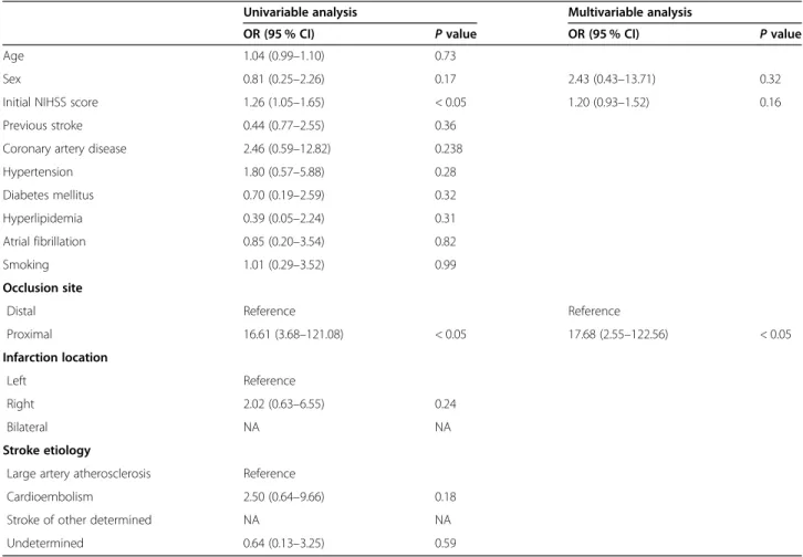
Among the 40 patients with a good outcome at 3 months, 25 (62.5 %) had distinctive symptoms with ACA territory infarctions. Among them, 16 had features of in- difference and 9 had behavioural changes. In addition, two patients had aphasia, and one suffered from urinary incontinence and voiding difficulty; 23 (92.0 %) had oc- clusion of the proximal ACA portion. No patient showed improvement of previously distinctive symptoms at three months or one year after discharge. The initial NIHSS score and proximal ACA segment occlusion were signifi- cantly associated in cases developing distinctive symp- toms with ACA territory infarction. On multivariable analysis, occlusion of the proximal ACA portion (odds ratio, 12.37; 95 % confidence interval, 2.63 –92.04; p <
0.05) was independently associated with the
Fig. 2 Flow chart of patients ’ selection
Table 1 Demographic features of patients with ACA territory infarction according to the presence of distinctive symptoms Symptom (-)
( N = 22) Symptom (+)
( N = 25) Demographics
Age, years 69.1 ± 11.9 73.9 ± 11.2 0.165
Sex, men 9 (40.9) 9 (36.0) 0.964
Initial NIHSS score 1 [0.75 –3.25] 4 [1-7] 0.013
Risk factors
Hypertension 10 (45.5) 15 (60.0) 0.481
Diabetes mellitus 5 (22.7) 8 (32.0) 0.702
Hyperlipidemia 4 (18.2) 2 (8.0) 0.545
Atrial fibrillation 5 (22.7) 5 (20.0) 1.000
Smoking 7 (31.8) 8 (32.0) 1.000
Previous stroke 7 (31.8) 3 (12.0) 0.194
Coronary artery disease 3 (13.6) 7 (28.0) 0.399
Stroke etiology 0.579
Large artery atherosclerosis 6 (27.3) 10 (40.0)
Cardioembolism 5 (22.7) 5 (20.0)
Stroke of other determined etiology 1 (4.5) 0 (0)
Small vessel disease 0 (0) 0 (0)
Stroke of undetermined etiology
Two or more cause 1 (4.5) 0 (0)
Negative evaluation 9 (41.0) 10 (40.0)
Occlusion site, proximal 9 (40.9) 23 (92.0) 0.001
Infarction location 0.317
Right 13 (59.1) 10 (40.0)
Left 9 (40.9) 14 (56.0)
Bilateral 0 (0) 1 (4.0)
Data are shown as n (%), mean ± standard deviation, or median [interquartile range]. ACA anterior cerebral artery; NIHSS National Institutes of Health Stroke Scale
Fig. 3 Modified Rankin scale at discharge, 3 months after discharge, and 1 year after discharge
development of distinctive symptoms in ACA territory infarction (Table 2).
Dwelling, job status, and distinctive symptoms in ACA territory infarction
Prior to the qualifying stroke, all patients enrolled in this study dwelled in their home with their family and 12 (25.5 %) had a job. Among the 41 (87.2 %) patients clas- sified as having good mRS at 1 year, 17 (36.2 %) moved into chronic care facilities and 4 (33.3 %) lost their jobs.
Among the 21 patients who moved their residence or lost their jobs, 20 (95.2 %) had distinctive symptoms of ACA territory infarction, and all of them had proximal ACA segment occlusion.
Discussion/Conclusion
This study showed that (1) short- and long-term prog- noses in isolated ACA territory infarction were good, (2) proximal ACA segment occlusion was an independently associated factor for developing distinctive symptoms in ACA territory infarction, and (3) the distinctive symp- toms related to proximal ACA segment occlusion
influenced dwelling and job status, regardless of positive mRS.
Short-term mortality after isolated ACA territory in- farction can range between 0 and 8 %, which is much lower than the 17.3 % short-term mortality after MCA territory infarction [3, 8, 13]. Consistent with previous studies, only 1 patient (2.1 %) died during the follow-up period in our study. Regarding functional outcomes, ap- proximately 70 % of patients achieved functional inde- pendence at 3 months in previous studies [13, 14]. In our study, 85.1 % of patients had a good outcome at 3 months with isolated ACA territory infarction. Thus, short-term outcomes after isolated ACA territory infarc- tion seemed to be favourable, and were line with previ- ous research. However, little is known regarding the long-term prognosis after isolated ACA territory infarc- tion. We investigated the long-term outcomes, with 87.2 % of patients showing good outcomes at one year after discharge. Thus, similar to the short-term out- comes, the long-term outcomes in isolated ACA terri- tory infarction also seem to be favourable.
The most common symptom in isolated ACA territory infarction was motor deficit, typically involving the lower
Table 2 Factors associated with distinctive symptoms in anterior cerebral artery territory infarction
Univariable analysis Multivariable analysis
OR (95 % CI) P value OR (95 % CI) P value
Age 1.04 (0.99 –1.10) 0.73
Sex 0.81 (0.25 –2.26) 0.17 2.43 (0.43 –13.71) 0.32
Initial NIHSS score 1.26 (1.05 –1.65) < 0.05 1.20 (0.93 –1.52) 0.16
Previous stroke 0.44 (0.77 –2.55) 0.36
Coronary artery disease 2.46 (0.59 –12.82) 0.238
Hypertension 1.80 (0.57 –5.88) 0.28
Diabetes mellitus 0.70 (0.19 –2.59) 0.32
Hyperlipidemia 0.39 (0.05 –2.24) 0.31
Atrial fibrillation 0.85 (0.20 –3.54) 0.82
Smoking 1.01 (0.29 –3.52) 0.99
Occlusion site
Distal Reference Reference
Proximal 16.61 (3.68 –121.08) < 0.05 17.68 (2.55 –122.56) < 0.05
Infarction location
Left Reference
Right 2.02 (0.63 –6.55) 0.24
Bilateral NA NA
Stroke etiology
Large artery atherosclerosis Reference
Cardioembolism 2.50 (0.64 –9.66) 0.18
Stroke of other determined NA NA
Undetermined 0.64 (0.13 –3.25) 0.59
OR odds ratio; CI confidential interval; NIHSS National Institutes of Health Stroke Scale; NA Not available
extremity contralateral to the infarction site [3, 4, 8]. As suggested in a previous report, pure motor stroke is the most frequent lacunar syndrome; it is known to involve an upper motor neuron lesion secondary to a lacunar in- farct in the ACA or MCA territory [15]. In our study, the median NIHSS sub-score for the leg was also higher than that for the arm [3]. Similar to previous studies, the symptoms in our patients also involved the lower ex- tremity more than the upper extremity. This dominance of the involvement of the lower extremity originates in the paracentral lobule located in the ACA territory. On the contrary, the cortical area for the hand and arm is located in the MCA territory; the corona radiata, where the projection fibres from the cortex merge, is also lo- cated in the MCA territory. Therefore, the development of hemiparesis is relatively uncommon in isolated ACA territory infarction, and preserving motor function in the arm may help achieve functional independence. In addition, motor function of the lower extremity usually recovers faster and more completely than that of the upper extremity [16]. This may be the reason why pa- tients with isolated ACA territory infarction showed favourable functional independence as measured by mRS.
Distinctive symptoms in ACA territory infarction in- clude altered mental status, abulia, mutism, decreased verbal fluency, aphasia, and urinary incontinence [3, 4].
Abulia and mutism are associated with cingulate gyrus and supplementary motor area involvement; these areas are important for human behaviour [17–19]. Aphasia is associated with involvement of the supplementary motor area located in the superior medial frontal lobe [20, 21]. Urinary incontinence suggests involvement of the superior frontal gyrus, cingulate, and large infarc- tion lesions affecting the superior and medial parts of the frontal lobe [22]. In terms of distinctive symptom development in ACA territory infarction, infarct size may be an influencing factor. Moreover, the structures associated with distinctive symptoms in the ACA terri- tory are located at least above the A3 segment, and were classified as the proximal portion in our study.
Thus, the risk of developing distinctive symptoms in the ACA territory seems to be high in cases of proximal ACA segment occlusion. As mentioned above, proximal ACA segment occlusion was an independent factor for the development of distinctive symptoms in ACA terri- tory infarction.
Furthermore, the presence of distinctive symptoms in ACA territory infarction was closely associated with dwelling and job status in patients with good mRS. Ac- cording to the mRS, patients with a slight disability who were able to look after their own affairs without assist- ance, but unable to perform all previous activities, were assigned a score of 2. However, if patients had mild
aphasia or abulia with a mild degree of motor deficit, they could still take care of their own affairs as motor function was relatively good. In such cases, their out- come may have been classified as good, even if the pa- tient’s family suffered from the after-effects of the distinctive symptoms of ACA territory infarction. Thus, the real prognosis after ACA territory infarction may not be correctly assessed by mRS alone; it may be wrong to conclude, based on mRS, that patients with isolated ACA territory infarction had favourable outcomes.
Currently, mechanical thrombectomy (MT) is recom- mended as primary treatment for MCA and carotid ar- tery occlusion [23, 24]. Owing to its rarity, there are few studies on MT in ACA occlusion; the average A2 and A3 diameters were > 2.0 mm [25]. Thus, considering the minimal stent retriever diameter (3.00 mm), the prox- imal ACA segment was suitable for MT [26]. In a retro- spective study in 30 patients, MT on ACA occlusions led to a good recanalization rate with few complications [27]. In addition, recent studies have shown that the presence of ACA occlusion by secondary embolism dur- ing MT for MCA without recanalization was associated with poor functional outcomes [28, 29]. Thus, if patients with proximal ACA segment occlusion arrived within the right therapeutic window of time, MT could be con- sidered because proximal segment occlusion was associ- ated with the development of distinctive symptoms in ACA territory infarction; this could affect future pa- tient’s true prognosis, which could not be assessed with mRS alone.
This study had several limitations. First, this was a retrospective single-center study with a small sample size. We did not use a validated questionnaire or exam- ination to check for distinctive symptoms in ACA terri- tory infarction. To circumvent these limitations, we plan to perform a prospective study with a validated ques- tionnaire for prognosis in ACA territory infarction.
Both short- and long-term prognoses based on mRS in isolated ACA territory infarction were favourable. How- ever, despite good mRS, proximal ACA segment occlu- sion was associated with the development of distinctive symptoms in ACA territory infarction; this could affect the patients’ dwelling and job status. Acute treatment such as endovascular thrombectomy or administration of intravenous tissue plasminogen activator may be beneficial to patients with proximal occlusion.
Abbreviations
ACA: anterior cerebral artery; CT: computed tomography; MCA: middle cerebral artery; MRI: magnetic resonance imaging; MT: mechanical thrombectomy; NIHSS: National Institutes of Health Stroke Scale; TOAST: Trial of ORG 10172 in Acute Stroke Management
Acknowledgements
None.
Authors' contributions
Conceptualization, H.P. and J.Y.; Methodology, Y.S.J. and S.H.L.; Formal analysis, H.P. and J.Y.; Investigation: H.P., Y.S.J., S.H.L., D.H.K., S.H.J., J-H.H., S-I.S., and J.Y.; Writing-original draft preparation, H.P. and J.Y.; Writing-review and editing: H.P., Y.S.J., S.H.L., D.H.K., S.H.J., J-H.H., S-I.S., and J.Y. ; Read and ap- proved the final manuscript, all authors.
Funding
This work was supported by the National Research Foundation of Korea grant funded by the Korean government (MSIT) (No.2020R1G1A007100).
Availability of data and materials
The datasets generated and/or analysed during the current study are not publicly available due them containing information that could compromise research participant privacy/consent but are available from the
corresponding author on reasonable request.
Declarations
Ethics approval and consent to participate
This study was approved by the institutional review board of Keimyung University Dongsan Hospital, which waived the requirement for informed consent from the patients due to the retrospective nature of this study. All methods were performed in accordance with relevant guidelines and regulations.
Consent for publication Not applicable.
Competing interests
The authors declare no conflict of interest.
Author details
1