Address for correspondence : Tae Sung Kim, M.D.
Department of Radiology, Samsung Medical Center, Sungkyunkwan University School of Medicine 50, Ilwon-dong, Gangnam-gu, Seoul 135-710, Korea Phone : 822-3410-2518 Fax : 822-3410-2559 E-mail : [email protected]
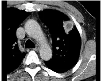
Figure 1. 50-year-old man with an early stage of pa
renchymal actinomycosis.
Delayed image of contrast-enhanced CT scan shows an irregularly marginated, peripheral pulmonary nodule in the left upper lobe. Note a central low-attenuation area suggestive of central necrosis. Localized pleural thickening is also noted in the adjacent pleura.
인 방선균 (Actinomyces israelii) 에 의한 만성적 화 농성 폐 감염이다. 이 세균은 그람 양성이고 혐기성인 부패균 (saprophytic organism) 으로서 구강내에서 발견된다1. 방선균증은 구강내 위생 상태가 좋지않은 사람에서 구인두내의 내인성 균주의 흡인에 의해 발 생한다. 폐 방선균증의 임상 증상은 객담성 기침, 미 열, 혈흔을 동반한 가래이다. 폐 감염을 확인하고 적 절한 항생제 치료를 시행하는 경우 대부분 예후는 좋 다. 지금까지 폐 실질에 발생하는 방선균증과2,3, 이물 질4 혹은 기관지결석과 동반된 기관지내 방선균증의 CT 소견을 기술한 몇 편의 논문들이 있다5,6. 본 종설 에서는 문헌 고찰을 통해 이들 질환의 CT 소견들을 정리하고, 전형적인 증례 영상들을 제시하고자 한다.
폐 실질 방선균증
천 등에 의하면3 폐 실질 방선균증은 CT상 특징적 으로 만성적인 분절성 폐 경화 (segmental airspace consolidation) 병변으로 나타나는데, 내부에 괴사성 저 음영 부분과 조영 증강되는 주변 부분으로 이루어 져 있고, 인접한 늑막에 비후를 동반한다. 감염 초기 에는 경계가 불분명한 작은 폐 결절로 시작하여 (Figure 1), 경과가 진행하면서 천천히 크기가 증가하
보이는 괴사성 저 음영 부분과 빈번한 공동화는 폐 방선균증의 전형적인 CT 소견이다.
부수적인 소견으로서, 폐문과 종격동의 임파절 비 후가 동반되고, 폐 경화 내부에 기관지 확장증을 볼 수 있으며, 부분적인 늑막 비후, 늑막 삼출을 동반하 기도 한다 (Figure 3). 때때로, 엽간 열을 통과하여 (transfissural extension) 인접한 폐엽을 침범하거나 (Figure 4), 늑막과 흉벽에까지 병변이 파급될 수도 있다.
병리조직 소견상, CT 에서 보이는 중심성 저 음영 부분은 미세농양이거나 혹은 확장된 기관지내에 충 만되어 있는 괴사성 물질이고, 조영 증강되는 주변부
Figure 4. 52-year-old woman with parenchymal acti
nomycosis manifesting as chronic necrotizing pne
umonia with transfissural extension.
A, Lung window image of chest CT scan shows a segmental consolidation in the lingular division of the left upper lobe. Note transfissural extension of the necrotic consolidation into the adjacent left lower lobe across the major interlobar fissure (arrow). B, Lung window image obtained at a lower level to A shows secondary involvement of parenchymal actinomycosis in the adjacent left lower lobe (arrow).
Figure 2. 48-year-old man with parenchymal actin
omycosis manifesting as a necrotic peripheral mass.
Delayed image of contrast-enhanced CT scan shows an irregularly marginated, subpleural mass with a necrotic low-attenuation area and a cavity (arrow) in the left upper lobe. Actinomycosis was diagnosed using percutaneous transthoracic core biopsy.
Figure 3. 57-year-old man with parenchymal actin
omycosis manifesting as a segmental consolidation.
Delayed image of contrast-enhanced CT scan shows a segmental consolidation in the right middle lobe.
Note an eccentric area of necrotic low attenuation (arrow). A small-sized pleural effusion is also noted.
Figure 5. 55-year-old woman with both parenchymal and endobronchial actinomycosis associated with bronchi
ectasis and broncholithiasis.
A, Contrast-enhanced CT scan shows complete atelectasis of the right middle lobe. Diffuse tubular bronchiectasis of the medial segmental bronchus (arrowheads) is seen, and a broncholith (arrow) is noted within the lateral segmental bronchus, which is also dilated with mucoid impaction. B, Photograph of specimen obtained from right middle lobectomy shows diffuse tubular (arrowheads) and cystic bronchiectasis (arrows) with bronchial wall thickening with parenchymal inflammation and fibrosis. C, Photomicrograph of histopathologic specimen shows acute suppurative inflammation containing an Actinomyces colony (sulfur granule) in the background of numerous neutrophils (H and E, x200).
경화 부분은 다양한 정도의 섬유화를 동반한 만성 염 증 조직이다. 조직 검체 내에서 무수한 방선균들의 집 락체인 황 과립 (sulfur granules; 군집된 균사체로 이 루어진 덩어리로서 황색을 띔.) 을 확인하거나 (Figure 5), Gomori methenamine silver 염색에 양성인 미세실 구조의 병원체를 찾으면 확진할 수 있다7.
방선균증의 감별 진단으로서 괴사성 폐암, 폐결핵, 반침습성 폐 아스페르길루스증, 아급성 괴사성 세균 성 폐렴 등이 포함된다.
기관지 확장증 형태의 방선균증
방선균은 사멸된 조직에 집락하는 경향이 있다5. 이전의 결핵 감염이나 다른 세균성 감염에 의해 파괴 된 폐 실질에 이차적으로 방선균이 감염되어 폐실질 파괴와 기관지 확장증을 더욱 심화시킬 수 있다. 방선 균에 의한 기관지 감염은 기존의 기관지 염증과 기관 지 확장증, 기관지주위 폐실질의 소실을 더욱 악화시 킨다. 이의 CT소견은 국소적인 기관지 확장증, 불규 칙한 기관지벽 비후, 농양 형성을 동반할 수 있는 기 관지주위 폐 경화이다 (Figure 5).
기관지 결석증과 동반된 기관지내 방선균증
기관지 결석증 혹은 기관지내 이물질과 동반된 기 관지내 방선균증에 관한 몇 편의 드문 증례 보고가 알려져 있다4,6,8. 기관지 결석증과 동반된 기관지내 방 선균증은 구인두로부터 흡인된 방선균이 기존에 존 재하던 기관지내 결석에 이차적으로 집락하여 발생 하는 것으로 여겨진다5. 기관지 결석의 이차 감염으로 인해 기도 폐색이 진행하고, 결국 원위부 폐에 후폐쇄 성 폐렴이 발생한다. 방선균이 기관지내 결석으로부 터 점차 떨어져 나와 염증반응을 초래하여, 원위부 폐 경화를 악화시키고, 폐 농양을 형성하게 된다.
기관지 결석증과 동반된 기관지내 방선균증의 CT 소견은 근위부 기관지내에 위치한 석회화된 결석과 원위부 폐엽 혹은 폐분절의 후폐쇄성 경화이다6 (Figures 6 and 7). 원위부 폐엽의 경화는 내부에 중 심성 저 음영 부분을 보이며, 공동화가 동반되기도 한 다. 폐문 혹은 종격동 임파절도 흔히 비후된다. 대부 분의 경우, 과거에 폐 결핵을 앓았던 흔적들 (석회화 된 폐문 종격동 임파절, 석회화된 폐결절들) 을 볼 수 있다. 그러므로 결핵 유병률이 높은 지역에서 흉부
Figure 6. 38-year-old man with endobronchial actinomycosis associated with broncholithiasis.
A, Contrast-enhanced CT shows a small broncholith obstructing the bronchial lumen of a basal segment of the right lower lobe (arrow). B, More distally, a necrotic subsegmental consolidation is seen with bronchial dilatation containing mucoid material and another broncholith (arrow). C, Photomicrograph of histopathologic specimen obtained from the endobronchial calcified nodule shows numerous filamentous structures representing Actin
omyces covering the broncholith (Gomori methenamine silver stain, x200).
Figure 7. 46-year-old man with endobronchial actin
omycosis associated with broncholithiasis.
Contrast-enhanced CT scan shows a proximal end
obronchial calcified nodule (a broncholith) with com
plete distal atelectasis of the right middle lobe. End
obronchial actinomycosis was diagnosed by broncho
scopic biopsy.
CT상 기관지 결석이 보일 때는 기관지 결석증과 동 반된 기관지내 방선균증의 가능성을 생각해야 한다.
병리조직 소견상, 기관지 결석 표면과 주위에 가지 치는 미세실 형태의 수많은 세균들의 집락체인 ,황 과
립들을 포함하는 화농성 조직을 확인할 수 있다. CT 상 보이는 원위부 폐렴성 경화 부분은 병리학적으로 는 농양 형성을 동반한 급성 화농성 염증 혹은 다양 한 정도의 섬유화를 동반한 기질화 폐렴으로 이루어 져 있다.
결론적으로, 폐 실질 방선균증의 전형적인 CT 소 견은 괴사성 저음영 부분을 포함한 만성적인 폐 분절 성 경화이다. 드물게 방선균 감염은 기관지 결석증과 동반된 기관지내 방선균증의 형태로 발현할 수 있으 며, 이 경우에는 CT상 근위부 기관지내 석회화 결석 과 이에 의한 기도 폐색 및 원위부 폐 경화로 보인다.
참 고 문 헌
1. Suzuki JB, Delisle AL. Pulmonary actinomycosis of periodontal origin. J Periodontol 1984;55:581-4.
2. Kwong JS, Muller NL, Godwin JD, Aberle D, Gry
maloski MR. Thoracic actinomycosis: CT findings in eight patients. Radiology 1992;183:189-92.
3. Cheon JE, Im JG, Kim MY, Lee JS, Choi GM, Yeon KM. Thoracic actinomycosis: CT findings. Radiology 1998;209:229-33.
4. Chouabe S, Perdu D, Deslee G, Milosevic D, Marque E, Lebargy F. Endobronchial actinomycosis associated with foreign body: four cases and a review of the literature. Chest 2002;121:2069-72.
5. Hirschfield LS, Graver LM, Isenberg HD. Broncholi